. 2024 Dec 31;26(1):282.
doi: 10.3390/ijms26010282.

Evaluation of the Anti-Amyloid and Anti-Inflammatory Properties of a Novel Vanadium(IV)-Curcumin Complex in Lipopolysaccharides-Stimulated Primary Rat Neuron-Microglia Mixed Cultures

Affiliations

Evaluation of the Anti-Amyloid and Anti-Inflammatory Properties of a Novel Vanadium(IV)-Curcumin Complex in Lipopolysaccharides-Stimulated Primary Rat Neuron-Microglia Mixed Cultures

Georgios Katsipis et al. Int J Mol Sci. .

Abstract

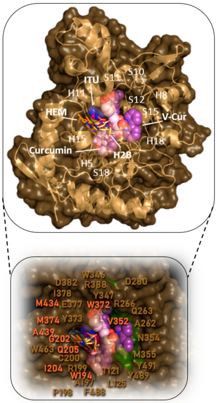
Lipopolysaccharides (LPS) are bacterial mediators of neuroinflammation that have been detected in close association with pathological protein aggregations of Alzheimer's disease. LPS induce the release of cytokines by microglia and mediate the upregulation of inducible nitric oxide synthase (iNOS)-a mechanism also associated with amyloidosis. Curcumin is a recognized natural medicine but has extremely low bioavailability. V-Cur, a novel hemocompatible Vanadium(IV)-curcumin complex with higher solubility and bioactivity than curcumin, is studied here. Co-cultures consisting of rat primary neurons and microglia were treated with LPS and/or curcumin or V-Cur. V-Cur disrupted LPS-induced overexpression of amyloid precursor protein (APP) and the in vitro aggregation of human insulin (HI), more effectively than curcumin. Cell stimulation with LPS also increased full-length, inactive, and total iNOS levels, and the inflammation markers IL-1β and TNF-α. Both curcumin and V-Cur alleviated these effects, with V-Cur reducing iNOS levels more than curcumin. Complementary insights into possible bioactivity mechanisms of both curcumin and V-Cur were provided by In silico molecular docking calculations on Aβ1-42, APP, Aβ fibrils, HI, and iNOS. This study renders curcumin-based compounds a promising anti-inflammatory intervention that may be proven a strong tool in the effort to mitigate neurodegenerative disease pathology and neuroinflammatory conditions.

Keywords: amyloid precursor protein; curcumin; lipopolysaccharides; mixed neuron-glia cultures; neuroinflammation; vanadium-curcumin complex.


Conflict of interest statement

The authors have no competing interests to declare that are relevant to the content of this article.

Figures

Figure 1
Figure 1
Levels of amyloid precursor protein (APP) in mixed cultures of primary neuron-microglia, in the absence or presence of 0.1, 1, or 10 μg/mL of LPS (a,b). Effect of LPS (1 μg/mL) in the presence or absence of 2 μΜ curcumin or V-Cur complex on APP levels (c,d). Analysis performed with Western blotting. Glyceraldehyde-3-phosphate dehydrogenase (GAPDH) was used to verify equal loading. The density of the blots was semi-quantified with ImageJ 1.54. Results are presented as fractional changes in comparison with the control sample and are the mean (±SD) of three independent biological experiments. One-way ANOVA was employed to compare untreated or LPS-treated samples. Statistical significance when compared with: * untreated sample (control); # LPS-treated sample; $ curcumin-treated: * p < 0.05; ** p < 0.01; *** p < 0.001; **** p < 0.0001; #### p < 0.0001; $$$$ p < 0.0001.
Figure 2
Figure 2
Docking poses orientation of curcumin and V-Cur in the crystal structure of the Kunitz protease inhibitor domain (APPI) of APP (PDB accession number 1AAP). The target protein is illustrated as a semi-transparent cartoon and surface colored in yellow orange and chocolate (chains A and B, respectively), while curcumin and V-Cur molecules are rendered in ball-and-stick mode and colored according to atom type in light pink and violet purple C atoms, respectively. The ligand binding site of both molecules depicting the architecture of the binding interactions is also illustrated (in the upper part) with an additional depiction of selected contacting amino acid residues of the binding pocket rendered in line and colored according to the cartoon. Binding interactions are illustrated in light pink (for curcumin) and violet (for V-Cur). Heteroatom color code: V: grey, N: blue, and O: red. Molecular docking simulations of both ligands were performed individually. Hydrogen atoms are omitted for clarity. The final structure was ray-traced and illustrated with the aid of PyMol Molecular Graphics.
Figure 3
Figure 3
In vitro fibrillation assay with insulin in the presence of several concentrations (0–100 μΜ) of either curcumin (■) or V-Cur (●). The insulin amyloid fibers formed in the absence or presence of either curcumin or V-Cur were semi-quantified by employing Thioflavin T fluorescence, with excitation at 450 nm and recording the emission spectrum at 490 nm. The results from three independent experiments are provided as mean normalized fibrillization rates (±SEM), setting the value of the control sample as 100%. Some error bars are not visible due to very small values (<1%).
Figure 4
Figure 4
Docking pose orientation of curcumin and V-Cur in the crystal structure of dimer (PDB ID 1GUJ) and hexamer (PDB ID 6GNQ) HΙ target proteins. In the hexameric structure of HI are also illustrated the six chain-stabilizing Zn2+ ions, the co-crystallized meta-cresol (CRS, depicted in gold sticks), and some critical to self-assembly and aggregation resides of HI (represented in stick mode colored in orange). Both HI proteins’ structures are depicted as cartoon colored in wheat and firebrick for A and B chains, respectively. Curcumin and V-Cur are rendered in sphere representation colored according to atom type in light pink and violet purple, respectively. The two Zn ions co-crystallized in the hexameric structure are depicted in sphere representation in lemon color and are shown to be connected with polar contact with Nε2 of His10 in the three double chains (A and B). Heteroatom color code: V: grey, N: blue, and O: red. Molecular docking simulations of both ligands were performed individually. Hydrogen atoms are omitted for clarity. The final structure was ray-traced and illustrated with the aid of PyMol Molecular Graphics.
Figure 5
Figure 5
Levels of (a) active inducible NO synthase (iNOS) (100 kDa), (b) inactive iNOS (50 and 75 kDa), and (c) total iNOS levels, after 24 h of treatment with LPS 1 μg/mL, in the presence or absence of 2 μΜ of curcumin or V-Cur complex, in mixed cultures of primary neurons-microglia. iNOS levels were determined by Western blotting (d). Glyceraldehyde-3-phosphate dehydrogenase (GAPDH) was employed to verify equal loading. The density of the blots was semi-quantified with ImageJ 1.54. Results are presented as fractional changes in comparison with the control sample and are the mean (±SD) of three independent biological experiments. One-way ANOVA was employed to compare untreated or LPS-treated samples. Statistical significance when compared with: * untreated sample (control); # LPS-treated sample; $ curcumin-treated: * p < 0.05; ** p < 0.01; *** p < 0.001; **** p < 0.0001; # p < 0.05; ## p < 0.01; ### p < 0.001; #### p < 0.0001; $ p < 0.05; $$ p < 0.01.
Figure 6
Figure 6
Docking pose orientation of curcumin and V-Cur in the crystal structure of iNOS monomer enzyme (PDB accession number 4NOS). The target protein is illustrated as cartoon colored in the sand along with a semi-transparent surface colored in the dark sand. Curcumin and V-Cur molecules, as well as the co-crystallized iNOS inhibitor ethylisothiourea (ITU) are rendered in sphere mode and colored according to atom type in light pink, violet purple, and hot pink C atoms, respectively. The co-crystallized molecules heme (HEM) (iron protoporphyrin IX) and H2B superimposed with the docked molecules are rendered in stick representation and colored according to atom type in orange and yellow-orange C atoms, respectively. H4B, essential for the dimerization of the protein, is not shown since it is located farther down the binding cavity, near the dimerization interface. The target protein structure model in the lower panel, depicting in a close-up view of the binding cavity of the target enzyme the architecture of the binding interactions, is illustrated as a semi-transparent surface colored in dark sand with an additional depiction of selected contacting amino acid residues of the binding pocket highlighted in the molecular surface in smudge green (for V-Cur) and white (for curcumin). Binding interaction residues are labeled in white (for curcumin) and smudge green (for V-Cur). Heteroatom color code: V: grey, N: blue, and O: red. Molecular docking simulations of both ligands were performed individually. Hydrogen atoms are omitted for clarity. The final structure was ray-traced and illustrated with the aid of PyMol Molecular Graphics.
Figure 7
Figure 7
Levels of (a) tumor necrosis factor-α (TNF-α), and (b) interleukin-1β (IL-1β), in mixed cultures of primary neurons-microglia after treatment with LPS (1 μg/mL) in the presence or absence of 2 μΜ curcumin or V-Cur. Cytokine levels were determined with Western blotting (c,d). Glyceraldehyde-3-phosphate dehydrogenase (GAPDH) was employed to verify equal loading. The density of the blots was semi-quantified with ImageJ 1.54. Results are presented as fractional changes in comparison with the control sample and are the mean (± SD) of three independent biological experiments. One-way ANOVA was employed to compare untreated or LPS-treated samples. Statistical significance when compared with: * untreated sample (control); # LPS-treated sample; $ curcumin-treated: ** p < 0.01; *** p < 0.001; **** p < 0.0001; # p < 0.05; #### p < 0.0001; $$ p < 0.01.

Similar articles

Cited by

References

    1. Alzheimer’s Association 2022 Alzheimer’s Disease Facts and Figures. Alzheimer’s Dement. 2022;18:700–789. doi: 10.1002/alz.12638. - DOI - PubMed
    1. Serrano-Pozo A., Frosch M.P., Masliah E., Hyman B.T. Neuropathological Alterations in Alzheimer Disease. Cold Spring Harb. Perspect. Med. 2011;1:a006189. doi: 10.1101/cshperspect.a006189. - DOI - PMC - PubMed
    1. Chen G.-F., Xu T.-H., Yan Y., Zhou Y.-R., Jiang Y., Melcher K., Xu H.E. Amyloid Beta: Structure, Biology and Structure-Based Therapeutic Development. Acta Pharmacol. Sin. 2017;38:1205–1235. doi: 10.1038/aps.2017.28. - DOI - PMC - PubMed
    1. Hardy J., Selkoe D.J. The Amyloid Hypothesis of Alzheimer’s Disease: Progress and Problems on the Road to Therapeutics. Science. 2002;297:353–356. doi: 10.1126/science.1072994. - DOI - PubMed
    1. Haass C., Selkoe D. If Amyloid Drives Alzheimer Disease, Why Have Anti-Amyloid Therapies Not yet Slowed Cognitive Decline? PLoS Biol. 2022;20:e3001694. doi: 10.1371/journal.pbio.3001694. - DOI - PMC - PubMed

MeSH terms

LinkOut - more resources