

3D Scanning of Surgical Specimens to Improve Communication Between Surgeon and Pathologist: A Head and Neck Pilot Study
- PMID: 39796645
- PMCID: PMC11718930
- DOI: 10.3390/cancers17010014
3D Scanning of Surgical Specimens to Improve Communication Between Surgeon and Pathologist: A Head and Neck Pilot Study
Abstract
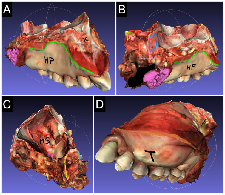
Background/Objectives Successful surgical outcomes in head and neck cancer depend on the accurate identification of resection margins. Effective communication between surgeons and pathologists is critical, but is often jeopardised by challenges in sampling and orienting anatomically complex specimens. This pilot study aims to evaluate the use of 3D scanning of surgical specimens as a tool to improve communication and optimise the pathology sampling process. Methods Two structured light 3D scanners, Cronos Dual and Optor Lab, were used to acquire 3D models of anatomical specimens in both preclinical (cadaver specimens) and clinical contexts (fresh surgical specimens). Surgical margins and critical points were annotated on the digital models. Acquisition quality, operating times and subjective feedback from surgeons and pathologists were evaluated. Results The Optor Lab scanner demonstrated superior image quality, shorter processing times and a more user-friendly interface than the Cronos Dual. Key challenges identified included specimen geometry, surface reflectivity and tissue stability. Feedback from both surgeons and pathologists was positive, highlighting the potential of 3D models to improve the surgical-pathology workflow. Conclusions 3D scanning of surgical specimens provides accurate, detailed digital models that can significantly enhance communication between surgeons and pathologists. This technology shows promise in improving pathological staging and clinical decision making, with further studies required to validate its integration into routine practice.
Keywords: 3D optical scanners; head and neck surgery; inter-specialist communication; surgical specimens.
Conflict of interest statement
The authors have no relevant financial or non-financial interests to disclose.
Figures


Similar articles
-
The computer-aided design margin: Ex vivo 3D specimen mapping to improve communication between surgeons and pathologists.Head Neck. 2023 Jan;45(1):22-31. doi: 10.1002/hed.27201. Epub 2022 Sep 26. Head Neck. 2023. PMID: 36156327 Free PMC article.
-
Visual pathology reports for communication of final margin status in laryngeal cancer surgery.J Pathol Inform. 2024 Oct 28;15:100404. doi: 10.1016/j.jpi.2024.100404. eCollection 2024 Dec. J Pathol Inform. 2024. PMID: 39640916 Free PMC article.
-
Virtual Resection Specimen Interaction Using Augmented Reality Holograms to Guide Margin Communication and Flap Sizing.Otolaryngol Head Neck Surg. 2023 Oct;169(4):1083-1085. doi: 10.1002/ohn.325. Epub 2023 Mar 19. Otolaryngol Head Neck Surg. 2023. PMID: 36934457
-
Frozen Section Analysis in Head and Neck Surgical Pathology: A Narrative Review of the Past, Present, and Future of Intraoperative Pathologic Consultation.Oral Oncol. 2023 Aug;143:106445. doi: 10.1016/j.oraloncology.2023.106445. Epub 2023 Jun 6. Oral Oncol. 2023. PMID: 37285683 Review.
-
Macroscopy of specimens from the head and neck.J Clin Pathol. 2024 Feb 19;77(3):185-189. doi: 10.1136/jcp-2023-208834. J Clin Pathol. 2024. PMID: 38373780 Review.
References
LinkOut - more resources
Full Text Sources
Miscellaneous
