

Prospects for Narrow Band Imaging Magnification Endoscopy in Oral Lesions: Recommendations from Oral and Maxillofacial Surgeons and a Gastroenterologist
- PMID: 39796652
- PMCID: PMC11718781
- DOI: 10.3390/cancers17010021
Prospects for Narrow Band Imaging Magnification Endoscopy in Oral Lesions: Recommendations from Oral and Maxillofacial Surgeons and a Gastroenterologist
Abstract
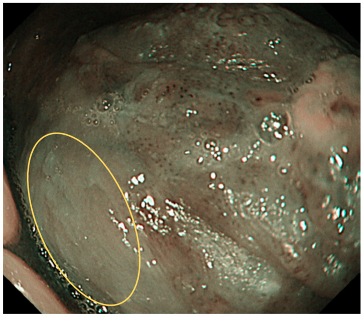
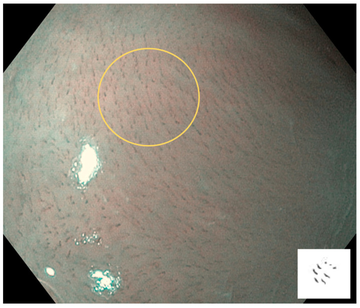
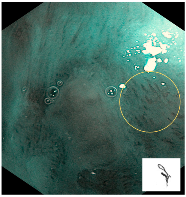
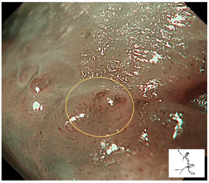
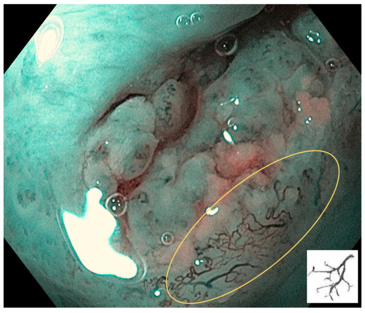
Narrow band imaging (NBI) magnification endoscopy for the diagnosis of early-stage oral cavity-related cancer and precancerous lesions can recognize oral lesions as brownish areas, and can observe intraepithelial papillary capillary loops (IPCLs) in the mucosa and submucosa to make a qualitative diagnosis of the lesion and highlight the mucosal surface microstructure to facilitate appropriate diagnosis and early treatment. IPCLs are classified from Type 0 to IV: Type 0 is normal mucosa or no blood vessels observed, e.g., in keratinization; Type I is mainly normal mucosa; Type II is mainly inflammatory sites or non-malignant lesions; Type III is mainly precancerous or suspected malignant lesions; and Type IV is cancerous or malignant lesions. NBI magnification endoscopy is a useful noninvasive method for identifying the malignant transformation of oral potentially malignant disorders (OPMDs). Oral lesions classified as IPCL Type II or higher are atypical epithelial or oral squamous cell carcinoma (OSCC); oral biopsy is recommended for early and accurate diagnosis, and is an indicator of the appropriate biopsy site in the follow-up for OPMDs. In the future, the accuracy of NBI magnification endoscopy for malignant transformation of OPMDs and OSCC will be further confirmed.
Keywords: intraepithelial papillary capillary loops; magnification endoscopy; narrow band imaging; oral potentially malignant disorders; oral squamous cell carcinoma.
Conflict of interest statement
The authors declare no conflicts of interest.
Figures
References
-
- Muto M., Minashi K., Yano T., Saito Y., Oda I., Nonaka S., Omori T., Sugiura H., Goda K., Kaise M., et al. Early detection of superficial squamous cell carcinoma in the head and neck region and esophagus by narrow band imaging: A multicenter randomized controlled trial. J. Clin. Oncol. 2010;28:1566–1572. doi: 10.1200/JCO.2009.25.4680. - DOI - PMC - PubMed
Publication types
LinkOut - more resources
Full Text Sources
