Review
. 2024 Dec 29;17(1):69.
doi: 10.3390/cancers17010069.

A Systematic Review of the Applications of Deep Learning for the Interpretation of Positron Emission Tomography Images of Patients with Lymphoma

Affiliations
Review

A Systematic Review of the Applications of Deep Learning for the Interpretation of Positron Emission Tomography Images of Patients with Lymphoma

Theofilos Kanavos et al. Cancers (Basel). .

Abstract

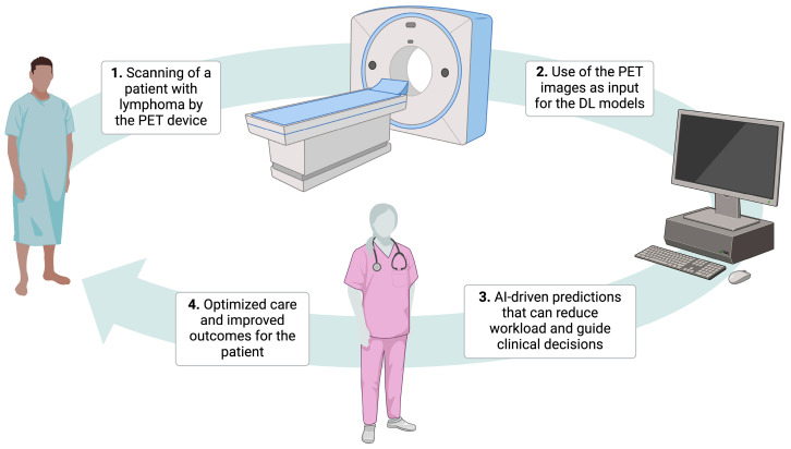
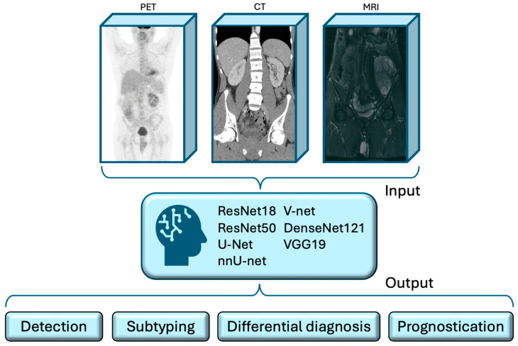
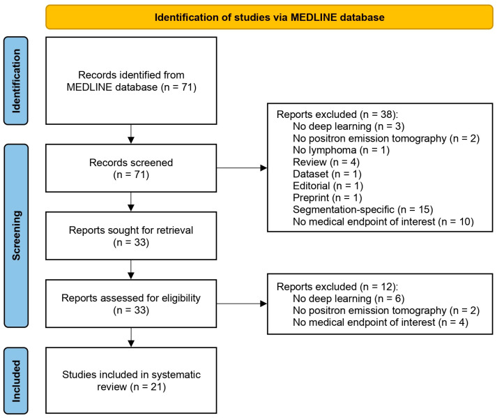
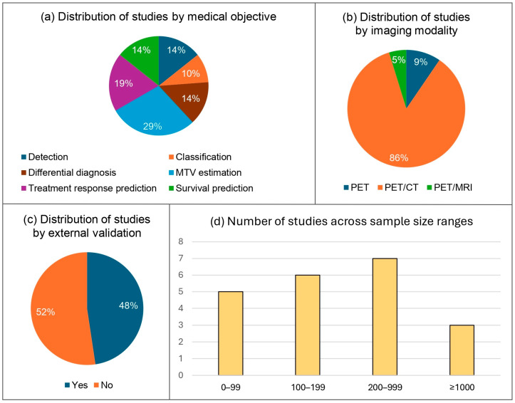
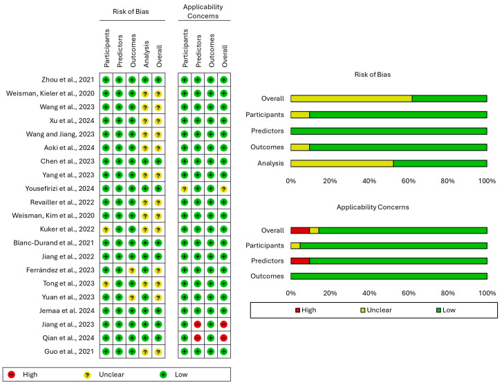
Background: Positron emission tomography (PET) is a valuable tool for the assessment of lymphoma, while artificial intelligence (AI) holds promise as a reliable resource for the analysis of medical images. In this context, we systematically reviewed the applications of deep learning (DL) for the interpretation of lymphoma PET images. Methods: We searched PubMed until 11 September 2024 for studies developing DL models for the evaluation of PET images of patients with lymphoma. The risk of bias and applicability concerns were assessed using the prediction model risk of bias assessment tool (PROBAST). The articles included were categorized and presented based on the task performed by the proposed models. Our study was registered with the international prospective register of systematic reviews, PROSPERO, as CRD42024600026. Results: From 71 papers initially retrieved, 21 studies with a total of 9402 participants were ultimately included in our review. The proposed models achieved a promising performance in diverse medical tasks, namely, the detection and histological classification of lesions, the differential diagnosis of lymphoma from other conditions, the quantification of metabolic tumor volume, and the prediction of treatment response and survival with areas under the curve, F1-scores, and R2 values of up to 0.963, 87.49%, and 0.94, respectively. Discussion: The primary limitations of several studies were the small number of participants and the absence of external validation. In conclusion, the interpretation of lymphoma PET images can reliably be aided by DL models, which are not designed to replace physicians but to assist them in managing large volumes of scans through rapid and accurate calculations, alleviate their workload, and provide them with decision support tools for precise care and improved outcomes.

Keywords: AI; CNN; DLBCL; PET; artificial intelligence; convolutional neural network; deep learning; lymphoma; machine learning; positron emission tomography.


Conflict of interest statement

The authors declare that there are no conflicts of interest.

Figures

Figure 1
Figure 1
The role of artificial intelligence in optimizing the management of patients with lymphoma using positron emission tomography images without replacing the human touch. Abbreviations: AI, artificial intelligence; DL, deep learning; PET, positron emission tomography. Created using BioRender. Kanavos, T. (2024) https://BioRender.com/w36u293, accessed on 23 December 2024.
Figure 2
Figure 2
Artificial intelligence-assisted workflow for the analysis of lymphoma medical images. Deep learning models can use positron emission tomography images with or without computed tomography or magnetic resonance imaging scans as inputs to deliver valuable clinical outputs. Abbreviations: CT, computed tomography; MRI, magnetic resonance imaging; PET, positron emission tomography.
Figure 3
Figure 3
Flow diagram presenting the study selection process along with the number of included and excluded papers.
Figure 4
Figure 4
Graphs illustrating the proportion of studies focusing on each different medical task (a), the proportion of studies that used each imaging modality (b), the proportion of studies that conducted external validation versus those that did not (c), and the distribution of studies by sample size (d). Abbreviations: CT, computed tomography; MRI, magnetic resonance imaging; MTV, metabolic tumor volume; PET, positron emission tomography.
Figure 5
Figure 5
Results of the evaluation of the risk of bias and concerns regarding applicability using the prediction model risk of bias assessment tool (PROBAST).

Similar articles

References

    1. Mugnaini E.N., Ghosh N. Lymphoma. Prim. Care. 2016;43:661–675. doi: 10.1016/j.pop.2016.07.012. - DOI - PubMed
    1. Munir F., Hardit V., Sheikh I.N., AlQahtani S., He J., Cuglievan B., Hosing C., Tewari P., Khazal S. Classical Hodgkin Lymphoma: From Past to Future-A Comprehensive Review of Pathophysiology and Therapeutic Advances. Int. J. Mol. Sci. 2023;24:10095. doi: 10.3390/ijms241210095. - DOI - PMC - PubMed
    1. Heneghan M.B., Belsky J.A., Milgrom S.A., Forlenza C.J. The Pediatric Approach to Hodgkin Lymphoma. Semin. Hematol. 2024;61:245–252. doi: 10.1053/j.seminhematol.2024.05.003. - DOI - PubMed
    1. Xu C., Feng J., Yue Y., Cheng W., He D., Qi S., Zhang G. A Hybrid Few-Shot Multiple-Instance Learning Model Predicting the Aggressiveness of Lymphoma in PET/CT Images. Comput. Methods Programs Biomed. 2024;243:107872. doi: 10.1016/j.cmpb.2023.107872. - DOI - PubMed
    1. Xu-Monette Z.Y., Young K.H. Therapeutic Vaccines for Aggressive B-Cell Lymphoma. Leuk. Lymphoma. 2020;61:3038–3051. doi: 10.1080/10428194.2020.1805113. - DOI - PMC - PubMed

LinkOut - more resources