

Second Versus First Molar Extractions in Class II Division 1 Malocclusion Treatment: A Retrospective Longitudinal Outcome Study into Maxillary Canine, Premolar, and Molar Movement
- PMID: 39797316
- PMCID: PMC11721531
- DOI: 10.3390/jcm14010225
Second Versus First Molar Extractions in Class II Division 1 Malocclusion Treatment: A Retrospective Longitudinal Outcome Study into Maxillary Canine, Premolar, and Molar Movement
Abstract
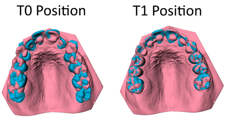
Background/objectives: This retrospective longitudinal outcome study comparing orthodontic extraction modalities, including extraction of maxillary first or second molars, aimed to compare the three-dimensional tooth movement of maxillary canines (C), premolars (P1, P2), and molars (M1, M2) in Class II division 1 malocclusion treatment with fixed appliances. Methods: A sample of 98 patients (mean age 13.20 ± 1.46 years) was selected for the M1 group, and 64 patients (mean age 13.20 ± 1.36 years) were chosen for the M2 group. Tooth movement was analyzed three-dimensionally on pre-treatment (T0) and post-treatment (T1) digital dental casts. Regression analyses compared the tooth movements (in mm) between the M1 and M2 groups. Results: The mean treatment duration for the M1 group was 2.51 ± 0.55 year, while, for the M2 group, it was 1.53 ± 0.37 year. The data showed limited distal movements of the C, P1, and P2 of approximately 2 mm in the M1 group and 1 mm in the M2 group during orthodontic treatment, but the M1 group exhibited significantly more distal movements than the M2 group (mean difference 1.11 to 1.24 mm). Vertical movements of the C, P1, and P2 in both groups were also minor (0.16 to 1.26 mm). The differences between groups did not exceed 0.2 mm and were not significant. Both treatment modalities resulted in a significant degree of anchorage loss with a distinct mesialization (8.40 ± 1.66 mm) of M2 in the M1 group and limited distalization (0.83 ± 0.98 mm) of M1 in the M2 group. Conclusions: The findings highlight the importance of thorough case evaluation when choosing between extraction modalities in Class II treatment. If a large distal movement of canines and premolars is required, additional anchorage mechanics should be considered.
Keywords: 3-D imaging; longitudinal studies; malocclusion Angle Class II; maxillary first molar extraction; maxillary second molar extraction; orthodontics; treatment outcome.
Conflict of interest statement
The author A.M.K.-J. is a member of the Section Board of JCM Section Stomatology. Author R.B. is a developer for 3DMedX®, a commercially available software package.
Figures


Similar articles
-
Three-dimensional dental model analysis of treatment outcomes for protrusive maxillary dentition: comparison of headgear, miniscrew, and miniplate skeletal anchorage.Am J Orthod Dentofacial Orthop. 2008 Nov;134(5):636-45. doi: 10.1016/j.ajodo.2007.05.017. Am J Orthod Dentofacial Orthop. 2008. PMID: 18984395
-
Class II malocclusion treatment changes with the Jones jig, Distal jet and First Class appliances.J Appl Oral Sci. 2020;28:e20190364. doi: 10.1590/1678-7757-2019-0364. Epub 2020 Apr 27. J Appl Oral Sci. 2020. PMID: 32348442 Free PMC article.
-
Comparative treatment outcomes after bilateral extractions of maxillary second molars or first premolars in patients with class II malocclusion: a retrospective study.Head Face Med. 2023 Mar 7;19(1):5. doi: 10.1186/s13005-023-00353-6. Head Face Med. 2023. PMID: 36882841 Free PMC article.
-
Extraction of maxillary first molars improves second and third molar inclinations in Class II Division 1 malocclusion.Am J Orthod Dentofacial Orthop. 2011 Sep;140(3):377-82. doi: 10.1016/j.ajodo.2010.06.026. Am J Orthod Dentofacial Orthop. 2011. PMID: 21889082
-
Treatment effects of intraoral appliances with conventional anchorage designs for non-compliance maxillary molar distalization: a literature review.Eur J Orthod. 2008 Dec;30(6):558-71. doi: 10.1093/ejo/cjn047. Epub 2008 Sep 27. Eur J Orthod. 2008. PMID: 18820306 Review.
References
-
- Proffit W.R., Fields H.W., Jr., Larson B. Contemporary Orthodontics. 6th ed. Elsevier; Philadelphia, PA, USA: 2018. pp. 140–202.
-
- Trevisi H., Trevisi Zanelato R. State-of-the-Art Orthodontics, Self-Ligation Appliances, Miniscrews and Second Molar Extractions. Elsevier; Philadelphia, PA, USA: 2018. pp. 156–223.
LinkOut - more resources
Full Text Sources
