Interstitial Lung Disease Associated with Anti-Ku Antibodies: A Case Series of 19 Patients
- PMID: 39797328
- PMCID: PMC11721168
- DOI: 10.3390/jcm14010247
Interstitial Lung Disease Associated with Anti-Ku Antibodies: A Case Series of 19 Patients
Abstract
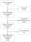
Background: Antibodies against Ku have been described in patients with various connective tissue diseases. The objective of this study was to describe the clinical, functional, and imaging characteristics of interstitial lung disease in patients with anti-Ku antibodies. Methods: This single-center, retrospective observational study was conducted at a tertiary referral institution. Patients with positive anti-Ku antibodies and interstitial lung disease identified between 2007 and 2022 were included. Clinical, immunological, functional, and imaging data were systematically reviewed. Results: Nineteen patients (ten females) with a mean age of 59 ± 12.6 years were included. The most frequent associated diagnosis was systemic sclerosis (42%), followed by rheumatoid arthritis (26%), Sjögren syndrome, undifferentiated connective tissue disease, and overlap between systemic sclerosis and idiopathic inflammatory myopathy (scleromyositis). Imaging revealed frequent septal and intralobular reticulations and ground-glass opacities, with nonspecific interstitial pneumonia as the predominant pattern (53%). The mean forced vital capacity was 82% ± 26 of the predicted value, and the mean diffusing capacity for carbon monoxide was 55% ± 21. Over the first year of follow-up, the mean annual forced vital capacity decline was 140 mL/year (range: 0-1610 mL/year). The overall survival rate was 82% at 5 years and 67% at 10 years. Conclusions: Most patients with interstitial lung disease and anti-Ku antibodies presented with dyspnea, a mild-to-moderate restrictive ventilatory pattern, and reduced diffusing capacity for carbon monoxide. The CT pattern was heterogeneous but was consistent with nonspecific interstitial pneumonia in half of the patients.
Keywords: autoimmunity; connective tissue disease; pulmonary fibrosis.
Conflict of interest statement
The authors declare no conflicts of interest.
Figures


References
-
- Joy G.M., Arbiv O.A., Wong C.K., Lok S.D., Adderley N.A., Dobosz K.M., Johannson K.A., Ryerson C.J. Prevalence, imaging patterns and risk factors of interstitial lung disease in connective tissue disease: A systematic review and meta-analysis. Eur. Respir. Rev. 2023;32:220210. doi: 10.1183/16000617.0210-2022. - DOI - PMC - PubMed
-
- Lynch D.A., Sverzellati N., Travis W.D., Brown K.K., Colby T.V., Galvin J.R., Goldin J.G., Hansell D.M., Inoue Y., Johkoh T., et al. Diagnostic criteria for idiopathic pulmonary fibrosis: A Fleischner Society White Paper. Lancet Respir. Med. 2018;6:138–153. doi: 10.1016/S2213-2600(17)30433-2. - DOI - PubMed
-
- Fischer A., Antoniou K.M., Brown K.K., Cadranel J., Corte T.J., du Bois R.M., Lee J.S., Leslie K.O., Lynch D.A., Matteson E.L., et al. An official European Respiratory Society/American Thoracic Society research statement: Interstitial pneumonia with autoimmune features. Eur. Respir. J. 2015;46:976–987. doi: 10.1183/13993003.00150-2015. - DOI - PubMed
LinkOut - more resources
Full Text Sources

