

Ruthenium-106 brachytherapy and central uveal melanoma
- PMID: 39798017
- PMCID: PMC11724772
- DOI: 10.1007/s10792-024-03381-6
Ruthenium-106 brachytherapy and central uveal melanoma
Abstract
Purpose: Uveal melanoma (UM) is the most common primary ocular malignancy. The size and location of the tumor are decisive for brachytherapy with the β-emitting ruthenium-106 (Ru-106) plaque. The treatment of juxtapapillary and juxtafoveolar UM may be challenging because of the proximity or involvement of the macula and optic nerve and high recurrence rates.
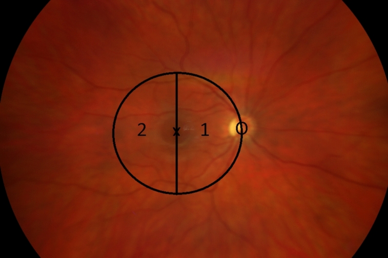
Methods: Central UMs were defined as lesions up to 5 mm off the optic disc or fovea radius of 5 mm. Between January 2011 and July 2020, we treated 56 patients with Ru-106-brachytherapy. The clinical outcomes for recurrence, visual acuity, and radiation-related toxicity were assessed. The follow-up was 66 (6-136) months.
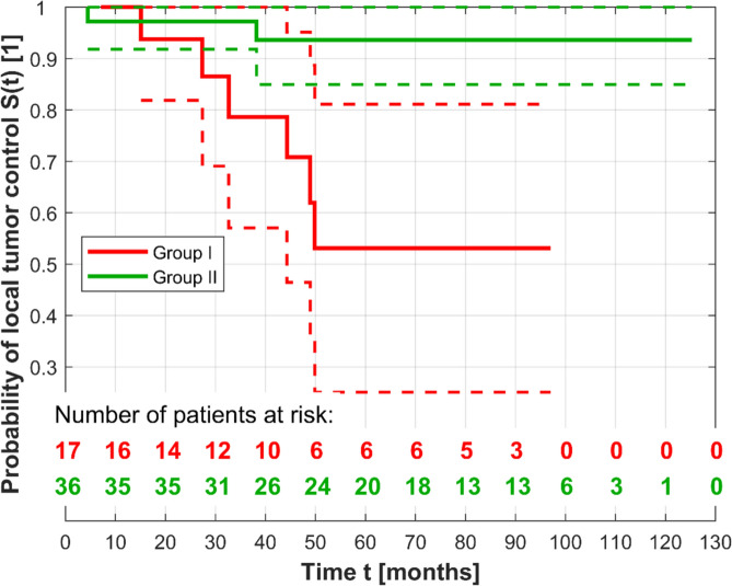
Results: Of the 56 patients (56 eyes), 8 (14%) suffered from local recurrence. Six relapsing UM in 19 (32%) patients were located close to the optic disc, and two patients had UM close to the macula (2/37, 5%) (p > 0.05). The overall eye-preservation rate was 89%. The pretreatment visual acuity (VA) was 0.45 and reduced to 0.26 after brachytherapy. Radiation retinopathy or optic neuropathy was detected in 7 (13%) patients and radiation maculopathy in 10 (17.9%). Six patients (11%) underwent enucleation for recurrence or radiation-induced ophthalmopathy.
Conclusion: Central UMs are challenging to treat. UMs should be categorized as lesions laterally or medially to the fovea because of different long-term control rates. Localization near the optic disc requires thoughtful management.
Keywords: Brachytherapy; Central; Ruthenium; Uveal melanoma.
© 2025. The Author(s).
Conflict of interest statement
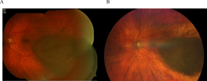
Declarations. Conflict of interests: The authors have no relevant financial or non-financial interests to disclose. Consent to participate: Informed consent was obtained from all individual participants included in the study. Consent to publish: The authors affirm that human research participants provided informed consent for publication of the images (Figs. 1, 5a and b). Ethics approval: This is an observational retrospective study. Therefore no ethical approval is required in Germany.
Figures


References
-
- Al-Wassia R, Dal Pra A, Shun K et al (2011) Stereotactic fractionated radiotherapy in the treatment of juxtapapillary choroidal melanoma: the McGill University experience. Int J Radiat Oncol 81:e455–e462. 10.1016/J.IJROBP.2011.05.012 - PubMed
-
- van Beek JGM, Ramdas WD, Angi M et al (2021) Local tumour control and radiation side effects for fractionated stereotactic photon beam radiotherapy compared to proton beam radiotherapy in uveal melanoma. Radiother Oncol 157:219–224. 10.1016/j.radonc.2021.01.030 - PubMed
-
- Bergman L, Nilsson B, Lundell G et al (2005) Ruthenium brachytherapy for uveal melanoma, 1979–2003: survival and functional outcomes in the Swedish population. Ophthalmology 112:834–840. 10.1016/J.OPHTHA.2004.11.038 - PubMed
MeSH terms
Substances
LinkOut - more resources
Full Text Sources
Medical
