. 2025 Jan 11;23(1):44.
doi: 10.1186/s12967-024-06065-y.

Molecule interacting with CasL-2 enhances tumor progression and alters radiosensitivity in cervical cancer

Affiliations

Molecule interacting with CasL-2 enhances tumor progression and alters radiosensitivity in cervical cancer

Yun Teng et al. J Transl Med. .

Abstract

Objective: Cervical cancer is a common malignancy among women, and radiotherapy remains a primary treatment modality across all disease stages. However, resistance to radiotherapy frequently results in treatment failure, highlighting the need to identify novel therapeutic targets to improve clinical outcomes.

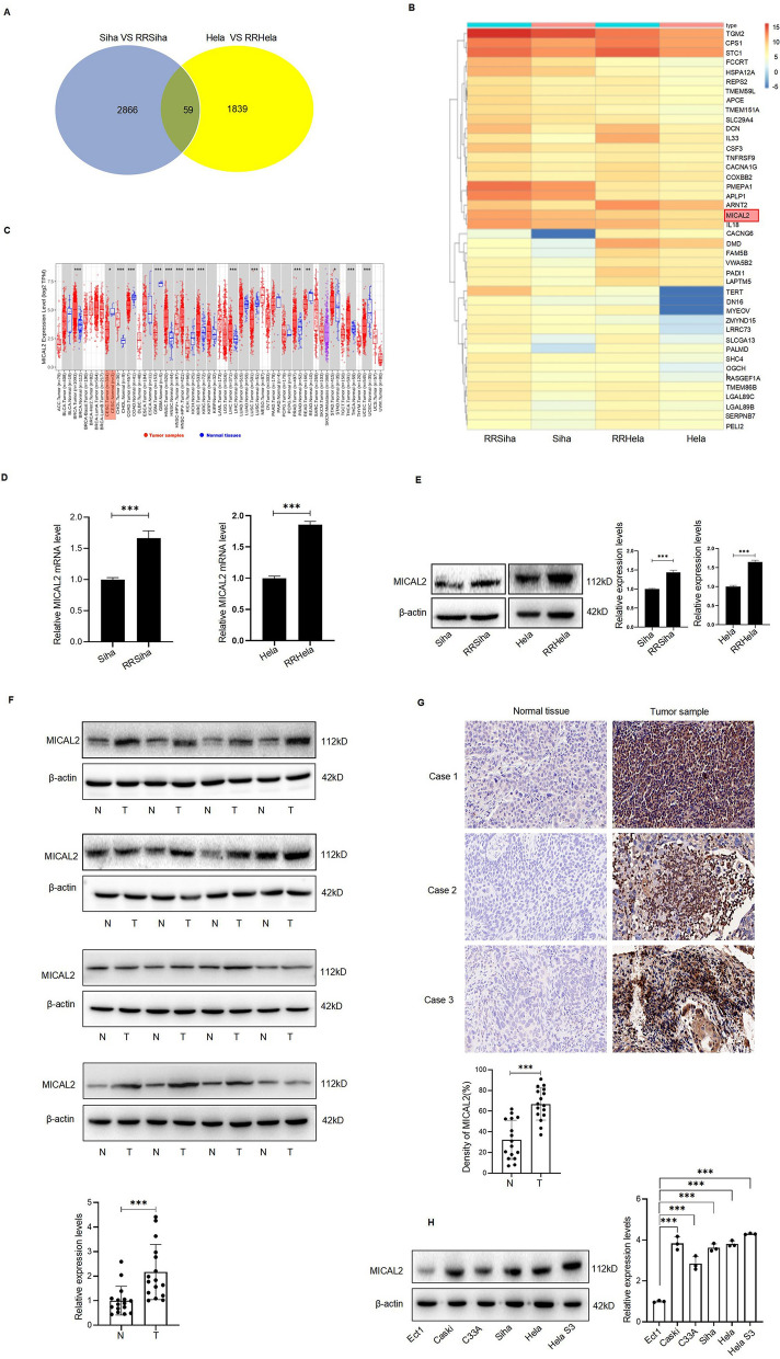
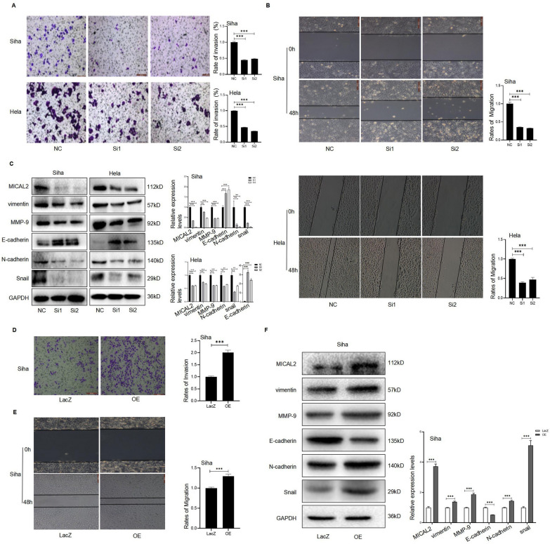
Methods: The expression of molecule interacting with CasL-2 (MICAL2) was confirmed in cervical cancer tissues and cell lines through western blotting (WB) and immunohistochemistry (IHC). Siha and Hela cells were used to examine the regulatory and biological functions of MICAL2 via knockdown and overexpression experiments. Assays including MTT, colony formation, wound healing, transwell migration, and sphere formation were employed, along with WB analysis. DNA damage in irradiated cells with MICAL2 knockdown or overexpression was evaluated using the comet assay, while γ-H2AX and Rad51 protein levels were detected by WB. In vivo experiments validated the tumorigenic and radioresistance functions of MICAL2. Additionally, the relationship between MICAL2 expression and radiotherapy response was analyzed in 62 patients with cervical cancer by assessing tumor regression and MICAL2 levels six months post-treatment.

Results: MICAL2 expression was significantly elevated in cervical cancer tissues and cells. Functional analyses demonstrated that MICAL2 promotes cell proliferation, migration, and invasion by activating the MAPK and PI3K/AKT pathways, as confirmed through both in vitro and in vivo experiments. Silencing MICAL2 increased DNA damage, impeded DNA repair, and enhanced radiosensitivity. Among the 62 patients with cervical cancer, elevated MICAL2 expression was associated with a lower complete response rate to radiotherapy (25.6% vs. 60.9% in those with low expression), reduced progression-free survival, and advanced cancer stage (*p < 0.05).

Conclusion: MICAL2 plays a critical role in tumor progression and radiotherapy resistance in cervical cancer. These findings provide a foundation for developing targeted therapies to improve treatment outcomes in this population.

Keywords: Cervical cancer; DNA damage; Irradiation; MICAL2; Radioresistance.


Conflict of interest statement

Declarations. Ethics approval and consent to participate: This study was conducted in accordance with the Declaration of Helsinki and received approval from the Ethics Committee of The Second Affiliated Hospital of Dalian Medical University (No 2023067). All animal care and experimental procedures adhered to the National Institutes of Health Guidelines for the Care and Use of Laboratory Animals and were approved by the Animal Care and Ethics Committee of Dalian Medical University (authorization code: 20233228). Consent for publication: Not applicable. Competing interests: The author declares that they have no competing interests.

Figures

Fig. 1
Fig. 1
MICAL2 exhibits high expression levels in cervical cancer cell lines and tumor tissues. A RNA sequencing analysis demonstrates MICAL2 as one of the 59 differential genes between cervical cancer cells and radioresistant cervical cancer cells. B Heatmap reveals that RRSiHa and RRHeLa share 42 significantly upregulated genes, including MICAL2, when compared to their parental Siha and Hela cell lines (n = 3). C Analysis of TCGA pan-cancer data demonstrates MICAL2 expression across various cancer types, with CESC highlighted in red. D WB analysis confirms elevated MICAL2 expression in tumor tissues (T) compared to normal tissues (N) from 16 patients with cervical cancer. E Immunohistochemical (IHC) analysis further demonstrates MICAL2 expression in cancerous and adjacent tissues from 16 patients with cervical cancer, with representative morphograms presented for three cases and quantitative analysis provided for all 16 cases in a histogram. F WB analysis reveals MICAL2 protein expression in one normal cervical cell line and five cervical cancer cell lines (n = 3). Data are presented as mean ± standard deviation, with statistical significance indicated as *p < 0.05, **p < 0.01, and ***p < 0.001 compared to the control group
Fig. 2
Fig. 2
MICAL2 promotes the growth and survival of cervical cancer cells in vitro, associated with apoptosis inhibition and activation of the MAPK and PI3K/AKT signaling pathways. A WB analysis was used to assess MICAL2 expression in Siha and Hela cells transfected with MICAL2-specific siRNAs (si-MICAL2-1 and si-MICAL2-2). B Colony formation assays were performed on Hela and Siha cells transfected with MICAL2 siRNA or non-specific control siRNA, with colonies greater than 50 μm counted after 14 days. C Cell viability of Hela and Siha cells was measured using the MTT assay following MICAL2 knockout. D WB analysis of proteins involved in the MAPK and PI3K/AKT pathways was conducted 48 h after transfection with MICAL2 siRNA or control siRNA. E AO/EB fluorescence staining was used to detect apoptotic cells in Siha and Hela cells transfected with MICAL2-specific siRNAs. F WB analysis assessed the effects of MICAL2 siRNA (siRNA1, siRNA2) on apoptosis-related proteins Bcl-2, Bax, cleaved caspase-9 (Cl-casp-9), and cleaved PARP (Cl-PARP) in Siha and Hela cell lines. GI Following transfection with a MICAL2 overexpression plasmid, WB, colony formation assays, and MTT assays were conducted to confirm MICAL2 protein overexpression in Siha cells and assess the number of cell colonies and cell viability. J WB analysis was used to verify the effects of MICAL2 overexpression on MAPK and PI3K/AKT pathway proteins in Siha cells. KL AO/EB staining and WB analysis was performed to detect apoptotic cells and apoptosis-related proteins (Bcl-2, Bax, Cl-casp-9, Cl-PARP) in MICAL2-overexpressing Siha cells, respectively. Data are presented as the mean ± standard deviation from three independent experiments, with statistical significance indicated as *p < 0.05, **p < 0.01, and ***p < 0.001 compared to the control group
Fig. 3
Fig. 3
MICAL2 enhances the migration and invasion of cervical cancer cells, with involvement of EMT signaling. A Hela and Siha cells transfected with MICAL2 siRNA1 or siRNA2 seeded in chambers coated with a diluted Matrigel matrix. The invasive cells were stained and imaged at 40 × magnification, with quantitative analysis of the invading cells presented in the histogram. B Cell migration assessed using a wound-healing assay. Hela and Siha cells were plated in 6-well plates, scratched with a 100 μL pipette tip, and cultured for 48 h before photographic recording. The histogram reveals the calculated cell mobility. C WB analysis used to measure the expression levels of vimentin, MMP-9, E-cadherin, N-cadherin, and Snail in Hela and Siha cells. D, E The invasion and migration abilities of Siha cells transfected with a MICAL2 overexpression plasmid assessed by transwell assay and wound-healing assay (× 40). F The impact of MICAL2 overexpression on the protein levels of vimentin, MMP-9, E-cadherin, N-cadherin, and Snail in the Siha cervical cancer cell line assessed by WB. Data are presented as the mean ± standard deviation. Statistical significance is indicated as *p < 0.05, **p < 0.01, and ***p < 0.001 compared to the control group
Fig. 4
Fig. 4
MICAL2 enhances stem cell-like properties and tumor formation in cervical cancer cells. A The impact of MICAL2 knockdown on stemness assessed using a spheroid formation assay. Cells with MICAL2 knockdown produced significantly smaller spheroids compared to controls, indicating a reduced spheroidization capability. B WB analysis employed to assess the expression levels of stemness-related proteins CD44, CD133, and Nanog in Hela and Siha cells following MICAL2 knockdown. C Stable overexpression of MICAL2 in Siha cells verified by WB after transfection with either a non-specific control LacZ plasmid or a MICAL2 overexpressing plasmid (OE). D, E The stemness of Siha cells transfected with the MICAL2 overexpressing plasmid assessed by spheroid formation assay and WB analysis of stemness-related proteins (CD44, CD133, and Nanog). F Tumor cells from the LacZ control group and the MICAL2 overexpression group implanted into the right axilla of female nude mice. Xenografts were collected 15 days post-implantation, and images of the tumor tissues from both groups were captured. G Volume and weight of xenografts in nude mice (n = 8) measured and compared. H, I WB and immunohistochemical analysis performed to detect the expression of key proteins Ki67, CD133, and P110α in the xenografts. Data are presented as the mean ± standard deviation. Statistical significance is indicated as *p < 0.05, **p < 0.01, and ***p < 0.001 compared to the control group
Fig. 5
Fig. 5
MICAL2 reduces both the DNA damage and the radiosensitivity of cervical cancer cells exposed to irradiation. A Cervical cancer Siha and Hela cell lines with stable MICAL2 knockdown established using MICAL2 lentiviral particles shRNA1 and shRNA2, with successful knockdown confirmed using WB analysis. B A colony formation assay conducted to assess the proliferation ability of Siha and Hela cells in both control and MICAL2 knockdown groups following 4 Gy X-ray irradiation. C, D Following MICAL2 shRNA transfection and 4 Gy X-ray exposure, cells were collected 6 h post-irradiation. DNA damage assessed using the comet assay, and expression levels of DNA damage marker (γ-H2AX) and repair protein (Rad51) analyzed using WB. E MICAL2 shRNA-treated Hela and Siha cells exposed to varying doses of X-rays (0, 2, 4, 6, and 8 Gy) for 48 h, and clonogenic survival curves generated. F MICAL2 overexpression in cervical cancer Siha cells achieved through MICAL2 plasmid transfection and confirmed using WB. GI The effects of MICAL2 overexpression on cell proliferation, DNA damage, γ-H2AX, and Rad51 expression in Siha cells exposed to 4 Gy X-ray irradiation assessed using colony formation assays, comet assays, and WB. J Siha and Hela cells subjected to continuous low-dose X-ray irradiation (2 Gy/day for 15 days, totaling 30 Gy) to generate radioresistant cell lines (RRSiha and RRHeLa). MICAL2 expression assessed in both parental and radioresistant cells using WB. K Stable MICAL2 knockdown in RRSiha and RRHeLa cells was confirmed using WB. LN The proliferation, DNA damage, and expression levels of γ-H2AX and Rad51 in RRSiha and RRHeLa cells with and without MICAL2 knockdown, following 4 Gy X-ray irradiation for 12 h, assessed using colony formation assays, comet assays, and WB. Data are presented as the mean ± standard deviation. Statistical significance is indicated as *p < 0.05, **p < 0.01, and ***p < 0.001 compared to the control group
Fig. 6
Fig. 6
MICAL2 knockdown impedes cervical cancer progression in a mouse model. Four types of cells used for subcutaneous implantation into nude mice: radiotherapy-resistant cervical cancer RRSiha cells (RR + NC), RRSiha cells with stable MICAL2 knockdown (RR + Sh1), RRSiha cells subjected to ionizing radiation (RR + NC + IR), and RRSiha cells with stable MICAL2 knockdown receiving radiotherapy (RR + Sh1 + IR). Xenograft tumors harvested 15 days post-treatment. A, B Tumor size measurements and images of xenografts are provided for each group (n = 5). C Tumor weight and volume comparisons across the different treatment groups. D, E WB and immunohistochemical analyses performed to assess the expression of γ-H2AX and Rad51 in xenograft tissues (n = 3). Data are expressed as the mean ± standard deviation. Statistical significance is indicated as *p < 0.05, **p < 0.01, and ***p < 0.001 compared to the control group
Fig. 7
Fig. 7
Patients with cervical cancer having high MICAL2 expression exhibited poor clinical response to radiotherapy. A Among the 62 patients with cervical cancer who underwent radiotherapy, 38 patients achieved a complete response (CR), 17 patients had a partial response (PR), and 7 patients either had stable disease (SD) or experienced disease progression (PD) six months post-radiotherapy. B MICAL2 expression levels in cervical cancer tissues from 2 cases with CR and 1 case with SD, along with their corresponding MRI images (transverse and median sagittal sections) taken before and after radiotherapy. C Kaplan–Meier survival analysis with log-rank test used to assess disease-free progression (DFS) in patients with cervical cancer based on different MICAL2 expression levels (n = 62)

Similar articles

References

    1. Ali I, Wani WA, Haque A, Saleem K. Glutamic acid and its derivatives: candidates for rational design of anticancer drugs. Future Med Chem. 2013;5(8):961–78. - PubMed
    1. Bray F, Laversanne M, Sung H, Ferlay J, Siegel RL, Soerjomataram I, et al. Global cancer statistics 2022: GLOBOCAN estimates of incidence and mortality worldwide for 36 cancers in 185 countries. CA Cancer J Clin. 2024; 74(3):229–263. - PubMed
    1. Chen S, Cao Z, Prettner K, Kuhn M, Yang J, Jiao L, et al. Estimates and projections of the global economic cost of 29 cancers in 204 countries and territories from 2020 to 2050. JAMA Oncol. 2023;9(4):465–72. - PMC - PubMed
    1. Guida F, Kidman R, Ferlay J, Schüz J, Soerjomataram I, Kithaka B, et al. Global and regional estimates of orphans attributed to maternal cancer mortality in 2020. Nat Med. 2022;28(12):2563–72. - PMC - PubMed
    1. Sung H, Ferlay J, Siegel RL, Laversanne M, Soerjomataram I, Jemal A, et al. Global Cancer Statistics 2020: GLOBOCAN estimates of incidence and mortality worldwide for 36 cancers in 185 countries. CA Cancer J Clin. 2021;71:209–49. - PubMed

MeSH terms