. 2025 Jan;15(1):e70180.
doi: 10.1002/ctm2.70180.

CMPK2 promotes NLRP3 inflammasome activation via mtDNA-STING pathway in house dust mite-induced allergic rhinitis

Affiliations

CMPK2 promotes NLRP3 inflammasome activation via mtDNA-STING pathway in house dust mite-induced allergic rhinitis

YaoMing Zheng et al. Clin Transl Med. 2025 Jan.

Abstract

Background: House dust mite (HDM) is the leading allergen for allergic rhinitis (AR). Although allergic sensitisation by inhaled allergens renders susceptible individuals prone to developing AR, the molecular mechanisms driving this process remain incompletely elucidated.

Objective: This study aimed to elucidate the molecular mechanisms underlying HDM-induced AR.

Methods: We examined the expression of cytidine/uridine monophosphate kinase 2 (CMPK2), STING and the NLRP3 inflammasome in both AR patients and mice. Additionally, we investigated the role of CMPK2 and STING in the activation of the NLRP3 inflammasome in AR.

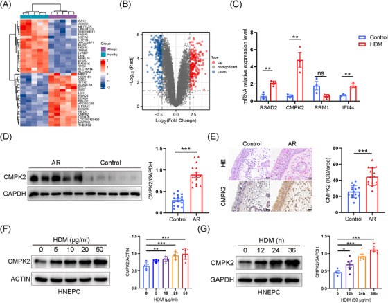
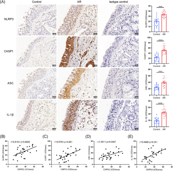
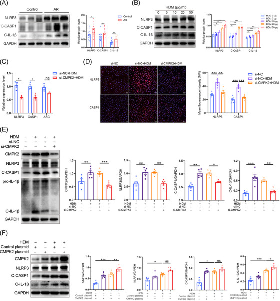
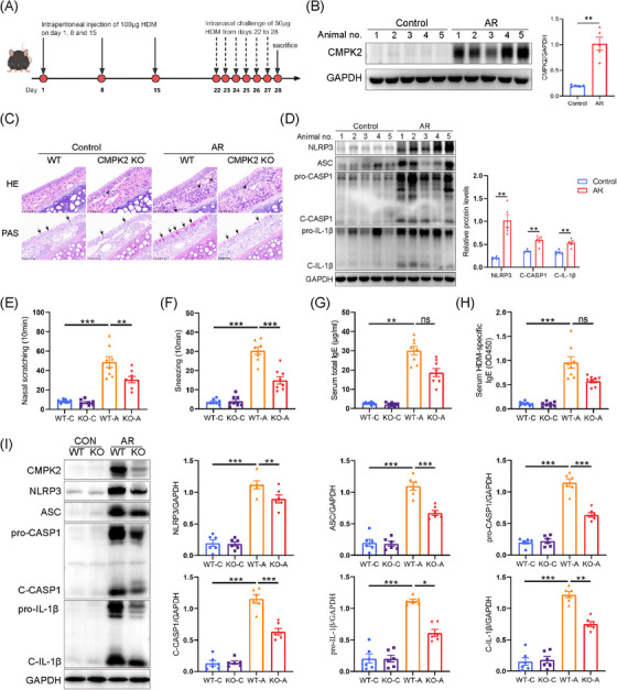
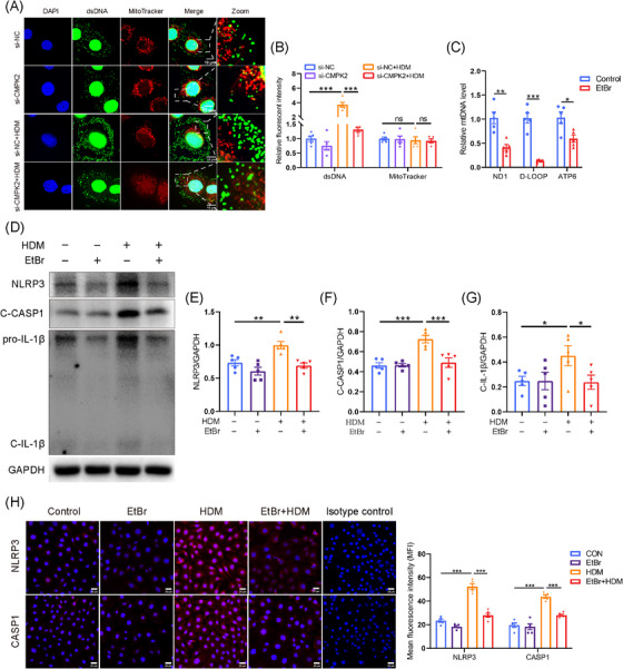
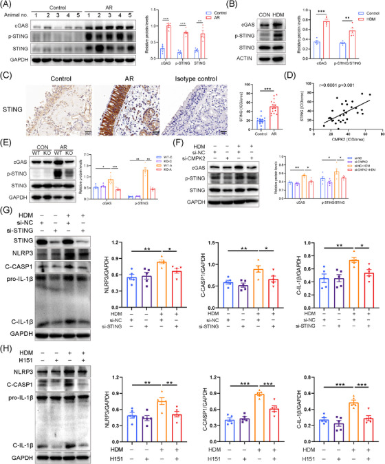
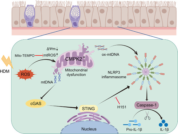
Results: The expression of CMPK2, STING and the NLRP3 inflammasome was significantly increased in the nasal mucosa of AR patients compared to non-AR controls. A positive correlation was found between CMPK2 expression and the levels of STING, NLRP3, ASC, CASP1 and IL-1β. HDM treatment up-regulated the expression of CMPK2, and CMPK2 overexpression enhanced NLRP3 inflammasome activation in human nasal epithelial cells (HNEPCs). Additionally, mitochondrial reactive oxygen species (mtROS) production following HDM exposure contributed to mitochondrial dysfunction and the release of mitochondrial DNA (mtDNA), which activated the cyclic GMP-AMP synthase (cGAS)-STING pathway. Remarkably, depletion of mtDNA or inhibition of STING signalling reduced HDM-induced NLRP3 inflammasome activation in HNEPCs. In vivo, genetic knockout of CMPK2 or STING alleviated NLRP3 inflammasome activation and ameliorated clinical symptoms of AR in mice.

Conclusions: Our results suggest that HDM promotes the activation of NLRP3 inflammasome through the up-regulation of CMPK2 and ensuing mtDNA-STING signalling pathway, hence revealing additional therapeutic target for AR.

Key points: Cytidine/uridine monophosphate kinase 2 (CMPK2) expression is up-regulated in the nasal mucosa of patients and mice with allergic rhinitis (AR). CMPK2 caused NLRP3 inflammasome activation via mitochondrial DNA (mtDNA)-STING pathway. Blocking CMPK2 or STING signalling significantly reduced the activation of NLRP3 in house dust mite (HDM)-challenged mice and human nasal epithelial cells (HNEPCs).

Keywords: CMPK2; NLRP3 inflammasome; STING; allergic rhinitis; mitochondrial DNA.


Conflict of interest statement

The authors declare no conflicts of interest.

Figures

FIGURE 1
FIGURE 1
Identification and validation of the haematologic/immune system‐specific expressed hub genes. (A) Heatmap of the top 20 up‐regulated DEGs and the top 20 down‐regulated DEGs (red: high expression; blue: low expression). (B) Volcano plot of DEGs (red: up‐regulated DEGs; blue: down‐regulated DEGs). (C) Expression of mRNA levels of NLRP3, ASC and CASP1 was assessed by quantitative polymerase chain reaction (PCR) in human nasal epithelial cells (HNEPCs) after house dust mite (HDM; 50 µg/mL) treatment for 24 h. (D) The protein levels of cytidine/uridine monophosphate kinase 2 (CMPK2) in nasal mucous tissue in the different study groups was detected by Western blotting. Control, n = 15; allergic rhinitis (AR), n = 15. (E) Representative photomicrographs and quantitative analysis of CMPK2 in nasal tissue of control subjects (n = 15) and patients with AR (n = 15; ×400 magnification). (F) HNEPCs were stimulated with HDM at the indicated concentration for 24 h, CMPK2 protein levels were measured by Western blotting (n = 5). (G) HNEPCs were stimulated with HDM (50 µg/mL) at the indicated hours. CMPK2 protein levels were measured by Western blotting (n = 5). *p < .05, **p < .01 and ***p < .001. DEG, differentially expressed gene; H&E, haematoxylin and eosin; IOD, integral optical density.
FIGURE 2
FIGURE 2
NLRP3 inflammasome‐related proteins increased in patients with allergic rhinitis (AR) and correlated with high expression of cytidine/uridine monophosphate kinase 2 (CMPK2; control, n = 15; AR, n = 15). (A) Representative photomicrographs and quantitative analysis of NLRP3, ASC, CASP1 and IL‐1β in nasal tissue of control subjects and patients with AR (×400 magnification). (B) Relationship between CMPK2 and NLRP3 in AR. (C) Relationship between CMPK2 and CASP1 in AR. (D) Relationship between CMPK2 and ASC in AR. (E) Relationship between CMPK2 and IL‐1β in AR. *p < .05, **p < .01 and ***p < .001. IOD, integral optical density.
FIGURE 3
FIGURE 3
Cytidine/uridine monophosphate kinase 2 (CMPK2) regulates the activation of NLRP3 inflammasome in human nasal epithelial cells (HNEPCs). (A) The protein levels of NLRP3, C‐CASP1 and C‐IL‐1β in nasal mucous tissue in the different study groups were detected by Western blotting. Control, n = 15; allergic rhinitis (AR), n = 15. (B) HNEPCs were stimulated with house dust mite (HDM) at the indicated concentration for 24 h. The protein levels of NLRP3, C‐CASP1 and C‐IL‐1β were measured by Western blotting (n = 5). (C) After si‐CMPK2 transfection, HNEPCs were treated with HDM (50 µg/mL) for 24 h. Cells were collected for reverse transcription polymerase chain reaction (RT‐PCR; n = 3). (D) Representative immunofluorescence (IF) staining of NLRP3 and CASP1 (n = 5). (E) HNEPCs were transfected with si‐CMPK2 or control small interfering RNA (siRNA) for 24 h and further stimulated with HDM (50 µg/mL) for another 24 h. Cells were collected for Western blotting (n = 5). Representative blots are shown, and densitometric analysis of blots was performed. (F) HNEPCs were transfected with CMPK2 plasmid and subsequently stimulated with HDM (50 µg/mL) for 24 h. Cell lysates were harvested for Western blotting. Representative blots are shown, and densitometric analysis of blots was performed (n = 5). *p < .05, **p < .01 and ***p < .001. ns = not significant.
FIGURE 4
FIGURE 4
Cytidine/uridine monophosphate kinase 2 (CMPK2) deficiency suppresses the activation of NLRP3 inflammasome in house dust mite (HDM)‐challenged allergic rhinitis (AR) mice. (A) Schematic representation of the experimental protocol. (B) The CMPK2 protein levels in the nasal tissues of mice (n = 5) with or without AR were determined by Western blotting. (C) Eosinophil infiltration and goblet cell metaplasia were evaluated in haematoxylin and eosin (H&E)‐ or PAS‐stained sections of wild‐type (WT) and CMPK2–/– mice with and without AR, respectively (×400 magnification). (D) Expression of protein levels of NLRP3 inflammasome in AR mice (n = 5). (E, F) Number of nasal scratching (E) and sneezing (F) in AR mice (n = 8). (G) Total immunoglobulin E (IgE) levels in serum (n = 8). (H) HDM‐specific IgE levels in serum (n = 8). (I) Expression of protein levels of CMPK2 and NLRP3 inflammasome in WT and CMPK2–/– mice with and without AR (n = 6). *p < .05, **p < .01 and ***p < .001. ns = not significant.
FIGURE 5
FIGURE 5
Mito‐TEMPO can inhibit the activation of NLRP3 inflammasome by inhibiting the accumulation of mitochondrial reactive oxygen species (mtROS) in human nasal epithelial cells (HNEPCs). HNEPCs pretreated with Mito‐TEMPO (50 µmol/L) for 3 h and then stimulated with house dust mite (HDM; 50 µg/mL) for 24 h. The HDM group was treated with HDM (50 µg/mL) for 24 h. (A, B) Representative fluorescence images of mitochondrial ROS staining using the MitoSOX probe (n = 5). Scale bar = 20 µm. (C, D) Representative fluorescence images of JC‐1 staining to evaluate MMP in HNEPCs (n = 5). Scale bar = 50 µm; red, aggregates; green, monomers. (E–H) Representative blots and quantitative analysis of NLRP3, C‐CASP1 and C‐IL‐1β relative protein expression in HNEPCs (n = 5). (I, J) Representative blots of cytidine/uridine monophosphate kinase 2 (CMPK2) expression in HNEPCs treated with HDM and Mito‐TEMPO (n = 5). (K, L) Expression of immunofluorescence (IF) costaining of 8‐OHdG/NLRP3 in HNEPCs (n = 5). Scale bar = 10 µm. *p < .05, **p < .01 and ***p < .001.
FIGURE 6
FIGURE 6
Cytosolic mitochondrial DNA (mtDNA) is necessary for the house dust mite (HDM)‐induced activation of NLRP3 inflammasome in human nasal epithelial cells (HNEPCs). (A, B) HNEPCs were transfected with si‐cytidine/uridine monophosphate kinase 2 (CMPK2) or control small interfering RNA (siRNA) for 24 h and further stimulated with HDM (50 µg/mL) for another 24 h. Representative fluorescence images of dsDNA (green) and mitochondria (red) in HNEPCs. Scale bar = 10 µm. (C) mRNA levels of ND1, D‐LOOP and ATP6 were assessed by quantitative real‐time polymerase chain reaction (qPCR) in HNEPCs after EtBr stimulation (2 µg/mL, 24 h; n = 5). (D–G) HNEPCs pretreated with EtBr (2 µg/mL) for 24 h and then stimulated with HDM (50 µg/mL) for another 24 h. The HDM group was treated with HDM (50 µg/mL) for 24 h (n = 5). (H) Representative immunofluorescence (IF) staining and quantitative analysis of NLRP3 and CASP1. Scale bar = 25 µm. *p < .05, **p < .01 and ***p < .001.
FIGURE 7
FIGURE 7
House dust mite (HDM)‐driven NLRP3 inflammasome activation relies on the cyclic GMP‐AMP synthase (cGAS)‐STING pathway. (A) The expression of cGAS, STING and p‐STING in the nasal tissues of mice (n = 5) with or without allergic rhinitis (AR) was determined by Western blotting. (B) The expression of cGAS, STING and p‐STING in human nasal epithelial cells (HNEPCs) after stimulation with HDM (50 µg/mL) for 24 h was determined by Western blotting (n = 5). (C) Immunohistochemical staining for STING in nasal tissue of control subjects and patients with AR (n = 15; ×400 magnification). (D) Correlations between cytidine/uridine monophosphate kinase 2 (CMPK2) and STING in nasal mucosa of AR patients. Spearman test. IOD, integrated optical density. (E) Western blotting for cGAS, STING and p‐STING in the nasal tissues of wild‐type (WT) and CMPK2–/– mice with and without AR, respectively (n = 6). (F) After knockdown of CMPK2 in HNEPCs, the effect of HDM (50 µg/mL) on the activation of cGAS‐STING pathway was detected (n = 5). (G) After si‐STING transfection, HNEPCs were treated with HDM (50 µg/mL) for 24 h. Representative blots and quantitative analysis of STING, NLRP3, C‐CASP1 and C‐IL‐1β relative protein expression in HNEPCs (n = 5). (H) HNEPCs pretreated with H151 (10 µM) for 3 h and then stimulated with HDM (50 µg/mL) for 24 h. The HDM group was treated with HDM (50 µg/mL) for 24 h. Representative blots and quantitative analysis of NLRP3, C‐CASP1 and C‐IL‐1β relative protein expression in HNEPCs (n = 5). *p < .05, **p < .01 and ***p < .001.
FIGURE 8
FIGURE 8
Activation of NLRP3 inflammasome in wild‐type (WT) and STING–/– mice with and without allergic rhinitis (AR). (A) Eosinophil infiltration and goblet cell hyperplasia were evaluated in haematoxylin and eosin (H&E)‐ or PAS‐stained sections of WT and STING–/– mice with and without AR, respectively (n = 5; ×400 magnification). (B–H) Expression of protein levels of STING and NLRP3 inflammasome in WT and STING–/– mice with and without AR (n = 6). (I) Immunohistochemical staining for STING and NLRP3 inflammasome in nasal tissue of WT and STING–/– mice with and without AR, respectively (n = 5; ×400 magnification). *p < .05, **p < .01 and ***p < .001. ns = not significant.
FIGURE 9
FIGURE 9
The diagram encapsulates our hypothesis that cytidine/uridine monophosphate kinase 2 (CMPK2) promotes the progression of allergic rhinitis by activating the NLRP3 inflammasome via the mitochondrial DNA (mtDNA)‐STING signalling pathway. Stimulation by house dust mite (HDM) results in the up‐regulation of CMPK2 expression and induces mitochondrial dysfunction, consequently causing the release of mtDNA into the cytoplasm. This release activates the STING signalling pathway, which subsequently leads to the activation of the NLRP3 inflammasome. Inhibition of CMPK2 and the STING signalling pathway can significantly mitigate the activation of the NLRP3 inflammasome, thereby alleviating allergic rhinitis. cGAS, cyclic GMP‐AMP synthase; HDM, house dust mite; ROS, reactive oxygen species.

Similar articles

Cited by

References

    1. Wise SK, Damask C, Roland LT, et al. International consensus statement on allergy and rhinology: allergic rhinitis – 2023. Int Forum Allergy Rhinol. 2023;13(4):293‐859. - PubMed
    1. Zhang Y, Zhang L. Increasing prevalence of allergic rhinitis in China. Allergy Asthma Immunol Res. 2019;11(2):156‐169. - PMC - PubMed
    1. Bernstein JA, Bernstein JS, Makol R, Ward S. Allergic rhinitis: a review. JAMA. 2024;331(10):866‐877. - PubMed
    1. Bousquet J, Anto JM, Bachert C, et al. Allergic rhinitis. Nat Rev Dis Prim. 2020;6(1):95. - PubMed
    1. Zuberbier T, Lötvall J, Simoens S, Subramanian SV, Church MK. Economic burden of inadequate management of allergic diseases in the European Union: a GA(2) LEN review. Allergy. 2014;69(10):1275‐1279. - PubMed

LinkOut - more resources