Review
. 2025 Jan;39(1):e70133.
doi: 10.1002/jbt.70133.

Oxidative/Nitrosative Stress, Apoptosis, and Redox Signaling: Key Players in Neurodegenerative Diseases

Affiliations
Review

Oxidative/Nitrosative Stress, Apoptosis, and Redox Signaling: Key Players in Neurodegenerative Diseases

Nuray Üremiş et al. J Biochem Mol Toxicol. 2025 Jan.

Abstract

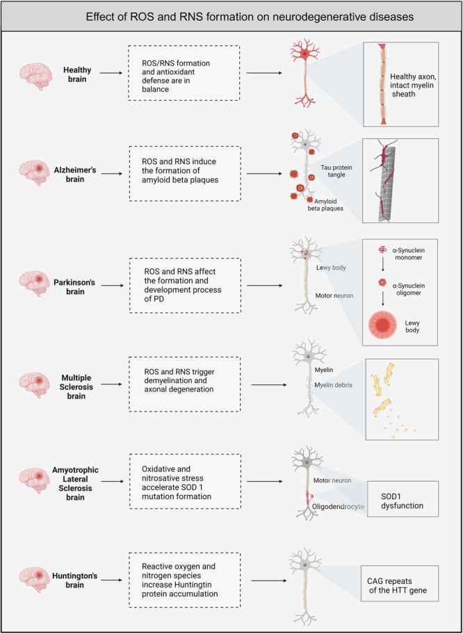
Neurodegenerative diseases are significant health concerns that have a profound impact on the quality and duration of life for millions of individuals. These diseases are characterized by pathological changes in various brain regions, specific genetic mutations associated with the disease, deposits of abnormal proteins, and the degeneration of neurological cells. As neurodegenerative disorders vary in their epidemiological characteristics and vulnerability of neurons, treatment of these diseases is usually aimed at slowing disease progression. The heterogeneity of genetic and environmental factors involved in the process of neurodegeneration makes current treatment methods inadequate. However, the existence of common molecular mechanisms in the pathogenesis of these diseases may allow the development of new targeted therapeutic strategies. Oxidative and nitrosative stress damages membrane components by accumulating ROS and RNS and disrupting redox balance. This process results in the induction of apoptosis, which is important in the pathogenesis of neurodegenerative diseases through oxidative stress. Studies conducted using postmortem human samples, animal models, and cell cultures have demonstrated that oxidative stress, nitrosative stress, and apoptosis are crucial factors in the development of diseases such as Alzheimer's, Parkinson's, Multiple Sclerosis, amyotrophic lateral sclerosis, and Huntington's disease. The excessive production of reactive oxygen and nitrogen species, elevated levels of free radicals, heightened mitochondrial stress, disturbances in energy metabolism, and the oxidation and nitrosylation of cellular macromolecules are recognized as triggers for neuronal cell death. Challenges in managing and treating neurodegenerative diseases require a better understanding of this field at the molecular level. Therefore, this review elaborates on the molecular mechanisms by which oxidative and nitrosative stress are involved in neuronal apoptosis.

Keywords: Oxidative stress; apoptosis; molecular mechanisms; neurodegenerative diseases; nitrosative stress; redox signaling.


Conflict of interest statement

The authors declare no conflicts of interest.

Figures

Figure 1
Figure 1
Mitochondrial Electron Transport Chain and Reactive Oxygen Species Production. The mitochondrial electron transport chain (ETC) and reactive oxygen species (ROS) production. The ETC consists of four complexes (I–IV) located in the inner mitochondrial membrane, where electron transfer occurs through redox reactions. Complex I: Electrons from NADH are transferred to Ubisemiquinone via FMN, reducing it to Ubiquinol. Complex II: Electrons from succinate are transferred to Ubiquinone, which is reduced to Ubiquinol. Complex III: Ubiquinol transfers electrons through the complex to cytochrome c, and Ubisemiquinone, a free radical, is formed as an intermediate product. Complex IV: Electrons from cytochrome c are transferred to molecular oxygen and reduced to water. Some electrons leak early from electron carriers and react with molecular oxygen to form superoxide anions during these processes. Superoxide dismutase converts superoxide to hydrogen peroxide, which can be reduced to water by glutathione peroxidase using reduced glutathione as substrate. However, hydrogen peroxide can also form more reactive species that contribute to oxidative stress if not adequately managed by cellular antioxidant systems. Nicotinamide Nucleotide Transhydrogenase enzyme maintains the balance of NADPH required to regenerate glutathione from its oxidized form, thus aiding in the detoxification of ROS. Created with Biorender. com.
Figure 2
Figure 2
Schematic representation of nitric oxide metabolism. Dimeric NOS facilitates NO production by collaborating with various cofactors, activators, regulators, and substrates. The resultant NO plays a role in diverse cellular reactions. It can interact with the O2 •− anion to generate ONOO. Both ONOO and NO can react with tyrosine residues in proteins, forming 3‐nitrotyrosines (3‐NT). Furthermore, NO can undergo S‐nitrosylation reactions with proteins containing cysteine residues, such as GSH. Created with Biorender. com.
Figure 3
Figure 3
Cellular sources and detoxification pathways of reactive oxygen and nitrogen species. Through diverse pathways, these mechanisms regulate superoxide and nitric oxide formation by cellular organelles and enzymes. Mitochondria, peroxisomes, and the endoplasmic reticulum play pivotal roles in hydrogen peroxide production. Additionally, the enzymes NOX and XO facilitate the generation of superoxide (O2 •−) radicals, while the NOS enzyme catalyzes NO production. Enzymatic and nonenzymatic antioxidant defense mechanisms are activated to counteract the detrimental effects of augmented free radicals, oxidants, and nitrogen compounds within the cell. Created with Biorender. com.
Figure 4
Figure 4
Schematic representation of the impact of ROS and RNS on the apoptosis mechanism triggered by the three primary death receptors. The extrinsic pathway, one of the two main apoptosis pathways, is initiated by binding death receptors such as TNFR, FAS, and TRAIL‐R to ligands. This initiation can be enhanced by NOX, which contributes to the accumulation of oxidant compounds outside the cell. The intrinsic pathway can activate caspase‐3 through two distinct mechanisms, with DISC activating caspase‐8. Additionally, factors such as ROS and RNS, generated by enzymes and organelles, are also involved in this initiation process, facilitating the progression of apoptosis. Activation of initiator caspases, apoptotic proteins, and oxidant/nitrogen compounds triggers the initiation of the mitochondrial apoptotic pathway. Consequently, as the apoptosis signal involving ROS and RNS compounds advances, effector caspase enzymes are activated, ultimately leading to programmed cell death. Created with Biorender. com.
Figure 5
Figure 5
Effect of ROS and RNS Formation on Neurodegenerative Diseases. Impact of ROS and RNS on the development and progression of various neurodegenerative diseases compared to a healthy brain. Healthy Brain: In a healthy brain, ROS and RNS formation is balanced by antioxidant defenses, maintaining neuronal health and intact myelin sheaths. Alzheimer's Brain: Excess ROS and RNS contribute to the pathology of Alzheimer's disease by inducing the formation of amyloid beta plaques and tau protein tangles. Parkinson's Brain: ROS and RNS influence the genesis and development of Parkinson's disease by promoting alpha‐synuclein aggregation into Lewy bodies, leading to motor neuron damage. Multiple Sclerosis Brain: ROS and RNS disrupt normal neural function in multiple sclerosis by triggering demyelination and axonal degeneration. Amyotrophic Lateral Sclerosis Brain: Oxidative and nitrosative stress accelerates mutation formation of superoxide dismutase 1, leading to motor neuron damage and oligodendrocyte dysfunction in amyotrophic lateral sclerosis. Huntington's brain: Reactive oxygen and nitrogen species contribute to the pathology of Huntington's disease by increasing the accumulation of the Huntingtin protein associated with the CAG repeats of the HTT gene. Created with Biorender. com.

Similar articles

Cited by

References

    1. Mayne K., White J. A., McMurran C. E., Rivera F. J., and de la Fuente A. G., “Aging and Neurodegenerative Disease: Is the Adaptive Immune System a Friend or Foe?,” Frontiers in Aging Neuroscience 12 (2020): 572090. - PMC - PubMed
    1. Fereshtehnejad S. M., Vosoughi K., Heydarpour P., et al., “Burden of Neurodegenerative Diseases in the Eastern Mediterranean Region, 1990‐2016: Findings From the Global Burden of Disease Study 2016,” European Journal of Neurology 26, no. 10 (2019): 1252–1265. - PubMed
    1. Rekatsina M., Paladini A., Piroli A., Zis P., Pergolizzi J. V., and Varrassi G., “Pathophysiology and Therapeutic Perspectives of Oxidative Stress and Neurodegenerative Diseases: A Narrative Review,” Advances in Therapy 37, no. 1 (2020): 113–139. - PMC - PubMed
    1. de la Fuente A. G., Pelucchi S., Mertens J., Di Luca M., Mauceri D., and Marcello E., “Novel Therapeutic Approaches to Target Neurodegeneration,” British Journal of Pharmacology 180, no. 13 (2023): 1651–1673. - PMC - PubMed
    1. Kumar A. and Singh A., “A Review on Mitochondrial Restorative Mechanism of Antioxidants in Alzheimer's Disease and Other Neurological Conditions,” Frontiers in Pharmacology 6 (2015): 206. - PMC - PubMed

Substances

LinkOut - more resources