. 2025 Dec;22(1):1-12.
doi: 10.1080/15476286.2024.2448391. Epub 2025 Jan 13.

Evolution of the RNA alternative decay cis element into a high-affinity target for the immunomodulatory protein Roquin

Affiliations

Evolution of the RNA alternative decay cis element into a high-affinity target for the immunomodulatory protein Roquin

Jan-Niklas Tants et al. RNA Biol. 2025 Dec.

Abstract

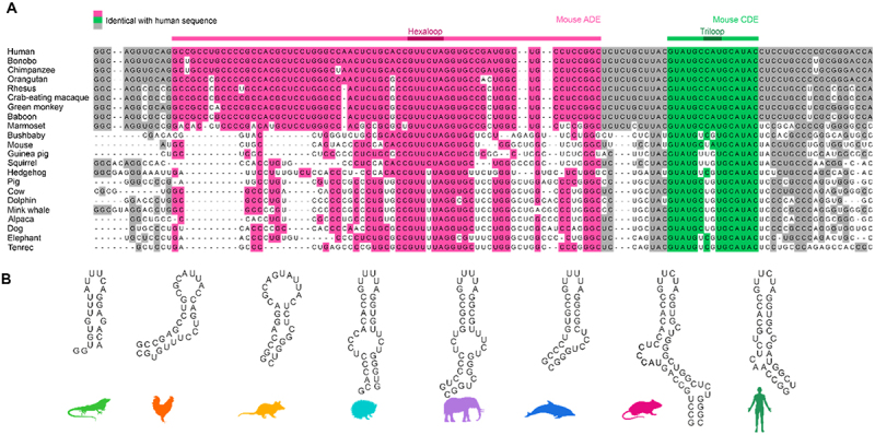
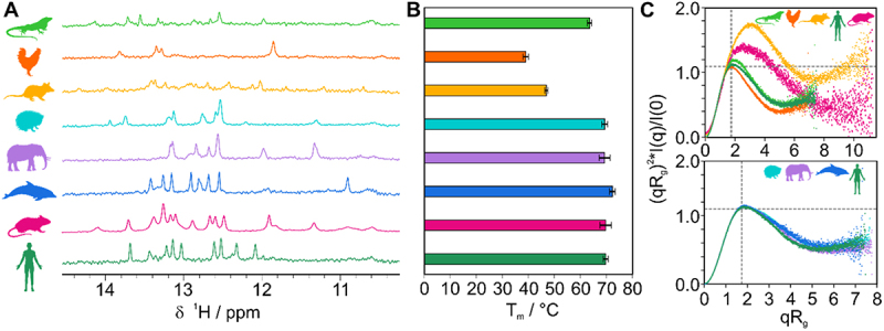
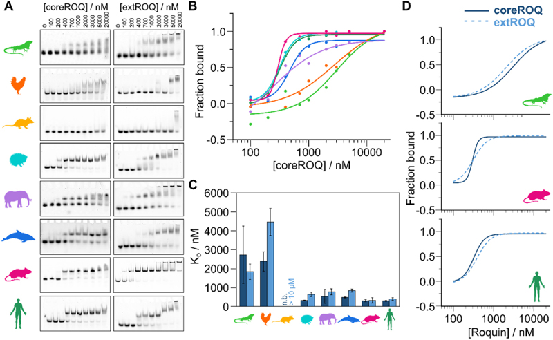
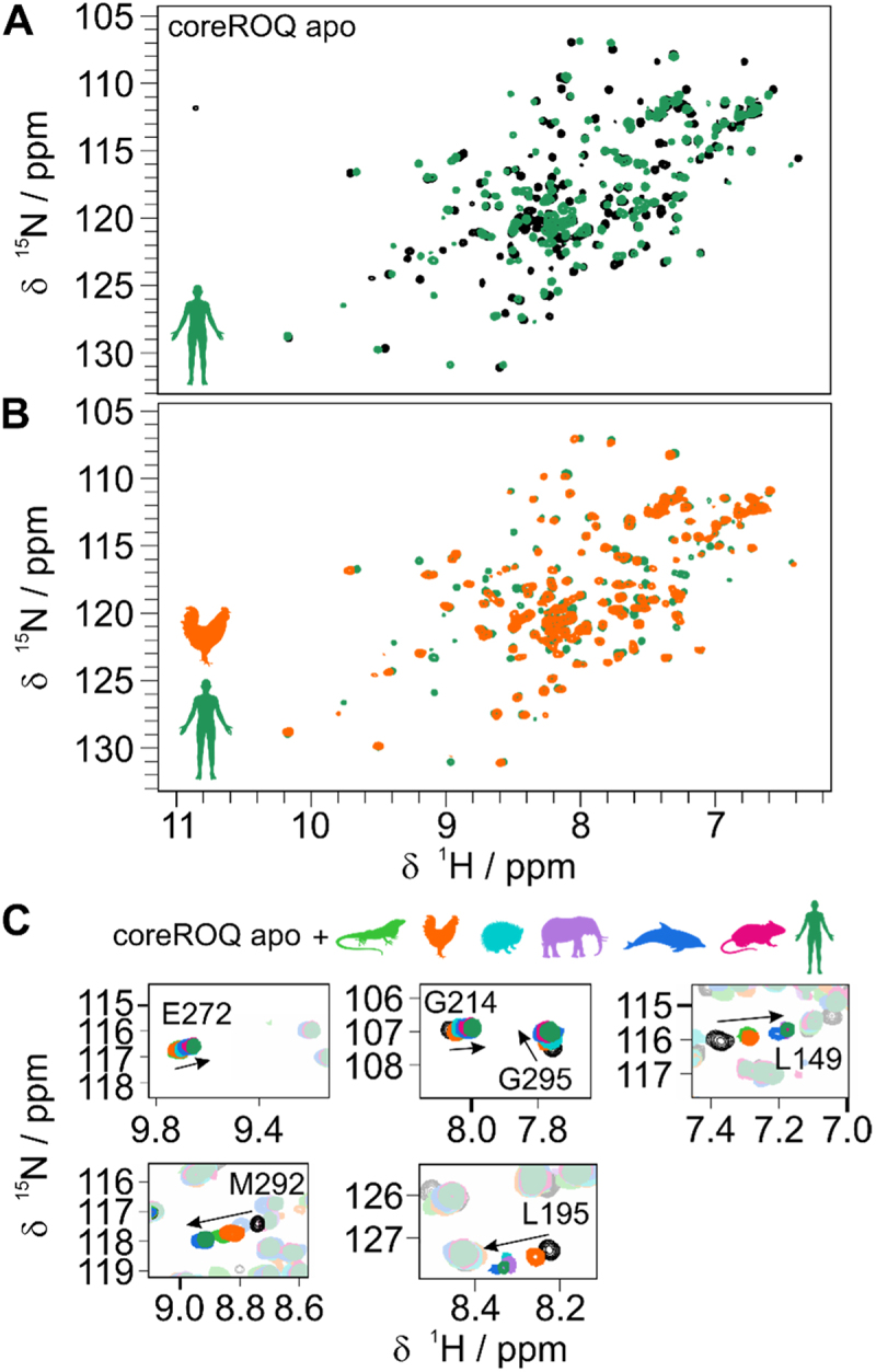
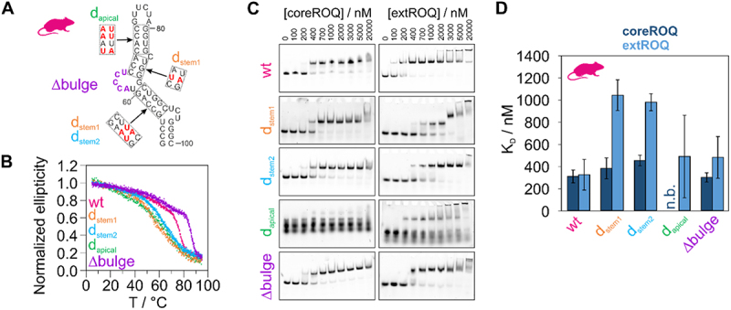
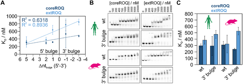
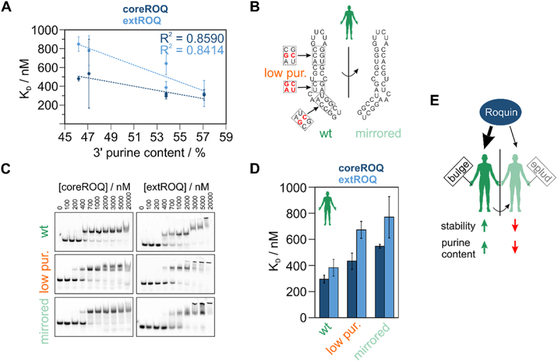
RNA cis elements play pivotal roles in regulatory processes, e.g. in transcriptional and translational regulation. Two stem-looped cis elements, the constitutive and alternative decay elements (CDE and ADE, respectively) are shape-specifically recognized in mRNA 3' untranslated regions (UTRs) by the immune-regulatory protein Roquin. Roquin initiates mRNA decay and contributes to balanced transcript levels required for immune homoeostasis. While the interaction of Roquin with several CDEs is described, our knowledge about ADE complex formation is limited to the mRNA of Ox40, a gene encoding a T-cell costimulatory receptor. The Ox40 3'UTR comprises both a CDE and ADE, each sufficient for Roquin-mediated control. Opposed to highly conserved and abundant CDE structures, ADEs are rarer, but predicted to exhibit a greater structural heterogeneity. This raises the question of how and when two structurally distinct cis elements evolved as equal target motifs for Roquin. Using an interdisciplinary approach, we here monitor the evolution of sequence and structure features of the Ox40 ADE across species. We designed RNA variants to probe en-detail determinants steering Roquin-RNA complex formation. Specifically, those reveal the contribution of a second RNA-binding interface of Roquin for recognition of the ADE basal stem region. In sum, our study sheds light on how the conserved Roquin protein selected ADE-specific structural features to evolve a second high-affinity mRNA target cis element relevant for adaptive immune regulation. As our findings also allow expanding the RNA target spectrum of Roquin, the approach can serve a paradigm for understanding RNA-protein specificity through back-tracing the evolution of the RNA element.

Keywords: EMSA; NMR; RNA; Roquin; SAXS; cis element; geometry; sequence; structure evolution.


Conflict of interest statement

No potential conflict of interest was reported by the author(s).

Figures

Figure 1.
Figure 1.
Evolutionary conservation of Ox40 tandem ADE-CDE cassette. A) Sequence alignment of Ox40 tandem ADE-CDE from multiple mammals. Bases identical with the human sequence are coloured. Residues corresponding to the murine ADE and CDE are coloured in magenta and green, respectively. Dark magenta and green highlight the hexa- and triloop, respectively. B) Secondary structure predictions of ADEs used in this study. These ADE constructs were designed to be comparable and small in size, to contain the central bulge and the basal stem and start with a stable GC or GU basepair. Note that these constructs were derived from Supplementary Figure S1D, where the extended ADE context is shown.
Figure 2.
Figure 2.
Ox40 ADEs from multiple species form stable structures. A) Imino 1H spectra of Ox40 species ADEs (from top to bottom: lizard, chicken, opossum, hedgehog, elephant, dolphin, mouse, and human). B) CD-derived melting points of ADEs aligned and colour coded as in panel A). Melting points are given as average from triplicates with standard deviations (error bars). C) Dimensionless Kratky plots derived from SAXS measurements of ADEs. A curve maximum at the intersection of the two grey dotted lines corresponds to a globular (compact) fold. Curves with a shift to the upper right indicate partially unfolded structures (e.g. as observed for opossum) or elongated shapes (e.g. as for the murine ADE due to its increased construct and stem size).
Figure 3.
Figure 3.
Affinity of Roquin for ADEs increased during evolution. A) Representative EMSAs of coreROQ and extROQ with species ADEs. Protein concentrations are given on top. Triplicates are shown in supplementary figure S6. B) CoreROQ binding curves derived from EMSAs. Plotted are averaged curves from triplicates. C) KD values of coreROQ (dark blue) and extROQ (light blue) for species ADEs as obtained from EMSAs shown as bar plot. KD values are averages from triplicates and errors are standard deviations (see Table 2). n.b. = no binding D) Comparison of coreROQ (dark blue, solid line) and extROQ (light blue, dashed line) binding curves with lizard, mouse and human ADEs.
Figure 4.
Figure 4.
Evolution of the conserved Roquin ADE binding mode. A) 1H,15N-HSQC spectra of apo coreROQ (black) and in complex with the human Ox40 ADE (green). B) 1H,15N-HSQC spectra of coreROQ in complex with the putative chicken (orange) or human (green) ADE. For all species comparisons refer to supplementary figure S7. C) Zoom-ins of 1H,15N-HSQC overlays of apo coreROQ (black) and in presence of species ADEs (colour).
Figure 5.
Figure 5.
ADE stability mutants modulate Roquin binding affinity. A) Secondary structure of murine Ox40 ADE wt. Red nucleotides in dashed boxes indicate sites of mutations. The corresponding constructs, in which these mutations occur are denoted besides the boxes. Nucleotides deleted in a ∆bulge version are coloured in purple. B) Normalized CD melting curves of ADE variants shown in A). C) Representative EMSAs of coreROQ and extROQ with stability variants of the murine Ox40 ADE from A). Protein concentrations are given on top. Triplicates are shown in supplementary figure S8. D) KD values of coreROQ (dark blue) and extROQ (light blue) for ADE stability variants as obtained from EMSAs shown as bar plot. KD values are averages from triplicates and errors are standard deviations (see Table 2). n.b. = no binding.
Figure 6.
Figure 6.
The central ADE bulge affects Roquin binding affinity. A) Correlation plot of coreROQ (dark blue) and extROQ (light blue) affinities for mammalian ADEs (taken from Figure 3C) with the number of unpaired nucleotides in the central bulge of the respective ADE (see supplementary table 3). Note that the difference between 5’ and 3’ unpaired nucleotides is plotted. Positive values thus indicate a bulge within the 5’ stem and negative values within the 3’ stem. B) Representative EMSAs of coreROQ and extROQ with human and murine wt ADEs and a version with a 3’ bulge (see Supplementary Figure S9). Protein concentrations are given on top. Triplicates are shown in Supplementary Figure S9. Wt EMSAs are the same as in Figure 3A. C) KD values of coreROQ (dark blue) and extROQ (light blue) for wt and 3’ bulge ADEs as obtained from EMSAs shown as bar plot. KD values are averages from triplicates and errors are standard deviations (see Table 2).
Figure 7.
Figure 7.
The impact of ADE geometry and purine content on Roquin binding affinity. A) Correlation of coreROQ and extROQ affinity with the 3’ purine content of ADEs. B) Secondary structure of human Ox40 ADE wt and a mirrored version. Note that the central stem is mirrored, while the basal stem and the loop orientation are maintained with respect to the wildtype. Red nucleotides in dashed boxes indicate sites of mutations within an ADE mutant with low purine content in the 3’ stem. C) Representative EMSAs of coreROQ and extROQ with human ADE variants from A). Protein concentrations are given on top. Triplicates are shown in supplementary figure 10. Wt EMSAs are the same as in Figure 3A. D) KD values of coreROQ (dark blue) and extROQ (light blue) for human ADE variants as obtained from EMSAs shown as bar plot. KD values are averages from triplicates and errors are standard deviations (see Table 2). E) Roquin shows a binding preference for ADEs with a bulge in the 5’ stem. Increased stability and a high purine content in the 3’ stem increase affinity. Overall, the interplay of geometric factors, stability and sequence fine tunes Roquin-ade complex formation.

Similar articles

Cited by

References

    1. Shine J, Dalgarno L.. The 3′-terminal sequence of Escherichia coli 16S ribosomal RNA: complementarity to nonsense triplets and ribosome binding sites. Proc Natl Acad Sci USA. 1974;71(4):1342–1346. doi: 10.1073/pnas.71.4.1342 - DOI - PMC - PubMed
    1. Shaw G, Kamen R. A conserved AU sequence from the 3′ untranslated region of GM-CSF mRNA mediates selective mRNA degradation. Cell. 1986;46(5):659–667. doi: 10.1016/0092-8674(86)90341-7 - DOI - PubMed
    1. Caput D, Beutler B, Hartog K, et al. Identification of a common nucleotide sequence in the 3’-untranslated region of mRNA molecules specifying inflammatory mediators. Proc Natl Acad Sci USA. 1986;83(6):1670–1674. doi: 10.1073/pnas.83.6.1670 - DOI - PMC - PubMed
    1. Bao C, Zhu M, Nykonchuk I, et al. Specific length and structure rather than high thermodynamic stability enable regulatory mRNA stem-loops to pause translation. Nat Commun. 2022;13(1):988. doi: 10.1038/s41467-022-28600-5 - DOI - PMC - PubMed
    1. Costantino DA, Pfingsten JS, Rambo RP, et al. tRNA–mRNA mimicry drives translation initiation from a viral IRES. Nat Struct Mol Biol. 2008;15(1):57–64. doi: 10.1038/nsmb1351 - DOI - PMC - PubMed

LinkOut - more resources