. 2025 Jan 7;9(1):016102.
doi: 10.1063/5.0231273. eCollection 2025 Mar.

Stem cell mechanoadaptation. I. Effect of microtubule stabilization and volume changing stresses on cytoskeletal remodeling

Affiliations

Stem cell mechanoadaptation. I. Effect of microtubule stabilization and volume changing stresses on cytoskeletal remodeling

Vina D L Putra et al. APL Bioeng. .

Abstract

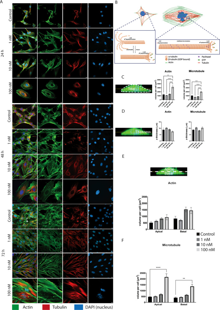
Here, we report on the first part of a two-part experimental series to elucidate spatiotemporal cytoskeletal remodeling, which underpins the evolution of stem cell shape and fate, and the emergence of tissue structure and function. In Part I of these studies, we first develop protocols to stabilize microtubules exogenously using paclitaxel (PAX) in a standardized model murine embryonic stem cell line (C3H/10T1/2) to maximize comparability with previously published studies. We then probe native and microtubule-stabilized stem cells' capacity to adapt to volume changing stresses effected by seeding at increasing cell densities, which emulates local compression and tissue template formation during development. Within the concentration range of 1-100 nM, microtubule-stabilized stem cells maintain viability and reduce proliferation. PAX stabilization of microtubules is associated with increased cell volume as well as flattening of the cell and nucleus. Compared to control cells, microtubule-stabilized cells exhibit thick, bundled microtubules and highly aligned, thicker and longer F-actin fibers, corresponding to an increase in the Young's modulus of the cell. Both F-actin and microtubule concentration increase with increasing PAX concentration, whereby the increase in F-actin is more prominent in the basal region of the cell. The corresponding increase in microtubule is observed more globally across the apical and basal region of the cell. Seeding at increasing target densities induces local compression on cells. This increase in local compression modulates cell volume and concomitant increases in F-actin and microtubule concentration to a greater degree than microtubule stabilization via PAX. Cells seeded at high density exhibit higher bulk modulus than corresponding cells seeded at low density. These data demonstrate the capacity of stem cells to adapt to an interplay of mechanical and chemical cues, i.e., respective compression and exogenous microtubule stabilization; the resulting cytoskeletal remodeling manifests as evolution of mechanical properties relevant to development of multicellular tissue constructs.


Conflict of interest statement

M.L.K.T. has co-founded startup companies to commercialize the intellectual property she and her collaborators developed and patent protected over the past decades. The topic of the current manuscript is fundamental in nature and not directly related to M.L.K.T.'s innovation translation and commercialization projects.

Figures

FIG. 1.
FIG. 1.
Concentration-dependent response to PAX and its effect in modulating C3H/10T1/2 murine embryonic stem cell proliferation, as well as cell and nucleus volume, shape, and/or size. Cell viability measured using the CyQuant assay after exposure to PAX ranging from 1 to 100 nM, where cell viability was maintained after 24 h (a), and significantly decreased at 48 (b), and 72 h (c) with 100 nM PAX. Metabolic activity measured via MTT assay revealed a significant reduction in MTT absorbance across the 24 (d), 48 (e), and 72 h (f) PAX exposure at 100 nM. All values are normalized to the control (DMSO treated). (g) Cell proliferation measured against the standard curve of known cell number used in the CyQuant assay. (h) Micrographs of cells (green) and their nuclei (blue) demonstrate gross and increasing changes in cell and nucleus size and shape, as well as nuclear fragmentation, with increasing PAX concentration and increasing cell number with increasing time in culture. Furthermore, for each PAX concentration studied, cell volume increased from 24 to 72 h in culture after which cell volume decreased relative to 48 h but remained significantly higher than cell volume at 24 h (scale bar = 50 μm). (I) PAX-induced changes in nucleus morphology include formation of multinuclear fragments and flatter morphology. (J) Quantification of cell volume (V) and surface area (SA) and shape (SA/V) demonstrates effects of increasing PAX concentration and time in culture. At 24, 48, and 72h, a linear increase in cell volume was observed with each tenfold increase in PAX dose (y = 31x + 4687, y = 93x + 6683, y = 97x + 3405). (K) Cell shape is defined as SA/V ratio, where relatively rounder cells have SA/V close to 1 and flatter cells with SA/V above 1. At 24 and 48 h, cells exhibit concentration-dependent flattening, which stabilized at 72 h. (l) Nucleus volume increases also occur in a time- and concentration-dependent manner, concomitant to cell volume increase. (m) Nucleus SA/V is visibly higher with increasing PAX concentration over time, indicative of their flatter and wider shape. Error bars represent ± standard error of mean. Significant differences are presented between groups (****p < 0.0001, **p < 0.01, *p < 0.05) and analyzed with one-way or two-way ANOVA repeated measures with Tukey's multiple correlation test. Detailed inter-group comparisons are included in the supplementary material (Fig. S6).
FIG. 2.
FIG. 2.
With increasing concentration of microtubule depolymerization inhibitor, PAX, microtubule and actin dynamics and spatial organization exhibit anisotropic and disparate effects. (a) Confocal images of actin (green), microtubules (red), and nuclei (blue) demonstrate structural changes in the respective cytoskeletal elements' length and organization (angle of orientation in space including alignment in the focal plane and distribution in space, as well as higher order architecture). (b) Schematic of cytoskeleton remodeling due to PAX. In unexposed cells, microtubule polymerization and depolymerization occur at an equal rate, thus maintaining the balance of pushing and pulling forces necessary to carry out protein transport and cell division. In PAX-exposed cells, microtubules are stabilized and depolymerization is inhibited, preventing further cell division. The continuation of microtubule polymerization results in a pushing force toward the cell periphery and increased cell volume. (c) Total actin and microtubule volume per cell are significantly higher in the 100 nM PAX-exposed cells, which show three- and twofold respective increases. No significant differences in total actin and/or microtubule were observed between control cells and microtubule-stabilized cells, i.e., exposed to less than 100 nM PAX. (d) Thickness of the actin and microtubule were not significantly different between the control and microtubule-stabilized groups. (e) Upon PAX exposure, the spatial distribution of actin was significantly higher in the basal region but not in the apical region. (f) In contrast, both the apical and basal distribution of microtubules were significantly higher in 100 nM PAX group than in all other groups. Scale bar: 10 μm. Error bars represent ± standard error of mean. Significant differences are presented between neighboring values (****p < 0.0001, ***p < 0.001, **p < 0.01, *p < 0.05) from non-parametric, two-way ANOVA and Tukey's multiple comparison. Detailed inter-group comparisons are included in the supplementary material (Fig. S6).
FIG. 3.
FIG. 3.
F-actin alignment and cell stifness increase with increasing PAX concentration. Actin orients in a predominant direction (0°–180°), as measured by noise-based segmentation (NoBS) and depicted on a color scale (a) and quantitatively at laser scanning radii of 1–100 μm (b). Alignment increases with increasing PAX concentration, with significant effects above 10 nM PAX concentration. (c) Actin alignment varies from the basal to the apical surface of the cell, with higher alignment in the basal region (up to 10 μm below the mid-plane) than in apical region (up to 20 μm above the mid-plane) with microtubule stabilization. Nucleus z-thickness is used to determine the mid-plane of the cell (dotted line marked at 0 μm) and to define the apical and basal regions. (d) AFM analysis of the mean Young's modulus of microtubule-stabilized cells, where exposure to 100 nM PAX for 72 h resulted in twice stiffer cells compared to the control cells. Each point depicts the mean stiffness of a single cell. (e) The height map of the cells (indicating the lower and higher topographies), determined from the contact point of each indentation measurement, reveals that stress fibers of microtubule-stabilized cells appear thicker and more defined, thus contributing to the overall increase in cell stiffness. (f) Deformability cytometry of microtubule-stabilized cells. After trypsinization, cells in suspension flowed through a 30 μm wide channel while measuring cell deformability and stiffness. Microtubule-stabilized cells exhibit a higher Young's modulus with (g) larger area and volume [Figs. S2(a) and S2(b)] compared to smaller and rounder control cells. Error bars represent ± standard error of mean. Significant differences are presented between neighboring values (****p < 0.0001, ***p < 0.001, **p < 0.01, *p < 0.05) from non-parametric, two-way ANOVA and Tukey's multiple comparison.
FIG. 4.
FIG. 4.
Multidimensional, multicellular constructs achieved via seeding at target seeding density. (Right) (a) C3H/10T1/2 cells treated with 100 nM PAX and seeded at (a) 5000 cells/cm2 (LD), (b) 15 000 cells/cm2 (HD), and (c) 45 000 cells/cm2 (VHD). (Left) 3D view of the image showing multidimensionality as cells overlap each other and adapt to the local compression. Scale bar = 50 μm.
FIG. 5.
FIG. 5.
Seeding at increased density modulates cell volume increases mediated by microtubule stabilization. Increasing seeding density, shown previously to introduce local compression, modulates stem cell and nucleus shape/volume, as well as mechanical properties. (a) Microtubule stabilization, after seeding at low (LD), high (HD), and very high density (VHD) and culture of cells over 72 h, exerts a profound influence on cell shape and volume (scale bar = 50 μm). PAX concentration-dependent cell volume increases are grossly observable after 24 h culture (b) in LD, HD, and VHD, and becomes less evident at later timepoints, i.e., 48 h (c) and 72 h (d). With time in culture, cells exhibit small increases in volume, and cells seeded at high-density generally flatten (e.g., exhibit a higher SA/V) compared to cells seeded at low-density cells in the earlier time points 24 h (e) and 48 h (f), eventually reaching a plateau at 72 h (g). Error bars represent ± standard error of mean. Significant differences are presented between neighboring values (****p < 0.0001, ***p < 0.001, **p < 0.01, *p < 0.05) from non-parametric, two-way ANOVA and Tukey's multiple comparison between PAX concentration groups. Detailed inter-group comparisons are included in the supplementary material (Fig. S6).
FIG. 6.
FIG. 6.
Exogenous microtubule stabilization results in increased cell volume. Top: Linear regression analysis of cell volume increase with increasing PAX concentration over 72h—showing that cell volume increases with increasing PAX concentration, and with increasing significance at later time points. Bands represent 95% confidence intervals. Error bars represent standard error of mean (SEM). Bottom: Two-way ANOVA, fixed effect type III analysis of cell volume increase with increasing PAX concentration, with Tukey's multiple comparison test, demonstrates that PAX-mediated cell volume increases correlate with time.
FIG. 7.
FIG. 7.
Seeding at increased density amplifies actin and microtubule emergent anisotropy and response to microtubule stabilization (PAX). (a) Initial seeding density is associated with significant effects on the concentration and spatial distribution of actin and microtubule (scale bar 10 μm), albeit no significant differences in total actin (b) and microtubule (c) thickness. The increase in total actin (d) and microtubule (e) concentration per cell is lower with increasing seeding density, so is the increase in the apical (f) and (g) and basal (h) and (i) regions. PAX concentration-dependent stiffening is significant in cells seeded at low density but not in those seeded at high density or in control cells (j). (k) RT-PCR of actin and microtubule in cells seeded at low (LD), high (HD), and very high density (VHD). Error bars represent ± standard error of mean. Asterisk(s) represent significant difference at ****p < 0.0001, ***p < 0.001, **p < 0.01, *p < 0.05, two-way ANOVA, with Tukey's multiple comparison test. Detailed inter-group comparisons are included in the supplementary material (Fig. S6).

Similar articles

References

    1. Putra V. D. L., Kilian K. A., and Knothe Tate M. L., “ Biomechanical, biophysical and biochemical modulators of cytoskeletal remodelling and emergent stem cell lineage commitment,” Commun. Biol. 6(1), 75 (2023).10.1038/s42003-022-04320-w - DOI - PMC - PubMed
    1. Chang H. and Knothe Tate M. L., “ Structure–function relationships in the stem cell's mechanical world B: Emergent anisotropy of the cytoskeleton correlates to volume and shape changing stress exposure,” Mol. Cell. Biomech. 8(4), 297–318 (2011).10.3970/mcb.2011.008.297 - DOI - PMC - PubMed
    1. Knothe Tate M. L., Falls T. D., McBride S. H., Atit R., and Knothe U. R., “ Mechanical modulation of osteochondroprogenitor cell fate,” Int. J. Biochem. Cell Biol. 40(12), 2720–2738 (2008).10.1016/j.biocel.2008.05.011 - DOI - PMC - PubMed
    1. Zimmermann J. A. and Knothe Tate M. L., “ Structure–function relationships in the stem cell's mechanical world A: Seeding protocols as a means to control shape and fate of live stem cells,” Mol. Cell. Biomech. 8(4), 275–296 (2011). - PubMed
    1. Knothe Tate M. L., Gunning P. W., and Sansalone V., “ Emergence of form from function—Mechanical engineering approaches to probe the role of stem cell mechanoadaptation in sealing cell fate,” Bioarchitecture 6(5), 85–103 (2016).10.1080/19490992.2016.1229729 - DOI - PMC - PubMed

LinkOut - more resources