Review
. 2025 Jan 7:18:1-13.
doi: 10.2147/MDER.S491331. eCollection 2025.

Dental Ultrasonography for Visualizing Osteoimmune Conditions and Assessing Jaw Bone Density: A Narrative Review

Affiliations
Review

Dental Ultrasonography for Visualizing Osteoimmune Conditions and Assessing Jaw Bone Density: A Narrative Review

Robert Huber et al. Med Devices (Auckl). .

Abstract

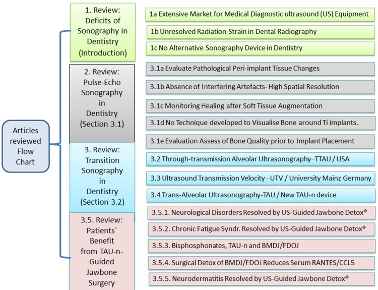
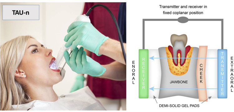
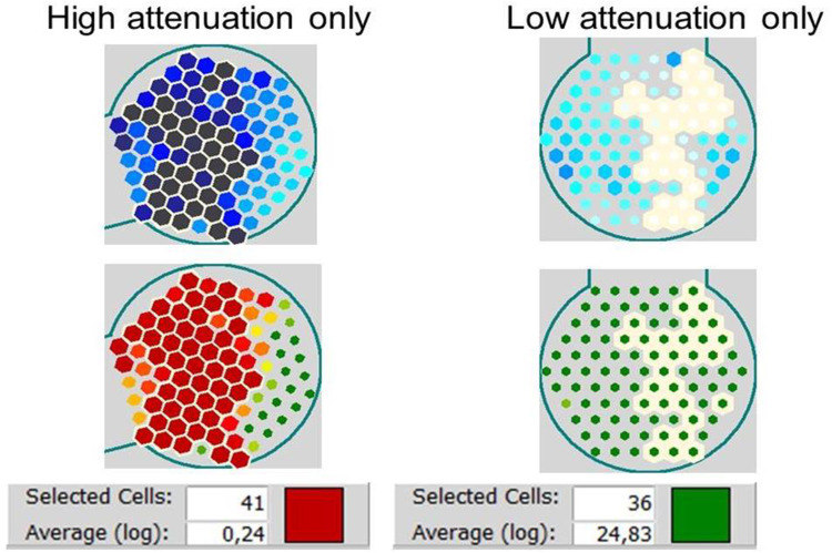
Despite the widespread use of ultrasonography (US) in medical diagnostics, there is no similar US device available for visualizing jawbone density. This study is a narrative review of the possible applications of US in dentistry. This review is divided as follows: (a) Pulse-echo ultrasonography: the applications offer new perspectives for periodontal and peri-implant assessment. (b) Through-transmission alveolar US (TTAU): this technique was a novel imaging modality until 2004, when TTAU devices were last available. Quantitative US scaling made the device useful for diagnosing chronic inflammatory conditions in the jaw. (c) Ultrasound transmission velocity (UTV): in 2008, this technique was introduced in German university dental clinics to analyze the mechanical properties of the jawbone without translating the scientific findings into a practical device. (d) Trans-alveolar US device (TAU): the growing importance of "osteoimmune focal bone marrow defects" has led practitioners to develop a new TAU device. The attenuation of US was used for imaging of jawbone density. (e) Patients who benefit from TAU-guided jawbone surgery: research has shown remarkable results in specific disease cases. This review concludes that US has been undervalued as a diagnostic tool in dentistry. The new TAU-n unit offers the opportunity to change this in the future.

Keywords: dentistry; jawbone density; osteonecrosis; radiation protection; trans-alveolar ultrasound; ultrasonography.


Conflict of interest statement

The authors report no conflicts of interest in this work.

Figures

None
Graphical abstract
Figure 1
Figure 1
Left window: areas of osteonecrosis on TAU-n measurement depicted in green (healthy) and red (inflamed medullary space), with normal radiographs. “Grd-0 to Grd-4” display the grading of the US attenuation by fatty degenerative tissue inside the bone marrow. 35 to 38 describes the alveolar areas of US transmission in the left lower jawbone (European scaling). Red circle describes the inflamed alveolar area and corresponds to the TAU-n scaling. Right window: example of characteristic osteonecrosis in the jawbone, also known in the literature as a bone marrow defect in the jawbone (BMDJ/FDOJ). Adapted from Lechner J, Von Baehr V. Silent inflammation in the jaw and neurological dysregulation – case study linking RANTES/CCL5 overexpression in jawbone with chemokine receptors in the central nervous system. J Dent Oral Health. 2017;3:68. Creative Commons.
Figure 2
Figure 2
Left window: The TAU-n transmitter and receiver are fixed in a parallel coplanar position with a single handpiece. The size of the TAU-n receiver is such that it can be easily placed in the patient’s mouth. The jawbone must be positioned between the two parts of the unit, sender outside and receiver inside the oral cavity. Right window: Acoustic coupling between the extraoral US transmitter and the intraoral US receiver is optimized and personalized with special demi-solid US gel pads for the parts of the unit to be placed inside and outside of the patient’s mouth. Adapted from Lechner J, Zimmermann B, Schmidt M. Focal bone-marrow defects in the jawbone determined by ultrasonography-validation of new trans-alveolar ultrasound technique for measuring jawbone density in 210 participants. Ultrasound Med Biol. 2021;47:3135–3146. https://creativecommons.org/licenses/by-nc-nd/4.0/.
Figure 3
Figure 3
Left window, top bar: TAU-n displays the results in images with different colors depending on the degree of attenuation (green is high bone density, ie, healthy jawbone, and red is low bone density, ie, bone marrow oedema). Left window, lower part: OPG of corresponding areas 37 to 39 (European scaling). Red circle displays the bone marrow defect in the jawbone (BMDJ/FDOJ) corresponding to the TAU-n pictures at the top bar. In area 38 (EU scaling) the blue rectangle indicates specific area of TAU-n imaging. Note: Area 39 shows the edentulous retromolar jawbone. Right window: example of characteristic osteonecrosis in the jawbone, also referred to in the literature as a bone marrow defect in the jawbone (BMDJ/FDOJ).
Figure 4
Figure 4
The CTU was measured by logarithmically averaging the sensor array values with the TAU instrument. Note: Numerical representation of TAU attenuation coefficients for reduced bone density (left panel) and dense material (right panel). Selected sensor cells (left panel: high attenuation; right panel: low attenuation) are indicated by a white outline. The evaluation is presented in the lower window for several selected sensor cells; the result is presented as a logarithmic mean value associated with a corresponding colour (ie, left panel: red corresponds to high attenuation; right panel: green corresponds to low attenuation). The TAU software allows the mean value to be calculated over a freely selected range of the 91 piezoelectric sensors. Reprinted from Ultrasound Med Biol, volume 47, Lechner J, Zimmermann B, Schmidt M. Focal bone-marrow defects in the jawbone determined by ultrasonography-validation of new trans-alveolar ultrasound technique for measuring jawbone density in 210 participants. 3135–3146, Copyright 2021. Creative Commons CC-BY-NC-ND.
None

References

    1. Surowiec RK, Does MD, Nyman JS. In Vivo assessment of bone quality without X-rays. Curr Osteoporos Rep. 2024. PMID: 38227178. doi:10.1007/s11914-023-00856-w - DOI - PMC - PubMed
    1. Ko TJ, Byrd KM, Kim SA. The chairside periodontal diagnostic toolkit: past, present, and future. Diagnostics. 2021;11(6):932. - PMC - PubMed
    1. Rodriguez Betancourt A, Samal A, Chan HL, Kripfgans OD. Overview of ultrasound in dentistry for advancing research methodology and patient care quality with emphasis on periodontal/peri-implant applications. Z Med Phys. 2023;33(3):336–386. PMID: 36922293; PMCID: PMC10517409. doi:10.1016/j.zemedi.2023.01.005 - DOI - PMC - PubMed
    1. Nguyen KT, Pacheco-Pereira C, Kaipatur NR, Cheung J, Major PW, Le LH. Comparison of ultrasound imaging and cone-beam computed tomography for examination of the alveolar bone level: a systematic review. PLoS One. 2018;13(10):e0200596. - PMC - PubMed
    1. Chan HL, Kripfgans OD. Ultrasonography for diagnosis of peri-implant diseases and conditions: a detailed scanning protocol and case demonstration. Dentomaxillofac Radiol. 2020;49(7):20190445. - PMC - PubMed

LinkOut - more resources