EARLY SURGERY IN RARE KNEE HETEROTOPIC OSSIFICATION LEADS TO SUCCESSFUL FUNCTIONAL OUTCOME: A CASE REPORT
- PMID: 39801832
- PMCID: PMC11711688
- DOI: 10.2340/jrm-cc.v8.41323
EARLY SURGERY IN RARE KNEE HETEROTOPIC OSSIFICATION LEADS TO SUCCESSFUL FUNCTIONAL OUTCOME: A CASE REPORT
Abstract
Background: Heterotopic ossification is a common complication after joint replacement surgery, such as hip or knee arthroplasty. In the intensive care unit, it is most commonly associated with traumatic brain injury or spinal cord injury. To prevent recurrence, surgical resection of heterotopic ossification is recommended once the ectopic bone has fully matured, which is estimated to occur after at least 12 months.
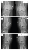
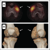
Case presentation: This case describes a young woman with no relevant previous medical history who developed severe bilateral heterotopic ossification on the anteromedial sides of her knees after an intensive care unit stay. Passive flexion of both knees was limited to 50°. X-ray was a simple diagnostic tool. Predisposing factors were extended immobilization, prolonged systematic inflammatory condition and mechanical ventilation. Due to the failure of initial conservative therapy, the heterotopic ossification was resected early, 4 months after onset of first symptoms. Following an intensive rehabilitation program, a normal, pain-free gait and full range of motion of both knees were achieved 9 months after surgery.
Conclusion: This case report demonstrates that early resection of heterotopic ossification can result in a good clinical and functional outcome.
Keywords: early surgery; functional outcome; heterotopic ossification; knee; rehabilitation.
Plain language summary
Heterotopic ossification is the formation of bone in soft tissues, typically around joints like hip, knee and shoulder, causing significant pain and loss of function in the affected limb. Generally, surgical resection of the excess bone is recommended once the heterotopic ossification is fully matured, which may take at least 12 months. In this case, a young woman developed severe heterotopic ossification on the inner sides of both knees after a prolonged intensive care unit stay. She experienced intense pain, limited knee bending and severely impaired walking. As initial non-surgical treatments failed, the heterotopic ossification was surgically removed just 4 months after the onset of the first pain symptoms. After surgery and an intensive rehabilitation program, she regained pain-free walking and full range of motion in her knees. This case demonstrates that early surgical intervention for heterotopic ossification can lead to good clinical and functional outcomes.
© 2025 The Author(s).
Conflict of interest statement
The authors have no conflicts of interest to declare.
Figures


Similar articles
-
Acquired heterotopic ossification in hips and knees following encephalitis: case report and literature review.BMC Surg. 2014 Oct 3;14:74. doi: 10.1186/1471-2482-14-74. BMC Surg. 2014. PMID: 25280472 Free PMC article. Review.
-
Surgical excision of heterotopic ossification associated with anti-N-methyl-d-aspartate receptor encephalitis: A case report.Int J Surg Case Rep. 2021 Dec;89:106643. doi: 10.1016/j.ijscr.2021.106643. Epub 2021 Dec 2. Int J Surg Case Rep. 2021. PMID: 34864268 Free PMC article.
-
Acceptable outcome following resection of bilateral large popliteal space heterotopic ossification masses in a spinal cord injured patient: a case report.J Orthop Surg Res. 2010 Jun 22;5:39. doi: 10.1186/1749-799X-5-39. J Orthop Surg Res. 2010. PMID: 20569483 Free PMC article.
-
Bilateral severe heterotopic ossification following primary total knee arthroplasty: a case report in Pakistan.Arthroplasty. 2021 Jan 8;3(1):5. doi: 10.1186/s42836-020-00057-1. Arthroplasty. 2021. PMID: 35236464 Free PMC article.
-
Severe Heterotopic Ossification After Revision Total Knee Arthroplasty: A Case Report and Review of the Literature.J Am Acad Orthop Surg Glob Res Rev. 2022 Nov 11;6(11):e22.00053. doi: 10.5435/JAAOSGlobal-D-22-00053. eCollection 2022 Nov 1. J Am Acad Orthop Surg Glob Res Rev. 2022. PMID: 36733984 Free PMC article. Review.
References
-
- Molligan J, Mitchell R, Schon L, Achilefu S, Zahoor T, Cho Y, et al. . Influence of bone and muscle injuries on the osteogenic potential of muscle progenitors: Contribution of tissue environment to heterotopic ossification. Stem Cells Transl Med 2016; 5(6): 745–753. 10.5966/sctm.2015-0082 - DOI - PMC - PubMed
-
- Shapira J, Yelton MJ, Chen JW, Rosinsky PJ, Maldonado DR, Meghpara M, et al. . Efficacy of NSAIDs versus radiotherapy for heterotopic ossification prophylaxis following total hip arthroplasty in high-risk patients: a systematic review and meta-analysis. Hip Int 2022; 32: 576–590. 10.1177/1120700021991115 - DOI - PubMed
LinkOut - more resources
Full Text Sources
Research Materials

