Review
. 2024 Nov 28;5(1):115-140.
doi: 10.1021/acsmaterialsau.4c00125. eCollection 2025 Jan 8.

Microneedles as a Promising Technology for Disease Monitoring and Drug Delivery: A Review

Affiliations
Review

Microneedles as a Promising Technology for Disease Monitoring and Drug Delivery: A Review

Rashmi Hulimane Shivaswamy et al. ACS Mater Au. .

Abstract

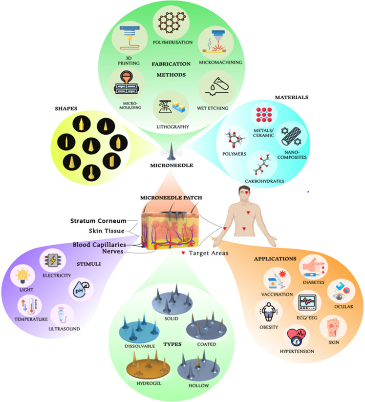
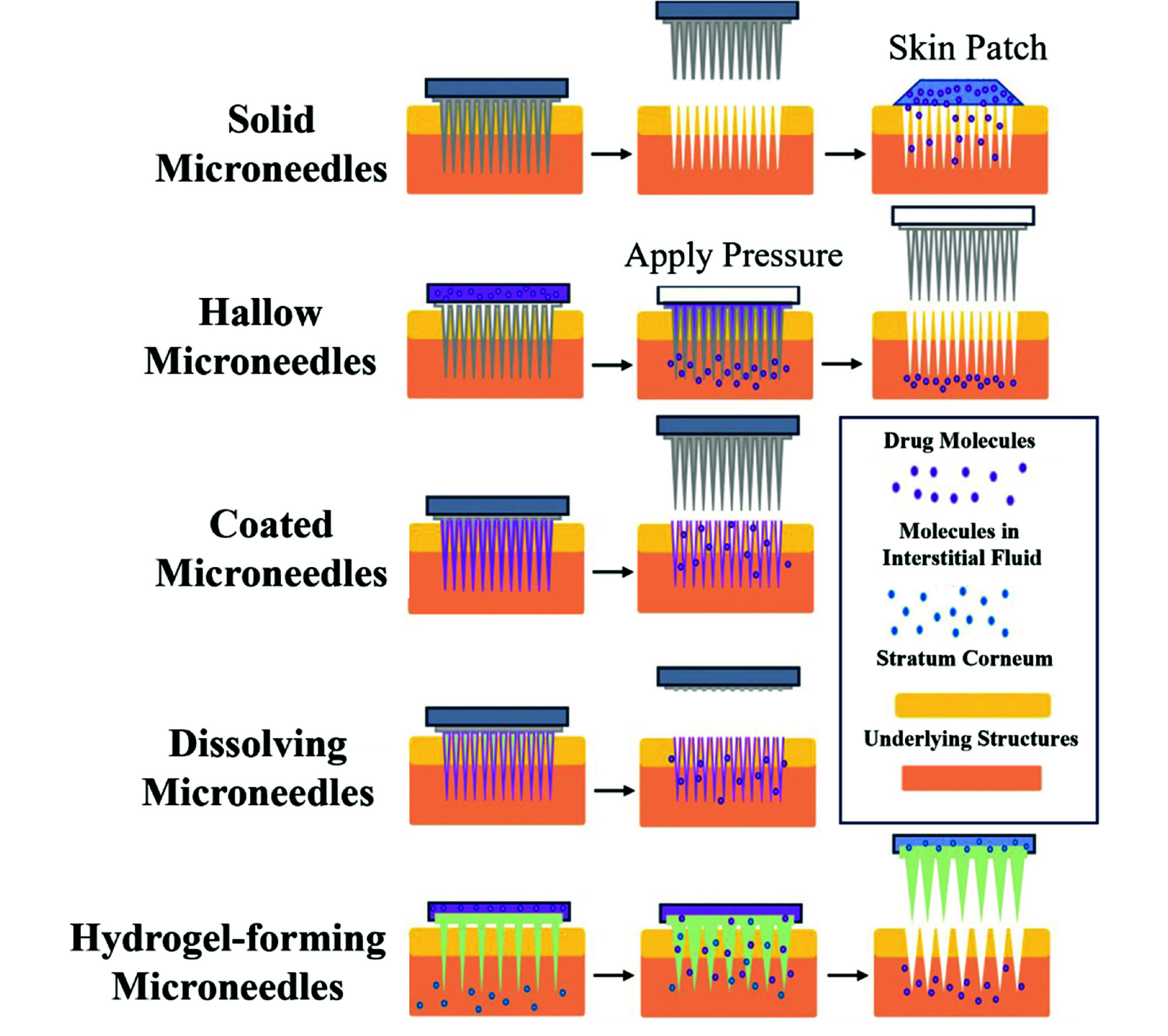
The delivery of molecules, such as DNA, RNA, peptides, and certain hydrophilic drugs, across the epidermal barrier poses a significant obstacle. Microneedle technology has emerged as a prominent area of focus in biomedical research because of its ability to deliver a wide range of biomolecules, vaccines, medicines, and other substances through the skin. Microneedles (MNs) form microchannels by disrupting the skin's structure, which compromises its barrier function, and facilitating the easy penetration of drugs into the skin. These devices enhance the administration of many therapeutic substances to the skin, enhancing their stability. Transcutaneous delivery of medications using a microneedle patch offers advantages over conventional drug administration methods. Microneedles containing active substances can be stimulated by different internal and external factors to result in the regulated release of the substances. To achieve efficient drug administration to the desired location, it is necessary to consider the design of needles with appropriate optimized characteristics. The choice of materials for developing and manufacturing these devices is vital in determining the pharmacodynamics and pharmacokinetics of drug delivery. This article provides the most recent update and overview of the numerous microneedle systems that utilize different activators to stimulate the release of active components from the microneedles. Further, it discusses the materials utilized for producing microneedles and the design strategies important in managing the release of drugs. An explanation of the commonly employed fabrication techniques in biomedical applications and electronics, particularly for integrated microneedle drug delivery systems, is discussed. To successfully implement microneedle technology in clinical settings, it is essential to comprehensively assess several factors, such as biocompatibility, drug stability, safety, and production cost. Finally, an in-depth review of these criteria and the difficulties and potential future direction of microneedles in delivering drugs and monitoring diseases is explored.


Conflict of interest statement

The authors declare no competing financial interest.

Figures

Figure 1
Figure 1
Schematic illustration depicting the numerous types of microneedles, the materials employed in their manufacture, the diverse forms and fabrication techniques utilized, the different stimuli that activate drug delivery as needed, and the wide range of uses for microneedle patches.
Figure 2
Figure 2
Different types of microneedles and their mode of action during drug delivery and ISF extraction. Adopted with permission under a Creative Commons CC BY-4.0 license from ref (14). Copyright 2020 The Authors.
Figure 3
Figure 3
(a) Schematic of BP/GT MPs and MPs-MN patch. Adopted with permission from ref (30). Copyright 2021 Wiley. (b) Assembled ePatch applied on human subjects. Adopted with permission under Creative Commons CC BY-4.0 license from ref (42). Copyright 2024 The Authors. (c) Dissolving microneedles composed of thermoresponsive material. Adopted with permission under a Creative Commons CC-BY-4.0 license from ref (50). Copyright 2024 The Authors. (d) Schematic illustration of drug-coated bubble generating MNs. Adopted with permission from ref (49). Copyright 2021 The Author(s).
Figure 4
Figure 4
(a) Schematic representation of coaxial electrospinning process. Adopted with permission under Creative Commons CC-BY-4.0 license from ref (92). Copyright 2020 The Author(s). (b) In situ fabrication and processing of PVDF-TrFE by corona poling. Adopted with permission under Creative Commons CC-BY-4.0 from ref (93). Copyright 2021 The Authors. (c) Template wetting process for PLLA nanowire formation. Adopted with permission under Creative Commons CC-BY-4.0 from ref (98). Copyright 2017 The Author(s). (d) Epitaxial growth of PVDF-TrFE on chitin. Adopted with permission under a Creative Commons CC-BY-4.0 license from ref (99). Copyright 2020 The Author(s).
Figure 5
Figure 5
(a) XRD patterns: (i) dexamethasone-loaded microparticles encapsulated in dissolving MN array. Adopted with permission under a Creative Commons CC-BY-4.0 license from ref (110). Copyright 2023 The Authors(s). (ii) Chitosan and chitosan formate. Adopted with permission under a Creative Commons attribution 3.0 unported license from ref (104). Copyright 2017 Royal Society of Chemistry. (b) FTIR spectra: (i) MNs containing inclusion complexes loaded with progesterone. Adopted with permission under a Creative Commons CC-BY-4.0 license from ref (111). Copyright 2023 The Author(s). (ii) Chitosan and chitosan formate. Adopted with permission under a Creative Commons attribution 3.0 unported license from ref (104). Copyright 2017 Royal Society of Chemistry. (c) SEM images: (i) PLA and P(3HB-co-3HV) polymer surface morphology before and after melting/cooling. Adopted with permission from ref (112). Copyright 2020 American Chemical Society. (ii) DS@PEG–PLGA microcapsules. Adopted with permission from ref (108). Copyright 2019 Royal Society of Chemistry. (d) (i) TEM images and (ii) diffraction pattern of MWCNTs dispersed in ethanol after a sonication time of 4 h. Adopted with permission under a Creative Commons CC-BY-4.0 license from ref (109). Copyright 2018 The Authors.
Figure 6
Figure 6
(a) Fabrication of microneedle array by photolithography combined with deep reactive ion etching. Adopted with permission under a Creative Commons CC-BY-4.0 license from ref (142). Copyright 2021 The Author(s). (b) Laser ablation: (i) CO2 laser cutting for acrylic mold fabrication by crossover lines (COL) technique and (ii) PDMS mold fabrication using acrylic mold. Adopted with permission under a Creative Commons CC-BY-4.0 license from ref (143). Copyright 2018 The Author(s). (c) Microneedles via direct ink drawing. Adopted with permission from ref (144). Copyright 2023 American Chemical Society. (d) 3D-printed microneedles. Adopted with permission from ref (145). Copyright 2018 Royal Society of Chemistry.
Figure 7
Figure 7
Microneedle-based devices for drug delivery. (a) (i) Optical image of microneedle without and with gold coating after various periods in artificial tissue. Gold needles can be seen to be intact. (ii) Image of the wireless flexible patch. (iii) Top view of the wireless patch with electronics visible. Adopted with permission under a Creative Commons CC-BY-4.0 license from ref (150). Copyright 2024 The Author(s). (b) An iontophoresis-driven microneedle patch for active delivery of charged macromolecular drugs. Adopted with permission from ref (151). Copyright 2020 Acta Materialia Inc. (c) An implantable and bioresorbable microneedle device that provides wireless electrostimulation and sustained drug delivery for tissue regeneration. Adopted with permission from ref (154). Copyright 2022 American Chemical Society. (d) Illustration of conductive microneedle patch using polylactic acid–platinum–polypyrrole (PLA-Pt-PPy) for controlled transdermal drug delivery for atopic dermatitis. Adopted with permission from ref (39). Copyright 2022 American Chemical Society. (e) Images of smartphone-powered microneedle patch that uses iontophoresis to deliver drugs through microholes created by the microneedle patch. Adopted with permission under a Creative Commons CC-BY-4.0 license from ref (155). Copyright 2020 The Authors.
Figure 8
Figure 8
Microneedle based devices for disease diagnosis. (a) Wearable hollow microneedle patch for monitoring glucose in interstitial fluid. Adopted with permission from ref (156). Copyright 2021 Wiley. (b) Three-electrode wearable microneedle continuous glucose monitoring system. Adopted with permission from ref (157). Copyright 2024 Elsevier. (c) Touch-activated microneedle glucose sensor using reverse iontophoresis. Adopted with permission from ref (158). Copyright 2022 Elsevier. (d) 3D printed microneedle device for continuous glucose monitoring: (i) illustration and (ii) camera image. Adopted with permission under a Creative Commons CC-BY-4.0 license from ref (159). Copyright 2021 The Author(s).
Figure 9
Figure 9
Microneedle-based dual-function devices (both drug delivery and disease monitoring). (a) (j) Closed-loop system that senses glucose and automatically delivers insulin, Adopted with permission from ref (70). Copyright 2023 American Chemical Society. (b) Wearable microneedle patch for continuous electrochemical monitoring and delivery of methotrexate through ISF. Adopted with permission from ref (160). Copyright 2023 American Chemical Society. (c) A fully integrated wearable closed-loop system (IWCS) based on a mini-invasive microneedle platform is developed for in situ diabetic sensing and treatment. Adopted with permission under a Creative Commons CC-BY-4.0 license from ref (161). Copyright 2021 The Authors.

Similar articles

References

    1. Waghule T.; Singhvi G.; Dubey S. K.; Pandey M. M.; Gupta G.; Singh M.; Dua K. Microneedles: A Smart Approach and Increasing Potential for Transdermal Drug Delivery System. Biomedicine & Pharmacotherapy 2019, 109, 1249–1258. 10.1016/j.biopha.2018.10.078. - DOI - PubMed
    1. Li Z.; Fang X.; Yu D. Transdermal Drug Delivery Systems and Their Use in Obesity Treatment. Int. J. Mol. Sci. 2021, 22 (23), 12754.10.3390/ijms222312754. - DOI - PMC - PubMed
    1. Schafer N.; Balwierz R.; Biernat P.; Ochędzan-Siodłak W.; Lipok J. Natural Ingredients of Transdermal Drug Delivery Systems as Permeation Enhancers of Active Substances through the Stratum Corneum. Mol. Pharmaceutics 2023, 20 (7), 3278–3297. 10.1021/acs.molpharmaceut.3c00126. - DOI - PMC - PubMed
    1. Sugumar V.; Hayyan M.; Madhavan P.; Wong W. F.; Looi C. Y. Current Development of Chemical Penetration Enhancers for Transdermal Insulin Delivery. Biomedicines 2023, 11 (3), 664.10.3390/biomedicines11030664. - DOI - PMC - PubMed
    1. Azagury A.; Khoury L.; Enden G.; Kost J. Ultrasound Mediated Transdermal Drug Delivery. Adv. Drug Delivery Rev. 2014, 72, 127–143. 10.1016/j.addr.2014.01.007. - DOI - PubMed

LinkOut - more resources