

Metastatic Sclerosing Epithelioid Fibrosarcoma at Diagnosis: A Case Report
- PMID: 39803080
- PMCID: PMC11722666
- DOI: 10.7759/cureus.75544
Metastatic Sclerosing Epithelioid Fibrosarcoma at Diagnosis: A Case Report
Abstract
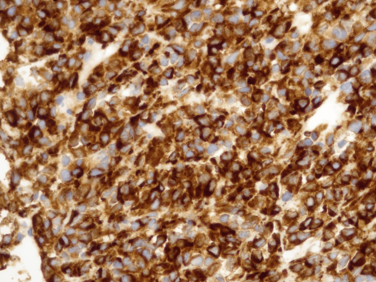
Sclerosing epithelioid fibrosarcoma (SEF) is a rare and aggressive neoplasm composed of epithelioid cells arranged in strands and nests embedded in a highly sclerotic collagenous stroma. We report a case of a 36-year-old man who started with lumbar pain, with extension to both legs, night sweats, and weight loss. He underwent magnetic resonance imaging (MRI) of the lumbar spine; computed tomography (CT) scan of the chest, abdomen, and pelvis; and [18F]-fluorodeoxyglucose positron emission tomography/computed tomography (18F-FDG PET/CT) scan. The CT scan revealed a 13 cm thoracic mass, the MRI presented with diffuse neoplastic invasion of the vertebrae, and the PET showed hepatic, bone, and possibly pulmonary metastases. The histological diagnosis was compatible with SEF. The disease progressed very quickly, namely, with an episode of spinal cord compression, which made the patient paraplegic. He underwent surgery and, subsequently, radiotherapy (RT). Due to the clinical and analytical evolution, it was not possible to initiate systemic treatment and the patient ultimately passed away. In conclusion, SEF is an aggressive type of sarcoma that affects middle-aged patients, with high rates of distant metastases and mortality. The usual treatment is surgery followed by either radiotherapy or chemotherapy. However, further clinical trials are needed to find more systemic target therapies.
Keywords: metastatic sarcoma; oncology; palliative care; radiation oncology; sclerosing epithelioid fibrosarcoma.
Copyright © 2024, Miguel et al.
Conflict of interest statement
Human subjects: Consent for treatment and open access publication was obtained or waived by all participants in this study. Health Ethics Committee of the Portuguese Institute of Oncology (CES-IPOP) issued approval 048/024. Conflicts of interest: In compliance with the ICMJE uniform disclosure form, all authors declare the following: Payment/services info: All authors have declared that no financial support was received from any organization for the submitted work. Financial relationships: All authors have declared that they have no financial relationships at present or within the previous three years with any organizations that might have an interest in the submitted work. Other relationships: All authors have declared that there are no other relationships or activities that could appear to have influenced the submitted work.
Figures


References
-
- Sclerosing epithelioid fibrosarcoma. A variant of fibrosarcoma simulating carcinoma. Meis-Kindblom JM, Kindblom LG, Enzinger FM. Am J Surg Pathol. 1995;19:979–993. - PubMed
-
- Fibrosarcoma: a review and update. Folpe AL. Histopathology. 2014;64:12–25. - PubMed
-
- Sclerosing epithelioid fibrosarcoma: in-depth review of a genetically heterogeneous tumor. Murshed KA, Al-Bozom I, Ammar A. APMIS. 2021;129:455–460. - PubMed
-
- The clinicopathological spectrum of sclerosing epithelioid fibrosarcoma: report of an additional series with review of the literature. Peng Y, Zhang D, Lei T, et al. Pathology. 2023;55:355–361. - PubMed
Publication types
LinkOut - more resources
Full Text Sources