

Vitrectomy for diabetic retinopathy: A review of indications, techniques, outcomes, and complications
- PMID: 39803397
- PMCID: PMC11717329
- DOI: 10.4103/tjo.TJO-D-23-00108
Vitrectomy for diabetic retinopathy: A review of indications, techniques, outcomes, and complications
Abstract
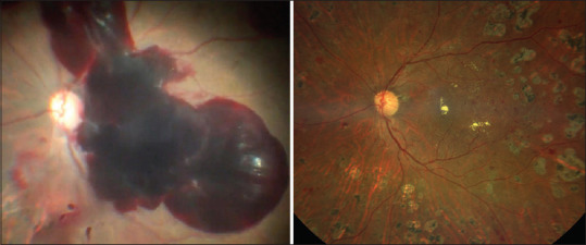
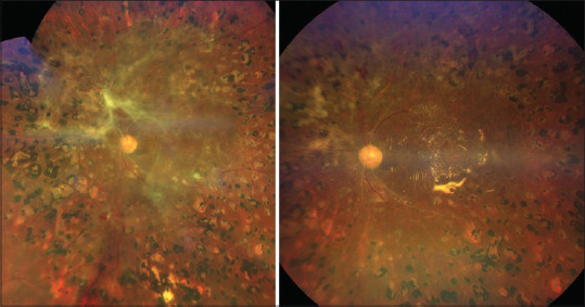
Diabetic retinopathy is one of the most severe forms of retinopathy and a leading cause of blindness all over the world. Of a greater concern is proliferative diabetic retinopathy which leads to vitreous haemorrhage and tractional retinal detachment in such cases. A majority of these cases require a surgical intervention to improve vision and prevent further vision loss. Surgical manouevers in these cases require a complex combination of vitrectomy, membrane dissection, judious usage of endodiathermy, endolaser, vital dyes, bimanual dissection and usage of intraoperative and post-operative tamponades. Each case presents a unique challenge and necessitates an appropriate combination of the steps mentioned above. In the current review we present the current understanding of the need for surgery in diabetic retinopathy, various surgical approaches and a summary of current literature on the same. Multiple surgical video clips demonstrating these steps are also included in this review.
Keywords: Diabetic vitrectomy; posterior hyaloid; proliferative diabetic retinopathy; subhyaloid hemorrhage; vitreous hemorrhage.
Copyright: © 2024 Taiwan J Ophthalmol.
Conflict of interest statement
The authors declare that there are no conflicts of interests of this paper.
Figures

References
-
- Leasher JL, Bourne RR, Flaxman SR, Jonas JB, Keeffe J, Naidoo K, et al. Global estimates on the number of people blind or visually impaired by diabetic retinopathy: A meta-analysis from 1990 to 2010. Diabetes Care. 2016;39:1643–9. - PubMed
-
- Sabanayagam C, Banu R, Chee ML, Lee R, Wang YX, Tan G, et al. Incidence and progression of diabetic retinopathy: A systematic review. Lancet Diabetes Endocrinol. 2019;7:140–9. - PubMed
Publication types
LinkOut - more resources
Full Text Sources