[Preprint]. 2025 Jan 3:2025.01.03.631205.
doi: 10.1101/2025.01.03.631205.

Crystal structure of MutYX: A novel clusterless adenine DNA glycosylase with a distinct C-terminal domain and 8-Oxoguanine recognition sphere

Affiliations

Crystal structure of MutYX: A novel clusterless adenine DNA glycosylase with a distinct C-terminal domain and 8-Oxoguanine recognition sphere

C H Trasviña-Arenas et al. bioRxiv. .

Abstract

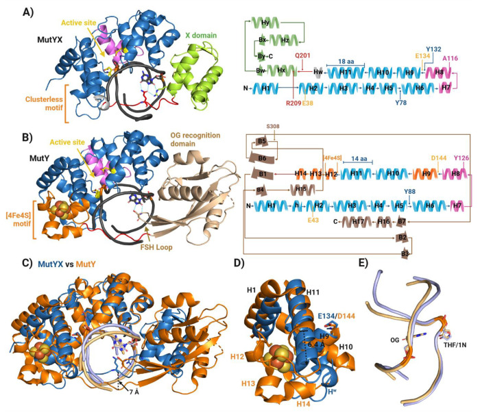
The [4Fe-4S] cluster is an important cofactor of the base excision repair (BER) adenine DNA glycosylase MutY to prevent mutations associated with 8-oxoguanine (OG). Several MutYs lacking the [4Fe-4S] cofactor have been identified. Phylogenetic analysis shows that clusterless MutYs are distributed in two clades suggesting cofactor loss in two independent evolutionary events. Herein, we determined the first crystal structure of a clusterless MutY complexed with DNA. On the basis of the dramatic structural divergence from canonical MutYs, we refer to this as representative of a clusterless MutY subgroup "MutYX". Interestingly, MutYX compensates for the missing [4Fe-4S] cofactor to maintain positioning of catalytic residues by expanding a pre-existing α-helix and acquisition of the new α-helix. Surprisingly, MutYX also acquired a new C-terminal domain that uniquely recognizes OG using residue Gln201 and Arg209. Adenine glycosylase assays and binding affinity measurements indicate that Arg209 is the primary residue responsible to specificity for OG:A lesions, while Gln201 bridges OG and Arg209. Surprisingly, replacement of Arg209 and Gln201 with Ala increases activity toward G:A mismatches. The MutYX structure serves as an example of devolution, capturing structural features required to retain function in the absence of a metal cofactor considered indispensable.

Keywords: Base Excision Repair; DNA glycosylase; Evolution; MutY; [4Fe-4S] cluster.


Figures

Figure 1.
Figure 1.
Overview of Eggerthella sp. MutYX structure. A) MutYX structure in complex with DNA. Each domain and motif are colored differently and indicated with labels. In orange is the region for the [4Fe-4S] cluster in canonical MutYs. B) and C) Region of MutYX structure highlighting important active site and OG recognition residues, respectively. The 2|Fo|–|Fc| map (green) was calculated to the 1.55 Å resolution limit and contoured at 1.0 rmsd. H-bonds are indicated with cyan dotted lines and water with red small spheres. D) Phylogenetic tree of MutY modified from Trasviña-Arenas et al, 2016. The clusterless MutY clades are highlighted in green and cyan. Only representative names for Bacillales, Lactobacillales and Anaerobic clades are included. For the full clade members please review reference (21).
Figure 2.
Figure 2.
Structural comparison between MutYX and canonical bacterial MutY. A) Structural overview of MutYX in complex with THF:OG-containing DNA. On the right panel a map of the secondary structure is displayed highlighting key residues for its activity. B) Crystal structure of G. stearothermophilus MutY in complex to the transition state analog 1N (PDB 6U7T). On the right panel a map of the secondary structure is displayed highlighting key residues for its activity. C) Structural alignment between MutYX (blue) and GsMutY (orange). The internalization of the IDC in MutYX is indicated with a black arrow. D) Magnified view of the MutYX vs MutY structural alignment showing the [4Fe-4S] cluster region. The elongation of the α-helix 11 in MutYX s indicated with a dotted arrow. E) Comparison of the DNA conformations excreted by their interaction with MutYX (light blue) and MutY (light orange). OG, AP site analogs (THF) and transition state analog (1N) are shown in sticks.
Figure 3.
Figure 3.
Representation of residues within the active site and the OG recognition sphere. A) Configuration of important residues within the active site of MutYX and canonical MutY (GsMutY; PDB 6U7T). Catalytic residues are colored in yellow, residue from the catalytic domain, pink from the Helix-hairpin-Helix (HhH) motif. Water molecules are shown in red small spheres, AP site analogs in black sticks and H-bonds in cyan dotted lines. B) Structural alignment of residues from MutYX’s and MutY’s active sites. Conserved water molecules within the active site are circled. Conserved H-bonds are indicated with black arrows and structural displacements or bending are shown with blue arrows. C) Residues involved in the OG recognition in MutYX and MutY. The residues from the catalytic domain that interact the Watson-crick face and Hoogsteen face of OG (gray) are in blue sticks. Q201 and R209 residues in MutYX which recognize exclusively the Hoogsteen face of OG from the IDC region and X domain are in red and green, respectively. The canonical S308 from the FSH loop in canonical MutYs are displayed in light brown. D) Structural alignment of residues of the OG recognition sphere from MutYX and MutY. Conserved H-bonds are indicated with black arrows while exclusive H-bonds of MutYX are blue arrows.
Figure 4.
Figure 4.
Structural analysis of the [4Fe-4S] cluster motif; its role in DNA-protein interactions and structural dispensability in MutYX. A) Close-up of the [4Fe-4S] cluster motif (orange) in MutY (Left panel) and corresponding region of MutYX (Right panel). The [4Fe-4S] cluster is indicated with dotted contour and R201 residue is showcased interacting with the phosphodiester backbone at the third nucleobase (−3) downstream the AP site analog. Similarly, the MutYX’s R193 from the α-helix Hw has the same interaction pattern as MutY’s R201. B) MutY α-helix H9 from the [4Fe-4S] cluster motif is stabilized by an intricated H-bond network in MutY (Left panel) mainly through 4 anchoring or stabilization points (red numbers with asterisks) which involves the cysteinyl ligand. In the right panel the reconfiguration of corresponding region of the [4Fe-4S] cluster motif in MutYX is shown. The intricate H-bond network is conserved where the novel α-helix Hw (R194) and the extended H11 (Y180) of MutYX participate as a fourth and fifth anchoring points for α-helix H9 stabilization. The 2|Fo|–|Fc| map (green) was calculated to the 1.55 Å resolution limit and contoured at 1.0 rmsd.
Figure 5.
Figure 5.. MutYX OG:A lesion affinity and specificity is dictated by Arg209 and Gln 201.
A) Fluorescence polarization experiments assess relative binding affinities of MutYX and mutants relative to EcMutY for 30-bp DNA duplex containing OG (top left) or G (top right) across from a non-cleavable 2’-fluorine-2’-deoxyadenosine analog (fA). B) Single turnover (STO) glycosylase assays determination of adenine excision rate constant (k2). The STO experiments were performed using 100 nM of active enzyme concentration and 20 nM of DNA substrates at 20 °C. C) Plot displaying substrate preference based on the k2-associated specificity (A:OG k2/A:G k2).
Figure 6.
Figure 6.
Logo sequence showing the conservation of important residues involved in catalysis and OG recognition in canonical MutY (Bacillale clade), clusterless MutY (Lactobacillus clade) and MutYX (Actinobacteria and Entamoeba/Chlorobi clade). The catalytic residues are indicated with red arrows, α-helix H9 stabilization residues with blue arrows, MutYX’s and MutY’s OG recognition residues with green and brown arrows, respectively. Double headed arrows indicate conservation on both MutYX and canonical MutY, and the direction of the single-headed arrows indicates exclusive conservation. Curved arrow shows the positions involved in the Tyr↔Ala swapping in the active site.
Scheme 1:
Scheme 1:

References

    1. Guan Y., Manuel R.C., Arvai A.S., Parikh S.S., Mol C.D., Miller J.H., Lloyd R.S. and Tainer J.A. (1998) MutY catalytic core, mutant and bound adenine structures define specificity for DNA repair enzyme superfamily. Nature Structural and Molecular Biology, 5, 1058. - PubMed
    1. Thayer M.M., Ahern H., Xing D., Cunningham R.P. and Tainer J.A. (1995) Novel DNA binding motifs in the DNA repair enzyme endonuclease III crystal structure. The EMBO journal, 14, 4108–4120. - PMC - PubMed
    1. Hinks J.A., Evans M.C., De Miguel Y., Sartori A.A., Jiricny J. and Pearl L.H. (2002) An iron-sulfur cluster in the family 4 uracil-DNA glycosylases. Journal of Biological Chemistry, 277, 16936–16940. - PubMed
    1. Mol C.D., Arvai A.S., Begley T.J., Cunningham R.P. and Tainer J.A. (2002) Structure and activity of a thermostable thymine-DNA glycosylase: evidence for base twisting to remove mismatched normal DNA bases. Journal of molecular biology, 315, 373–384. - PubMed
    1. Porello S.L., Cannon M.J. and David S.S. (1998) A substrate recognition role for the [4Fe-4S] 2+ cluster of the DNA repair glycosylase MutY. Biochemistry, 37, 6465–6475. - PubMed

Publication types