[Preprint]. 2024 Dec 31:2024.12.30.630837.
doi: 10.1101/2024.12.30.630837.

Immune Checkpoint Molecules as Biomarkers of Staphylococcus aureus Bone Infection and Clinical Outcome

Affiliations

Immune Checkpoint Molecules as Biomarkers of Staphylococcus aureus Bone Infection and Clinical Outcome

Motoo Saito et al. bioRxiv. .

Abstract

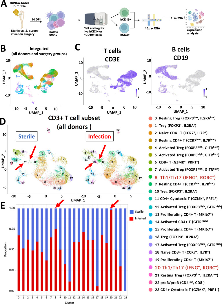
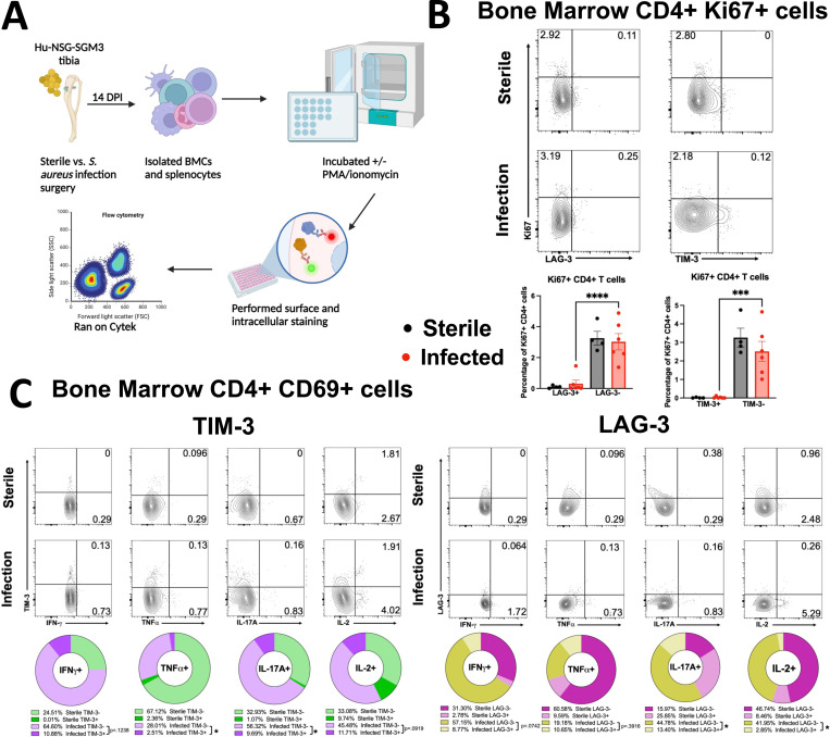
Staphylococcus aureus prosthetic joint infections (PJIs) are broadly considered incurable, and clinical diagnostics that guide conservative vs. aggressive surgical treatments don't exist. Multi-omics studies in a humanized NSG-SGM3 BLT mouse model demonstrate human T cells: 1) are remarkably heterogenous in gene expression and numbers, and 2) exist as a mixed population of activated, progenitor-exhausted, and terminally-exhausted Th1/Th17 cells with increased expression of immune checkpoint proteins (LAG3, TIM-3). Importantly, these proteins are upregulated in the serum and the bone marrow of S. aureus PJI patients. A multiparametric nomogram combining high serum immune checkpoint protein levels with low proinflammatory cytokine levels (IFN-γ, IL-2, TNF-α, IL-17) revealed that TIM-3 was highly predictive of adverse disease outcomes (AUC=0.89). Hence, T cell impairment in the form of immune checkpoint expression and exhaustion could be a functional biomarker for S. aureus PJI disease outcome, and blockade of checkpoint proteins could potentially improve outcomes following surgery.

Keywords: Staphylococcus aureus; T cell exhaustion; host-pathogen interactions; immune checkpoint proteins; osteomyelitis; scRNAseq.


Conflict of interest statement

Conflict of interest statement GM, EMS, and MS are inventors of a patent application filed by the University of Rochester and are currently under an exclusive licensing agreement with TEx Immunetics Inc. (TEX). GM and AG are co-founders of TEX and have stock in TEX. All other authors declare that no conflict of interest exists.

Figures

Figure 1.
Figure 1.. Humanized NSG-SGM3 BLT mice have exacerbated susceptibility to S. aureus osteomyelitis compared to Murinized NSG-SGM3 and C57BL/6 WT mice.
(A) Humanized NSG-SGM3 BLT mice were generated by engrafting with CD34+ human hematopoietic cells, autologous human fetal liver, and thymus from three different human donors. Murinized NSG-SGM3 BLT mice were generated with CD34+ murine hematopoietic cells derived from three different C57BL/6 WT mice. (B) Schematic illustration of the experimental design of in vivo experiments. 20-week-old humanized HuNSG-SGM3 BLT mice, murinized NSG-SGM3 and C57BL6 (WT) mice (n=25) were subjected to transtibial implant-associated osteomyelitis using bioluminescent MRSA (USA300 LAC::lux). (C) Longitudinal BLI images of representative mice with (D) statistical analysis of the groups demonstrate increased in vivo S. aureus growth in humanized NSG-SGM3 BLT mice. (E) In vivo BLI images of a representative NSG-SGM3 BLT mouse with local and disseminated MRSA infections, as evidenced by the focal BLI signal in the tibia and abdominal cavity from supine and prone views, respectively. Autopsy photograph confirmed S. aureus abscesses (yellow arrows) in the liver. (F-I) On day14 post-operation, implants, tibiae, surrounding soft tissues, and internal organs (heart, liver, kidneys, and spleen) were harvested for CFU assays and the data are presented with the mean for each group (n= 25, and differences between groups were assessed by ANOVA, *p<0.05, **p<0.01, ***p<0.001, ***p<0.0001). (J) Representative 10x images of Brown & Brenn (B&B) stained histology of infected tibia from each group are shown, highlighting the SACs (red arrows). (K) VisioPharm histomorphometry was performed to quantify the SAC area per tibia, and the value for each tibia is presented with the mean +/− SD (n≥4, ANOVA, *p<0.05).
Figure 2.
Figure 2.. Single-cell RNAseq reveals remarkable human T cell heterogeneity at the infection site in humanized BLT mice with S. aureus osteomyelitis.
(A) Schematic illustration showing the experimental overview of sc-RNAseq of humanized NSG-SGM3 BLT mice engrafted with three different human donor tissues. Bone marrow (BM) cells were collected from tibiae of humanized NSG-SGM3 BLT mice 14 days after transtibial implants surgery with or without USA300 LAC::lux, and the human CD45+CD19+ B cells and CD45+CD3+ T cells were isolated by FACS for scRNAseq. (B) UMAP of the unsupervised cluster analysis of ~30,000 BM cells with (C) Feature plots of the CD3+ T cells and CD19+ B cells. (D) UMAP and DEG clustering analyses of hCD45+/CD3+ T cells identified 24 T cell clusters with (E) bar graphs displaying the proportion of cell counts in each cluster between sterile implant and infected implant groups. Note the marked increase of Th1/Th17 cells (red arrows, Cluster 8,20) in the infected tibiae compared to unifected tibiae.
Figure 3:
Figure 3:. Immune checkpoint gene expression is elevated in CD4+ Th1/Th17 cells from S. aureus-infected humanized BLT tibiae.
(A) The scRNAseq data of the Th1/Th17 cells (clusters 8 and 20) identified in Figure 2 were subjected to UMAP and differential gene expression analyses (DEG) revealed 7 sub-clusters, and the relative proportions of these sub-clusters in uninfected (blue) and infected (red) tibiae are illustrated by the bar graph. (B) Violin plot analyses demonstrated that these cells were of the Th1/Th17 phenotype. Several Th1/Th17 clusters showed significantly increased expression of immune checkpoint molecules LAG-3, TIM-3 (HAVCR2), and, to a lesser extent, CTLA-4 and other immunosuppressive genes like TIGIT. (C) DEG analyses of transcriptional factors (TCF7, TOX1–2, EOMES, NR4A1), cytokines & chemokines, and chemokine receptor (IL-1, IL-17, CXCL13, CXCR5) associated with functional T cell exhaustion, chronic antigenic stimulation (CD40L) and proliferation (MKi67). Note that the lower expression of TCF7, MKi67, IL-1, and IL-17 genes and higher expression of CXCL13 and TOX 2 indicate transcriptional reprogramming of these cells to a terminally functionally exhausted state (*p<0.05). The Th1/Th17 subclusters were annotated based on the gene expression signatures into activated, progenitor-exhausted, and terminally-exhausted cells. The DEGs between the experimental groups within the Th1/Th17 cells were subjected to Ingenuity Pathway Analysis (IPA) to identify the (D) top significantly enriched canonical pathways and (E) predicted upstream regulators (cytokines, transcriptional factors and transmembrane receptors). Red indicates activation, while blue indicates suppression.
Figure 4.
Figure 4.. Immune checkpoint proteins are elevated in CD4+ T cells from S. aureus-infected humanized BLT tibiae.
(A) Immunofluorescent histochemistry analyses of tibia sections from uninfected and MRSA-infected humanized BLT mice 14 days post-op were performed with labeled antibodies against CD3, LAG-3, TIM-3, and PD-1 with DAPI counter stain, and representative images are shown at 4x. Note the increased numbers of T cells near the SAC (dashed yellow line) in the infected tibiae. (B) A multichromatic spectral flow cytometry analyses were performed on tibial bone marrow cells from uninfected and MRSA-infected BLT mice. Live human CD45+/CD3+/T cells and their subpopulations (CD4+, CD8+, Tregs) were analysed for immune checkpoint expression (LAG3, TIM-3, and PD-1) and proliferation (Ki67), and representative histograms are shown. Note the frequency of human CD3+/CD4+ T cells expressing TIM-3, LAG3 & PD-1) in the cells from MRSA-infected bone marrow (n=4-8 mice, *p<0.05, t-test).
Figure 5:
Figure 5:. Bone marrow CD4+ T cells from MRSA-infected tibiae expressing TIM-3 and LAG3 checkpoint proteins exhibit diminished proliferative capacity and altered cytokine production.
(A) Schematic illustration of the experimental design of ex-vivo experiments. 20-week-old humanized NSG-SGM3 BLT mice were subjected to aseptic or septic transtibial implant-surgery for 14 days, then their splenocytes and bone marrow cells were isolated, stimulated, stained with antibodies, and analyzed by flow cytometry. (B-C) Multichromatic spectral flow cytometry was performed on uninfected and MRSA-infected tibial bone marrow cells from BLT mice (B) on unstimulated cells (C) post-stimulation with PMA/ionomycin. (B) Live human CD45+/CD3+/CD4+ T cells expressing checkpoint molecules TIM-3 and LAG-3 were probed for their proliferative capacity using the cell surface marker Ki67. Note that CD4+TIM-3+ and CD4+LAG-3+ cells have lower amounts of proliferating Ki67+ cells in the bone marrow of infected BLT mice, suggesting functional exhaustion and dysfunction. (C) Live human CD45+/CD3+/CD4+/CD69+ T cells expressing checkpoint molecules TIM-3 and LAG-3 were probed for functional capacity using the cytokines IFN-𝛄, TNFɑ, IL-17A, and IL-2 (n=4–9 mice, *p<0.05, ANOVA).
Figure 6.
Figure 6.. T cells expressing immune checkpoint proteins accumulate in S. aureus infected bone tissue from PJI patients.
Bone tissues surgically removed from PJI patient with S. aureus osteomyelitis were processed for histology and immunohistochemistry. (A) Representative 100x image (bar =100 μm) of a H&E-stained section is shown to illustrate the inflammatory cells within the region of interest (box). (B-D) Parallel histology sections containing the region of interest were immunostained with labelled antibodies against CD3, PD1, S. aureus, TIM-3 (green), LAG-3, and CD66b, counter stained with DAPI, and representative fluorescent microscopy images are shown at 200x (bar = 100 μm). (B) Note CD3+/PD1+ T cells detected in areas of S. aureus infection (white arrows). (C) Note CD3+/TIM-3+ (white arrows) and CD3+/LAG-3+ (yellow arrows) T cells at the site of S. aureus infection. (D) Note TIM-3+/CD66b+ neutrophils at the site of infection (white arrows).
Figure 7.
Figure 7.. TIM-3 protein level in serum is highly prognostic of adverse outcomes in patients with S. aureus osteomyelitis.
(A) Serum samples were collected from healthy arthritis patients undergoing total hip/knee arthroplasty (n=15), and orthopaedic patients undergoing surgery for culture-confirmed S. aureus osteomyelitis whose clinical outcome at 1-year was adverse (n=12), infection controlled (n=11), or inconclusive (14). (B) Immune checkpoint proteins LAG-3, TIM-3, CTLA-4, PD-1 and cytokines (IFN-γ, IL-2, TNFα, IL-17A, IL-17F) were assessed by multiplex Luminex assay, and the data are presented for each patient with the mean +/− SEM for each group. The individual protein levels were utilized to perform receiver operating characteristic (ROC) curve analysis either singly or in combination to generate the area under the curve (AUC) for (C-D) differentiating acute vs. chronic S. aureus infections and (E-F) prognostic prediction of outcome. Interestingly, no correlation was observed between levels of immune checkpoint proteins and clinical time-based, anecdotal classification of acute vs. chronic classification. On the other hand, immune checkpoint proteins, especially TIM-3, were highly predictive of adverse in these patients (*p<0.05, **p<0.01, ****p<0.00001).

References

    1. Schwarz E. M. et al. 2018 International Consensus Meeting on Musculoskeletal Infection: Research Priorities from the General Assembly Questions. J Orthop Res 37, 997–1006 (2019). 10.1002/jor.24293 - DOI - PubMed
    1. Tande A. J. & Patel R. Prosthetic joint infection. Clin Microbiol Rev 27, 302–345 (2014). 10.1128/CMR.00111-13 - DOI - PMC - PubMed
    1. Van Hal S. J. et al. Predictors of Mortality in Staphylococcus aureus Bacteremia. Clinical Microbiology Reviews 25, 362–386 (2012). 10.1128/cmr.05022-11 - DOI - PMC - PubMed
    1. Cram P. et al. Total knee arthroplasty volume, utilization, and outcomes among Medicare beneficiaries, 1991–2010. Jama 308, 1227–1236 (2012). 10.1001/2012.jama.11153 - DOI - PMC - PubMed
    1. Masters E. A. et al. Evolving concepts in bone infection: redefining “biofilm”, “acute vs. chronic osteomyelitis”, “the immune proteome” and “local antibiotic therapy”. Bone Res 7, 20 (2019). 10.1038/s41413-019-0061-z - DOI - PMC - PubMed

Publication types