[Preprint]. 2025 Jan 1:2024.12.30.630810.
doi: 10.1101/2024.12.30.630810.

Fusobacterium nucleatum is enriched in invasive biofilms in colorectal cancer

Affiliations

Fusobacterium nucleatum is enriched in invasive biofilms in colorectal cancer

Jessica Queen et al. bioRxiv. .

Update in

Abstract

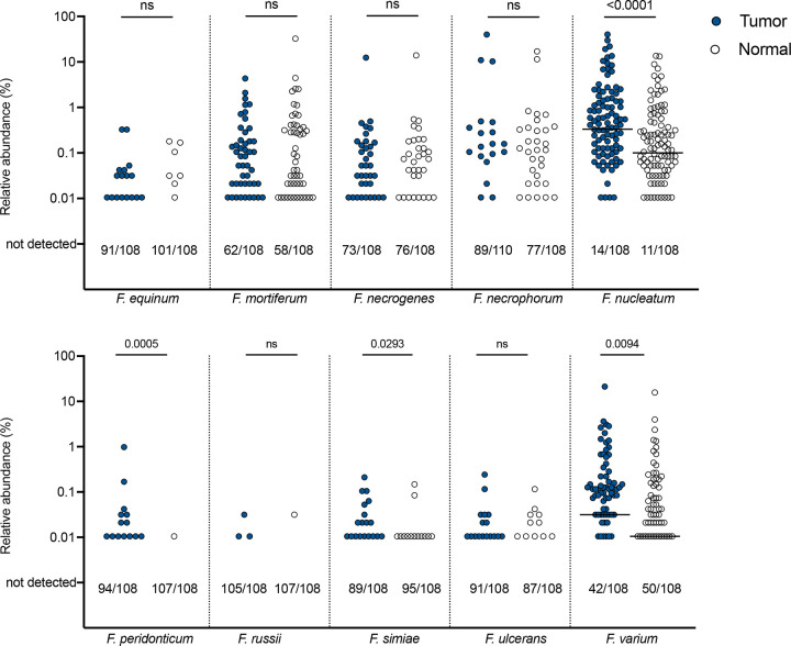
Fusobacterium nucleatum is an oral bacterium known to colonize colorectal tumors, where it is thought to play an important role in cancer progression. Recent advances in sequencing and phenotyping of F. nucleatum have revealed important differences at the subspecies level, but whether these differences impact the overall tumor ecology, and tumorigenesis itself, remain poorly understood. In this study, we sought to characterize Fusobacteria in the tumor microbiome of a cohort of individuals with CRC through a combination of molecular, spatial, and microbiologic analyses. We assessed for relative abundance of F. nucleatum in tumors compared to paired normal tissue, and correlated abundance with clinical and pathological features. We demonstrate striking enrichment of F. nucleatum and the recently discovered subspecies animalis clade 2 (Fna C2) specifically in colon tumors that have biofilms, highlighting the importance of complex community partnerships in the pathogenesis of this important organism.


Conflict of interest statement

C.L.S. has received research funding to Johns Hopkins University from Bristol-Myers Squibb and Janssen, and royalties from Up to Date outside the submitted work. J.R.W. reports equity ownership of Resphera Biosciences.

Figures

Figure 1.
Figure 1.. The CRC cohort.
A) Flow diagram of study participants and downstream analysis of samples. B) Demographic characteristics of the cohort (n=116), including age, sex, ethnicity, stage, and tumor location. EO-CRC: early-onset colorectal cancer, defined as age under 50. LO-CRC: late-onset colorectal cancer, defined as age 50 or above.
Figure 2.
Figure 2.. Biofilm analysis of the CRC cohort.
A) The location and biofilm status of CRC tumors overlaid on a diagram of a colon. One tumor marked as unknown had no fixed tissue suitable for screening. B) Biofilm status of CRC tumors classified as either positive or negative by FISH. C) Percent of left and right-sided tumors positive for biofilms. D) Percent concordance of biofilm status in paired tumor (T) and normal (N) tissue for biofilm-positive (BF+) tumors and biofilm-negative (BF-) tumors. E) Percent of tumors positive for biofilms stratified by tumor location and stage. C-E analyzed by Fisher’s exact test, with p<0.05 considered significant. f) Stacked bar graph depicting percent of tumor biofilms of each subtype screened by multi-probe FISH (n=70) and stratified by tumor stage. Analyzed by nonparametric Kruskal-Wallis one-way ANOVA, with p<0.05 considered significant. G) Representative 40X images of multi-probe FISH of each biofilm subtype stained for DAPI (blue), Bacteroidetes (green), Fusobacterium (yellow), Lachnospiraceae (red), and Proteobacteria (pink).
Figure 3.
Figure 3.. Abundance of Fusobacterium species in tumor/normal pairs.
Relative abundance on a log scale by 16S rRNA amplicon sequencing of each depicted Fusobacterium species in pairs of tumor (blue circle) and normal (clear circle) tissues. Bars indicated median. Numbers below each graph depict the number of samples out of the total in which each species was not detected by sequencing (i.e. 0% abundance). Analyzed by two-tailed Wilcoxon matched-pairs signed rank test, with p<0.05 considered significant. See Fig S5 for abundance of F. nucleatum as a sum of confident and ambiguous species assignments, and for F. varium abundance stratified by ethnicity.
Figure 4.
Figure 4.. Fusobacterium nucleatum abundance by individual and tumor characteristics.
Relative abundance by 16S rRNA amplicon sequencing of Fusobacterium nucleatum in pairs of tumor (blue circle) and normal (clear circle), stratified by A) sex, B) age, C) ethnicity, D) tumor location, E) tumor stage, F) biofilm status, and G) both biofilm status and tumor stage. Bars indicated median. Numbers below each graph depict the number of samples out of the total in which F. nucleatum was not detected by sequencing. Analyzed by two-tailed Wilcoxon matched-pairs signed rank test, with p<0.05 considered significant. EO-CRC: early-onset colorectal cancer. LO-CRC: late-onset colorectal cancer. See Fig S5 for abundance of F. nucleatum as a sum of confident and ambiguous species assignments, stratified by biofilm status. H) Matrix of F. nucleatum and other oral biofilm organism abundances in tumor samples, analyzed by non-parametric Spearman correlation coefficient. See Fig S7 for relative abundance of each organism in the matrix in tumor/normal pairs.
Figure 5.
Figure 5.. Fusobacterium nucleatum subspecies animalis abundance by clade and correlated with FISH.
A) Relative abundance by 16S rRNA amplicon sequencing of Fusobacterium nucleatum subspecies animalis clade 1 (Fna C1) and clade 2 (Fna C2) in pairs of tumor (blue circle) and normal (clear circle). Tumor/normal pairs analyzed by two-tailed Wilcoxon matched-pairs signed rank test, and tumor/tumor or normal/normal comparisons analyzed by two-tailed Mann Whitney test, with p<0.05 considered significant. B) Relative abundance of Fna C1 and C2 stratified by biofilm (BF) status and C-D) by both biofilm status and tumor stage. Bars indicated median. Numbers below each graph depict the number of samples out of the total in which each clade was not detected by sequencing. E) Heatmap depicting relative abundance by 16S rRNA amplicon sequencing for the Fusobacterium genus and each labeled Fusobacterium species or clade for n=29 tumors where dense blooms of Fusobacteria were visualized by FISH.
Figure 6.
Figure 6.. PICRUSt2 pathways associated with Fna C2 abundance in CRC tumors.
Heatmap of all statistically significant PICRUSt2 pathways with differences in gene content between low Fna C2 abundance (<1%) and high abundance (≥1%) in tumors (n=112), as analyzed by Mann Whitney test, with p<0.05 considered significant. Data are further stratified by biofilm (BF) status within each abundance category.

References

    1. Bray F. et al. Global cancer statistics 2018: GLOBOCAN estimates of incidence and mortality worldwide for 36 cancers in 185 countries. CA Cancer J Clin 68, 394–424 (2018). 10.3322/caac.21492 - DOI - PubMed
    1. Knippel R. J. & Sears C. L. The Microbiome Colorectal Cancer Puzzle: Initiator, Propagator, and Avenue for Treatment and Research. J Natl Compr Canc Netw 19, 986–992 (2021). 10.6004/jnccn.2021.7062 - DOI - PubMed
    1. Castellarin M. et al. Fusobacterium nucleatum infection is prevalent in human colorectal carcinoma. Genome Res 22, 299–306 (2012). 10.1101/gr.126516.111 - DOI - PMC - PubMed
    1. Kostic A. D. et al. Genomic analysis identifies association of Fusobacterium with colorectal carcinoma. Genome Res 22, 292–298 (2012). 10.1101/gr.126573.111 - DOI - PMC - PubMed
    1. Mima K. et al. Fusobacterium nucleatum in Colorectal Carcinoma Tissue According to Tumor Location. Clin Transl Gastroenterol 7, e200 (2016). 10.1038/ctg.2016.53 - DOI - PMC - PubMed

Publication types

LinkOut - more resources