[Preprint]. 2024 Nov 15:2024.11.13.623454.
doi: 10.1101/2024.11.13.623454.

Structural insights into SSNA1 self-assembly and its microtubule binding for centriole maintenance

Affiliations

Structural insights into SSNA1 self-assembly and its microtubule binding for centriole maintenance

Lorenzo Agostini et al. bioRxiv. .

Update in

Abstract

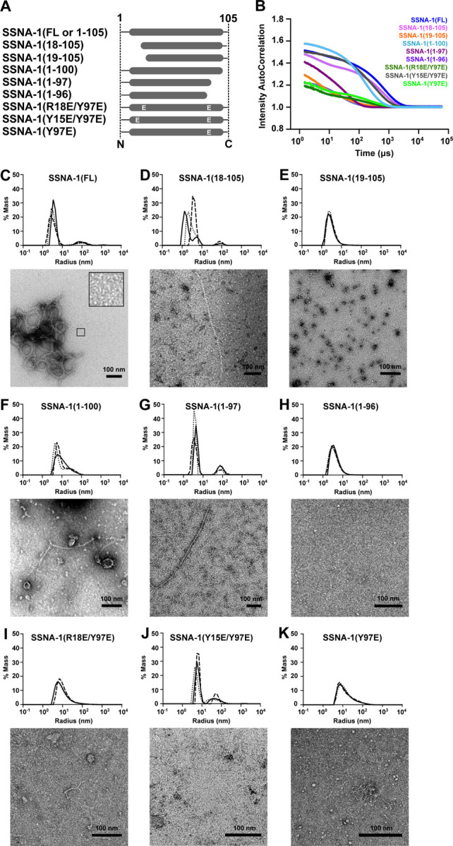
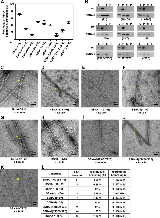
SSNA-1 is a fibrillar protein localized at the area where dynamic microtubule remodeling occurs including centrosomes. Despite the important activities of SSNA1 to microtubules such as nucleation, co-polymerization, and lattice sharing microtubule branching, the underlying molecular mechanism have remained unclear due to a lack of structural information. Here, we determined the cryo-EM structure of C. elegans SSNA-1 at 4.55 Å resolution and evaluated its role during embryonic development in C. elegans. We found that SSNA1 forms an anti-parallel coiled-coil, and its self-assembly is facilitated by the overhangs of 16 residues at its C-terminus, which dock on the adjacent coiled-coil to form a triple-stranded helical junction. Notably, the microtubule-binding region is within the triple-stranded junction, highlighting that self-assembly of SSNA-1 facilitates effective microtubule interaction by creating hubs along a fibril. Furthermore, our genetical analysis elucidated that deletion of SSNA-1 resulted in a significant reduction in embryonic viability and the formation of multipolar spindles during cell division. Interestingly, when the ability of SSNA-1 self-assembly was impaired, embryonic viability stayed low, comparable to that of the knockout strain. Our study provides molecular insights into the self-assembly mechanisms of SSNA-1, shedding light on its role in controlling microtubule binding and cell division through the regulation of centriole stability.


Figures

Figure 1.
Figure 1.. C. elegans SSNA-1 is required for embryonic development.
A Schematic of CRISPR/Cas-9-mediated genome editing of C. elegans and quantification of embryonic viability. B Embryonic viability of wild-type, ssna-1(Δ), and ssna-1::C-tag strains at 20oC. C Representative images of the gross phenotypes of wild-type and ssna-1(Δ) strains. Animals homozygous for ssna-1(Δ) presented either as wild-type (left), display vulva defects (center) or display body length anomalies (right). Scale bar 100 μm. D Representative time-lapse images from 2-cell to 8-cell stages of either wild-type or ssna-1(Δ) embryos expressing gfp::histone, mcherry::β-tubulin, and gfp::spd-2. Arrowheads indicate poles of multipolar spindles that form in ssna-1(Δ) embryos, with each color representing a different cell. Scale bars 10 μm. The corresponding full movies can be found as Supplementary Movies S1 and S2, respectively. E Immunofluorescent images of embryos at early anaphase of the zygote from either zyg-1::spot (left) or zyg-1::spot, ssna-1::c-tag (right) embryos probed with SPOT and C-tag nanobodies. SSNA-1-C-tag co-localized with the centriolar protein ZYG-1-SPOT. Scale bar 10 μm.
Figure 2.
Figure 2.. Cryo-EM analysis of SSNA-1(3E).
A Frontal and inner view (longitudinal section) of the SSNA-1(3E) reconstruction. B Cross-section view of SSNA-1(3E) filaments at two different slices. Top: Two-stranded coiled-coil region with protomers arranged in 8 fibrils. Bottom: Three-stranded helical junction region with two protomers stacking onto each other. Additional densities extend into the disordered inner lumen (green circle). The boxes indicate regions highlighted in panels C and D. The colored asterisks indicate different helices of protomers. C Frontal, inner, and side view of a single SSNA-1(3E) fibril unit (protomer) showing a periodicity of 112 Å. D Left: Frontal view of the density representing the core of the protomer (panel B slice 1). Center: inner view of the density representing the connection of two different protomers within the same fibril (three-stranded junction, panel B slice 2). Right: Side view of a three-stranded junction where the density extends to the disordered part (panel B slice 2). The colored asterisks indicate different helices of protomers. E Frontal view of densities connecting two different SSNA-1 fibrils. The colored asterisks indicate different helices of protomers.
Figure 3.
Figure 3.. Structural model of SSNA-1(3E).
A Individual fibrils are formed by antiparallel two-stranded coiled-coils that are connected through three-stranded helical junctions. The visible part of the protomers comprise residues 7–105. Individual helices are highlighted in different colors. N- and C-termini of each helix are indicated. B Areas connecting two individual fibrils near the three-stranded helical junctions. One fibril is multi-colored and another one is in gray. Additional densities are observed near residues Q98E, Y105 in one fibril and R24, S28 in another. C Blow-up view of a three-stranded helical junction with two protomers stacking onto each other. Key positions and interactions are highlighted. The point mutations R18E/R20E/Q98E were introduced to obtain the structurally amenable form SSNA-1(3E), which makes thin filaments. D Exploded view of the two terminal interaction interfaces of the three-stranded helical junctions. Key residues are highlighted with yellow circles for hydrophobic residues and turquoise circles for charged residues. The hydrophobic core is shielded from the solvent by neighbouring hydrophilic and charged residues. E Exploded view of the third interaction interface of the three-stranded helical junction created by antiparallelly oriented C-termini of two different protomers. Color are as in D. F The two-stranded coiled-coil regions are stabilized by a canonical hydrophobic core (yellow circles) between residues L19 and G71, respectively.
Figure 4.
Figure 4.. Biochemical characterization of SSNA-1.
A Schematic of SSNA-1 constructs generated to assess functional regions involved in the self-assembly process. B DLS autocorrelation curves (cumulative fits) of SSNA-1 constructs. The corresponding decays of the autocorrelation functions are SSNA-1(FL-WT): 5,324.8 μs, SSNA-1(18–105): 2,969.6 μs, SSNA-1(19–105): 332.8 μs, SSNA-1(1–100): 1,177.6 μs, SSNA-1(1–97): 3,481.6 μs, SSNA-1(1–96): 665.6 μs, SSNA-1(R18E/Y97E): 1,228.8 μs, SSNA-1(R15E/Y97E): 2,355.2 μs, SSNA-1(Y97E): 1,331.2 μs. The decay threshold is considered at an intensity autocorrelation equal to 1. C-K Top: Distribution of hydrodynamic radii of SSNA-1 particles calculated from DLS autocorrelation curves represented in B. Bottom: Representative negative-staining EM images of the respective SSNA-1 constructs. Scale bars: 100 nm.
Figure 5.
Figure 5.. Characterization of the microtubule-binding and -branching activity of SSNA-1 variants
A Quantification of co-sedimentation assays of pre-polymerized microtubules and different SSNA-1 constructs. The total concentration of each protein was 20 μM (1:1 ratio). The results indicate that the formation of fibrils is not essential for the binding of SSNA-1 to microtubules, however, the SSNA-1 N-terminus, including residue R18 and its neighbouring residues, is critical for microtubule-binding. The data is represented as whisker plots, with the central line indicating the mean value. Whiskers extend from the minimum to the maximum. Data points represent results from three independent experiments. B Representative SDS-PAGE images of the experiments quantified in panel A. C-K Representative negative-staining EM images of different SSNA-1 variants co-polymerized with tubulin at room temperature. The yellow asterisk represents observed microtubule-branching. L Summary of the characterization of SSNA-1 variants. The “++” represents the observation of filamentous bundles. The “+” represents the observation of filaments with no bundles. The “-” represents the lack of filaments or fibril formation. The “+/−” represents the observation of weak filament formation. These results indicate that microtubule-branching requires SSNA-1 to bind microtubules and form oligomeric or fibrillar assemblies.
Figure 6.
Figure 6.. SSNA-1’s ability to form fibrils dictates C. elegans viability during its embryonic development.
A Embryonic viability of strains carrying different ssna-1 alleles. Each data point shows the percentage of viable embryos from a single hermaphrodite. Bars depict mean and standard deviation. B Comparison of the viability observed in C. elegans for different SSNA-1 constructs with the characterization of SSNA-1 in vitro. Fibril formation is assessed as in Fig. 5L. Microtubule-binding ability was taken from Fig. 5A–B. These results show that residues Y15, R18, Y97, which are important for fibril formation, are also critical for the viability of C. elegans. C Model for the molecular function of SSNA-1 in stabilizing centrioles in C. elegans. Top: SSNA-1 stabilizes centrioles only when it is able to form filaments and when it can bind to microtubules. Filaments are depicted as pink lines with red dots indicating the position of key residue R18. Bottom: When residue R18 is mutated, fibril- and oligomer formation is impaired, and worms show low viability as centrioles lose their integrity.

References

    1. Ramos-Morales F, Infante C, Fedriani C, Bornens M, Rios RM. NA14 is a novel nuclear autoantigen with a coiled-coil domain. J Biol Chem. 1998. Jan 16;273(3):1634–9. - PubMed
    1. Pfannenschmid F, Wimmer VC, Rios RM, Geimer S, Kröckel U, Leiherer A, Haller K, Nemcová Y, Mages W. Chlamydomonas DIP13 and human NA14: a new class of proteins associated with microtubule structures is involved in cell division. J Cell Sci. 2003. Apr 15;116(Pt 8):1449–62. - PubMed
    1. Goyal U, Renvoisé B, Chang J, Blackstone C. Spastin-interacting protein NA14/SSNA1 functions in cytokinesis and axon development. PLoS One. 2014. Nov 12;9(11):e112428. - PMC - PubMed
    1. Basnet N, Nedozralova H, Crevenna AH, Bodakuntla S, Schlichthaerle T, Taschner M, Cardone G, Janke C, Jungmann R, Magiera MM, Biertümpfel C, Mizuno N. Direct induction of microtubule branching by microtubule nucleation factor SSNA1. Nat Cell Biol. 2018. Oct;20(10):1172–1180. - PMC - PubMed
    1. Lawrence EJ, Arpag G, Arnaiz C, Zanic M. SSNA1 stabilizes dynamic microtubules and detects microtubule damage. Elife. 2021. Dec 31;10:e67282. - PMC - PubMed

Publication types

LinkOut - more resources