[Preprint]. 2025 Jan 3:2025.01.02.631097.
doi: 10.1101/2025.01.02.631097.

Norditerpene natural products from subterranean fungi with anti-parasitic activity

Affiliations

Norditerpene natural products from subterranean fungi with anti-parasitic activity

Alexandra Kolas et al. bioRxiv. .

Abstract

Cryptosporidium is a common, waterborne gastrointestinal parasite that causes diarrheal disease worldwide. Currently there are no effective therapeutics to treat cryptosporidiosis in at-risk populations. Since natural products are a known source of anti-parasitic compounds, we screened a library of extracts and pure natural product compounds isolated from bacteria and fungi collected from subterranean environments for activity against Cryptosporidium parvum. Eight structurally related norditerpene lactones isolated from the fungus Oidiodendron truncatum collected from the Soudan Iron mine in Minnesota showed potent activity and were further tested to identify the most active compounds. The availability of a diverse suite of natural structural analogs with varying activities allowed us to determine some structure activity relationships for both anti-parasitic activity as well as cytotoxicity. The two most potent compounds, oidiolactones A and B, had EC50s against intracellular Cryptosporidium parvum of 530 and 240 nM respectively without cytotoxicity to confluent HCT-8 host cells. Both compounds also inhibited the related parasite Toxoplasma gondii. Oidiolactone A was active against asexual, but not sexual, stages of C. parvum, and killed 80% of the parasites within 8 hours of treatment. This compound reduced C. parvum infection by 70% in IFNγ-/- mice, with no signs of toxicity. The high potency, low cytotoxicity, and in vivo activity combined with high production, easy isolation from fungi, and synthetic accessibility make oidiolactones A and B attractive scaffolds for the development of new anti-Cryptosporidium therapeutics.


Conflict of interest statement

Competing interests: The authors declare that they have no competing interests.

Figures

Figure 1.
Figure 1.. Structures of oidiolactone norditerpenes (1–14) tested against intracellular, nanoluciferase expressing-C. parvum in vitro.
Active compounds are indicated in blue, and EC50s (μM) are shown in the table.
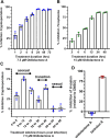
Figure 2:
Figure 2:. The oidiolactones have specific nM level activity against Cryptosporidium parvum and Toxoplasma gondii.
Compounds were tested at concentrations ranging from 7.5 uM to 117 nM against Cp-NLuc infected HCT-8 cells (A) or 10 uM to 156 nM against TgRH-Luc:GFP infected HFF cells (C). Results are from 3 biological replicates. Confluent HCT-8 (B) and HFF (D) cells were incubated with compounds (100 uM to 390 nM) for 72 hours or 48 hours respectively. Cell viability was determined by quantification of cellular ATP levels. Results are from 2 biological replicates (E) Summary of EC50s and confidence intervals. EC50s were determined in Graphpad Prism using the log (inhibitor) vs. response–variable slope (four parameters). Error bars show the standard deviation around each point.
Figure 3:
Figure 3:. Oidiolactone A inhibits growth of Toxoplasma gondii.
TgRH-Luc:GFP infected HFFs were treated with oidiolactone A for 24 hours. Cells were then fixed and labeled with anti-SAG1 antibody (red) to visualize the tachyzoite membrane and Hoescht (blue) to label host cell nuclei. Scale bar=10 μm.
Figure 4:
Figure 4:. Oidiolactone A rapidly kills intracellular, asexual parasite stages, but does not inhibit sporozoite invasion into host cells.
(A) Cp-NLuc was allowed to infect HCT-8 cells for 24 hours, at which point compound or DMSO vehicle control was added for the lengths of time indicated. At the end of each time point, compound was washed out and infection allowed to proceed until the plate was read at 72 hours post-infection. Results are the percent of growth inhibition relative to the DMSO control. (B) HFF cells infected with TgRH-Luc:GFP parasites were treated as described in (a) but the plate was read 48 hours post-infection. Results calculated as in A. (C) HCT-8 cells were infected with Cp-NLuc and the compound or DMSO added for 4 hour intervals as indicated. The plate was read at 72 hours post infection, and percent inhibition of parasite growth calculated based on DMSO control for that time interval. (D) Cp-NLuc oocysts were added to HCT-8 cells incubated in 7.5 μM oidiolactone A, 100 nM tartrolon E or DMSO and allowed to infect for 3 hours at which point plate was read for luciferase expression. Percent inhibition calculated by comparison to DMSO control. All data are compiled from 3 biological replicates. Error bars: +/−SD.
Figure 5:
Figure 5:. Oidiolactone A significantly reduces oocysts shedding in Cryptosporidium infected IFNγ-knockout mice.
(A) Mice were infected with Cp-NLuc and oocyst shedding monitored daily by luciferase expression. On days 5 and 6 post infection, 4 mg/kg oidiolactone A was administered every 12 hours by oral gavage (arrows). Controls received DMSO in parallel. Error bars represent standard error of the mean. (B) The area under the curve (AUC) of the oocysts shedding for each mouse was calculated. Each point is data from one mouse. A Mann-Whitney t-test was used to compare the groups. **p=0.0087
Figure 6.
Figure 6.. Structure activity relationship (SAR) summary for oidiolactones from O. truncatum tested against nluc C. parvum in HCT-8 cells.
”Activity” refers to inhibition of parasite growth, and ”cytotoxicity” refers to inhibition of host cell growth under sub-confluent conditions.

Similar articles

References

    1. Ma J-Y, Li M-Y, Qi Z-Z, Fu M, Sun T-F, Elsheikha HM, et al. Waterborne protozoan outbreaks: An update on the global, regional, and national prevalence from 2017 to 2020 and sources of contamination. Sci Total Environ. 2022;806: 150562. doi:10.1016/j.scitotenv.2021.150562 - DOI - PubMed
    1. Ali M, Ji Y, Xu C, Hina Q, Javed U, Li K. Food and waterborne cryptosporidiosis from a One Health perspective: A comprehensive review. Animals (Basel). 2024;14: 3287. doi:10.3390/ani14223287 - DOI - PMC - PubMed
    1. Tzipori S, Ward H. Cryptosporidiosis: biology, pathogenesis and disease. Microbes Infect. 2002;4: 1047–1058. doi:10.1016/s1286-4579(02)01629-5 - DOI - PubMed
    1. English ED, Guérin A, Tandel J, Striepen B. Live imaging of the Cryptosporidium parvum life cycle reveals direct development of male and female gametes from type I meronts. PLoS Biol. 2022;20: e3001604. doi:10.1371/journal.pbio.3001604 - DOI - PMC - PubMed
    1. Checkley W, White AC Jr, Jaganath D, Arrowood MJ, Chalmers RM, Chen X-M, et al. A review of the global burden, novel diagnostics, therapeutics, and vaccine targets for cryptosporidium. Lancet Infect Dis. 2015;15: 85–94. doi:10.1016/S1473-3099(14)70772-8 - DOI - PMC - PubMed

Publication types

LinkOut - more resources