[Preprint]. 2025 Jan 28:2025.01.02.630345.
doi: 10.1101/2025.01.02.630345.

Pathogenetic mechanisms of muscle-specific ribosomes in dilated cardiomyopathy

Affiliations

Pathogenetic mechanisms of muscle-specific ribosomes in dilated cardiomyopathy

Michael R Murphy et al. bioRxiv. .

Abstract

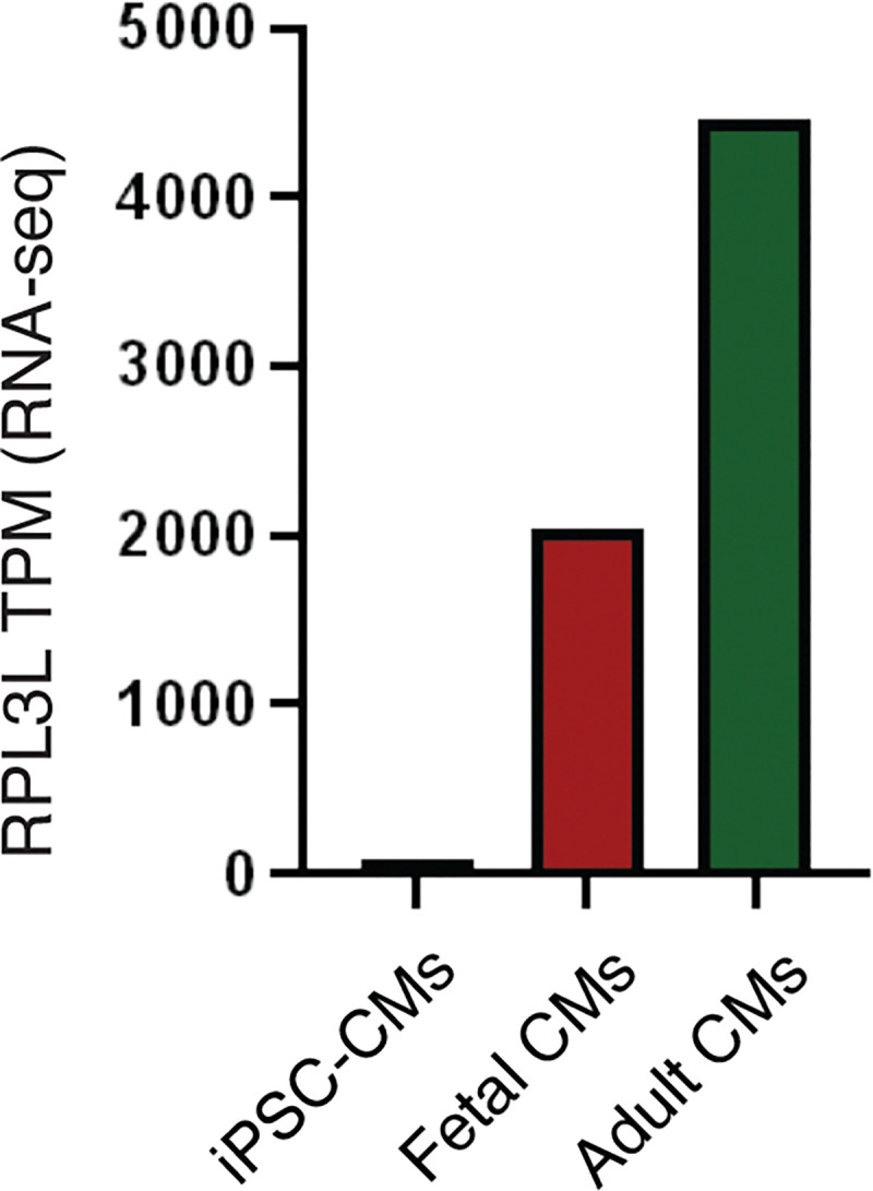
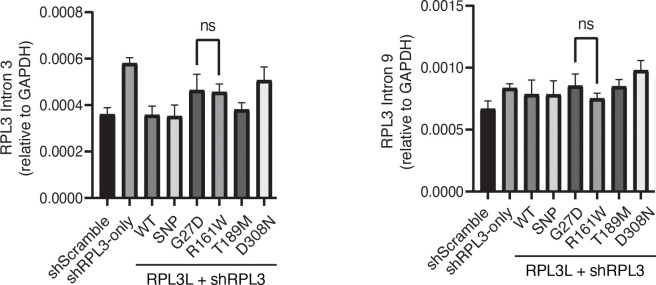
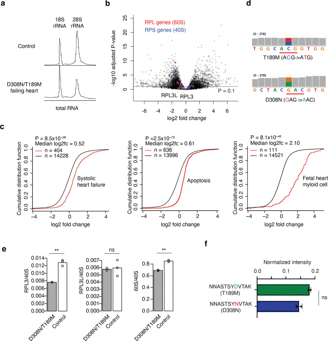
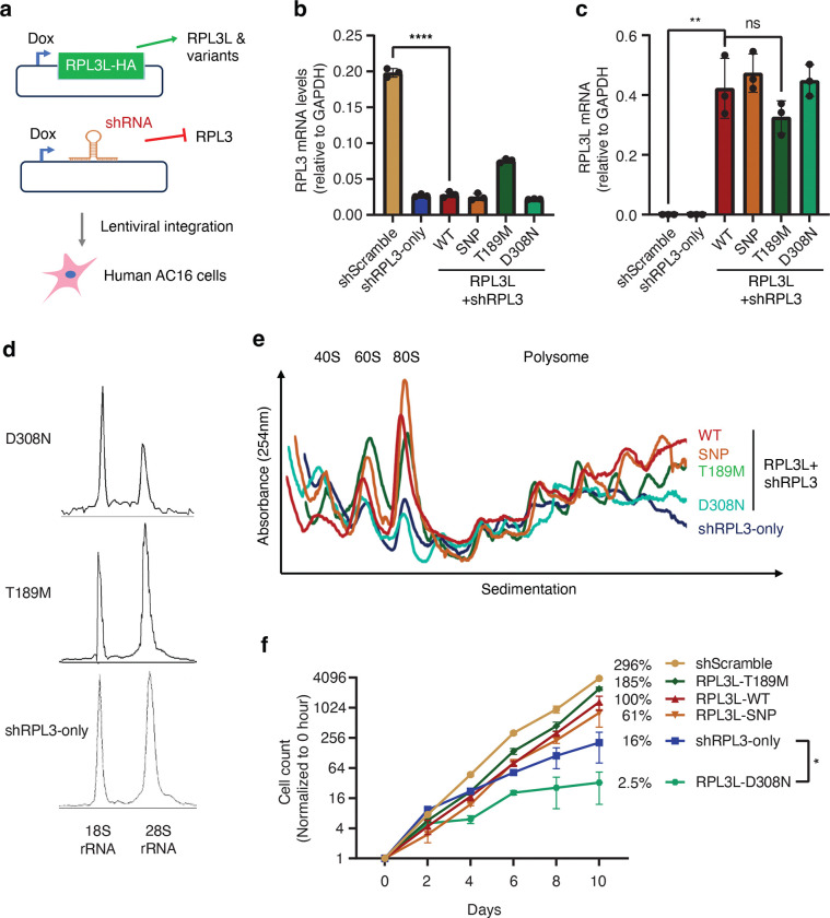
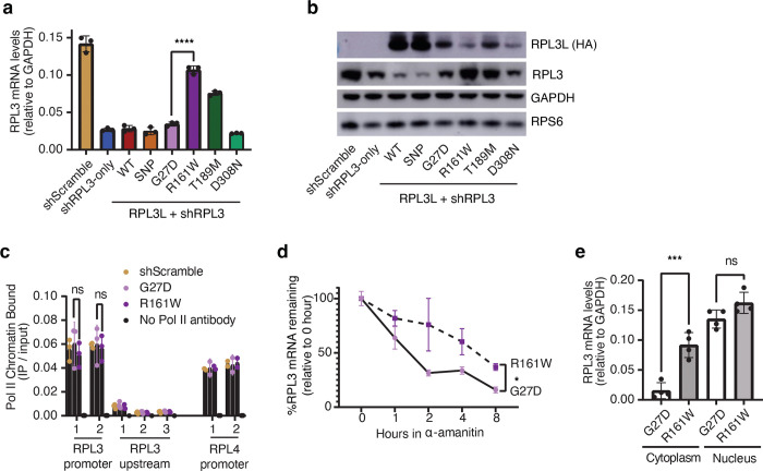
The heart employs a specialized ribosome in its muscle cells to translate genetic information into proteins, a fundamental adaptation with an elusive physiological role1-3. Its significance is underscored by the discovery of neonatal patients suffering from often fatal heart failure caused by severe dilated cardiomyopathy when both copies of the gene RPL3L are mutated4-9. RPL3L is a muscle-specific paralog1-3 of the ubiquitous ribosomal protein L3 (RPL3), which makes the closest contact of any protein to the ribosome's RNA-based catalytic center10. RPL3L-linked heart failure represents the only known human disease associated with tissue-specific ribosomes, yet the underlying pathogenetic mechanisms remain poorly understood. Intriguingly, disease is linked to a large number of mostly missense variants in RPL3L, and RPL3L-knockout resulted in no severe heart defect in either human or mice3, 11-13, challenging the prevailing view that autosomal recessive diseases are caused by loss-of-function mutations. Here, we report three new cases of RPL3L-linked severe neonatal heart failure and present a unifying pathogenetic mechanism by which a large number of variants in the muscle-specific ribosome led to disease. Specifically, affected families often carry one of two recurrent toxic gain-of-function variants alongside a family-specific putative loss-of-function variant. While the non-recurrent variants often trigger partial compensation of RPL3 similar to Rpl3l-knockout mice, both recurrent variants exhibit increased affinity for the RPL3/RPL3L chaperone GRWD114-16 and 60S biogenesis factors, sequester 28S rRNA in the nucleus, disrupt ribosome biogenesis, and trigger severe cellular toxicity that extends beyond the loss of ribosomes. These findings provide critical insights for genetic screening and therapeutic development of neonatal heart failure. Our results suggest that gain-of-toxicity mechanisms may be more prevalent in autosomal recessive diseases, and a combination of gain-of-toxicity and loss-of-function mechanisms could underlie many diseases involving genes with paralogs.


Conflict of interest statement

Declaration of interests X.W. is a member of the Scientific Advisory Board for Epitor Therapeutics. W.K.C. serves on the Board of Directors at Prime Medicine.

Figures

Extended Data Figure 1.
Extended Data Figure 1.. iPSC cardiomyocytes (iPSC-CMs) do not express RPL3L.
Data was analyzed from RNA-seq performed by Pozo et al (2022) comparing iPSC-CMs to fetal and adult human heart.
Extended Data Figure 2.
Extended Data Figure 2.. The hotspot G27D variant mirrors the effects of D308N.
a, Cell growth defect for G27D and R161W, as in Fig. 3f. b, Polysome fraction for G27D and R161W, as in Fig. 3e. Data are mean ± s.d.
Extended Data Figure 3.
Extended Data Figure 3.. Enrichment of 60S ribosome biogenesis factors in proteins co-immunoprecipitated with nuclear RPL3L variants.
Peptide intensities are normalized by the median of all samples and then log2-transformed. Error bars represent standard deviation.
Extended Data Figure 4.
Extended Data Figure 4.. R161W does not incorporate into polysomes.
Monosome and polysomal fractions from Fig. 5 were isolated by TCA precipitation and immunoblotted for HA-tag. Input (pre-ultracentrifugation) samples were loaded as controls.
Extended Data Figure 5.
Extended Data Figure 5.. No change in the expression of the RPL3-targeting shRNA in RPL3L-expressing cells.
A custom annotation was used to detect shRNA hairpin from RNA-seq data obtained from RPL3L variant-expressing cell lines. RPM, Reads per million (N = 3 biological replicates). Error bars represent standard deviation. ns, not significant.
Extended Data Figure 6.
Extended Data Figure 6.. R161W does not induce higher RPL3 intronic RNA as a proxy of transcription levels.
Total RNA was isolated and cDNA was synthesized using random primers. Primers targeting intron 3 and intron 9 of RPL3 were used for amplification (N = 3). Error bars represent standard deviation. ns, not significant.
Figure 1.
Figure 1.. Hotspot variants G27D and D308N/V as potential drivers in RPL3L-linked DCM.
a, Schematic representation of neonatal DCM-associated variants across the RPL3L protein. Each circle denotes a missense variant, while rectangles represent frameshift variants or the splice variant (Δexon9). Variants are color-coded by family, with lines connecting compound heterozygous variants identified in the same family. Variants identified in the three new cases are highlighted with a star. The protein domains within RPL3L are labeled. The hotspot variants G27D and D308N/V are emphasized in bold. b, Mapping of the variants (in red) onto the AlphaFold-predicted 3D structure of RPL3L. The region encoded by exon 9, deleted in the splice variant (Δexon9) is highlighted in salmon. Frameshift variants are not shown. c, Clustering of RPL3L missense variants based on allele frequency (gnomAD) and predicted pathogenicity (AlphaMissense). Variants identified in the same family are connected by dashed lines. d, Putative human RPL3L KO in the general population. LOF: loss-of-function.
Figure 2.
Figure 2.. Ribosome defects in explanted patient hearts.
a, Bioanalyzer electrophoresis of total RNA isolated from explanted patient heart ventricular tissue and a control sample (human AC16 cells). b, Differential gene expression analysis comparing explanted heart tissue (N=2)and age-matched healthy control heart ventricular tissue (N=3). Genes encoding 60S or 40S ribosomal proteins are colored red or blue, respectively. RPL3 and RPL3L are highlighted by yellow circles. c, Cumulative distribution function plots for three significant gene signatures. Red indicates genes of interest and black indicates all other genes. The median log2 fold change (log2fc) and Kolmogorov–Smirnov test P values are also shown. d, RNA-seq read coverage (BAM file visualized in IGV genome browser) showing that both D308N and T189M variants are equally expressed. e, Mass spectrometry quantification of RPL3 (left), RPL3L (center), and total 60S proteins (right) relative total 40S proteins in D308N/T189M patient ventricular tissue (N=2) and age-matched control tissue (N=3). ** P < 0.0001. f, Intensity of the peptide containing the D308 residue in the T189M variant and the peptide containing the N308 residue in the D308N variant normalized to the total intensity in RPL3L. ns: not significant.
Figure 3.
Figure 3.. Hotspot variants disrupt ribosome biogenesis and impairs cell viability.
a, Isogenic AC16 human cardiomyocyte-like cell lines were generated to simultaneously knockdown (KD) RPL3 and overexpress HA-tagged RPL3L (or its variants) via a dox-inducible promoter. b, RPL3 knockdown after dox treatment. Total RNA was isolated from indicated cell lines and analyzed for RPL3 levels. shScramble: cells expressing only a control scrambled shRNA. shRPL3: cells expressing only the RPL3 targeting shRNA without RPL3L or its variants. RPL3L-WT/SNP/T189M/D308N: cells with RPL3 KD and expresses either wild type or the indicated variant of RPL3L. c, Efficient expression of RPL3L variants in AC16 cells after 120 hours Dox treatment. Total RNA isolated from indicated cells was analyzed for RPL3L levels (N = 3 independent experiments). d, Bioanalyzer electrophoresis on total RNA from indicated cell lines was quantified by ImageJ. e, Loss of translation capacity in shRPL3 and D308N cells, but not control or T189M cells. RPL3+ (shScramble) and RPL3L-expressing cells were separated by ultracentrifugation and fractionated in the presence of cycloheximide (N = 3). f, Cell growth defects in D308N and shRPL3 cells. AC16-derived cells were seeded at equal density and counted every 2 days (N = 4). The y-axis is shown on a logarithmic scale. The percentage values represent the cell density at day 10 relative to the RPL3L-WT cell line. b, c, f, Data are mean ± s.d. *: P < 0.05.
Figure 4.
Figure 4.. Hotspot variants mislocalize and alter interactions with ribosome biogenesis factors.
a, Cells were stained for anti-HA antibody (RPL3L and variants) and 28S rRNA FISH probes and analyzed by 60x confocal microscopy. Scale bar = 50μm. b, Violin plot quantification of cytoplasmic and nuclear abundances of HA (RPL3L) signals for indicated cell lines, measured per cell (N = 26–41 cells). c, same as b but for 28S rRNA. One-way ANOVA was used for statistical analysis. d, PCA analysis of the proteins co-immunoprecipitated with RPL3L variants. N=3. e, Enrichment of GRWD1 and C7ORF50 in proteins co-immunoprecipitated with the hotspot variants D308N and G27D compared to other variants. Peptide intensities are normalized by the median of all samples and then log2-transformed. Error bars represent standard deviation. ns, not significant; ** P < 0.001; *** P < 0.0001; **** P < 0.00001.
Figure 5.
Figure 5.. Non-hotspot variants drive post-transcriptional RPL3 compensatory up-regulation.
a, Compensatory increase of RPL3 mRNA in R161W cells. DCM-associated RPL3L variants were expressed for 120 hours in doxycycline and total RNA was extracted and analyzed for RPL3 mRNA levels (N = 3 independent experiments). b, Western blotting shows compensatory increase of RPL3 protein levels in R161W cells. Cells were treated as in (a) and whole-cell lysates were immunoblotted with indicated antibodies (N = 3 independent experiments). c, No difference in transcription activity in RPL3 promoter assayed by ChIP-qPCR. Multiple primer pairs were used for each region. An intergenic region upstream of RPL3L was used as a negative control of non-transcribed region (N = 3). d, RPL3 mRNA half-life is enhanced in R161W cells. RPL3L-expressing variants were incubated with 50μg/ml RNA Polymerase II inhibitor α-amanitin for indicated timepoints prior to analysis for RPL3 mRNA relative to GAPDH, normalized to 0 hour (N = 3). e, Compensatory RPL3 mRNA increase occurs entirely in the cytoplasm in R161W cells. RNA underwent subcellular fractionation for amplification of RPL3 levels relative to GAPDH (N = 4). a,c,d,e, Data are mean ± s.d. ns, not significant; * P < 0.01; *** P < 0.0001; **** P < 0.00001.
Figure 6.
Figure 6.. Impaired protein synthesis in engineered compound heterozygous cells.
a, Cells stably integrated with G27D, R161W, or both G27D and R161W were induced with dox for 144hrs. Puromycin was added to 1uM final concentration for 30 minutes before cells were harvested for Western blotting (N = 4). b, Quantification of puromycin incorporation. c, Quantification of RPL3 protein abundance. Data are mean ± s.d. ns, not significant; * P < 0.01.

References

    1. Van Raay T.J., Connors T.D., Klinger K.W., Landes G.M. & Burn T.C. A novel ribosomal protein L3-like gene (RPL3L) maps to the autosomal dominant polycystic kidney disease gene region. Genomics 37, 172–176 (1996). - PubMed
    1. Chaillou T., Zhang X. & McCarthy J.J. Expression of Muscle-Specific Ribosomal Protein L3-Like Impairs Myotube Growth. J Cell Physiol 231, 1894–1902 (2016). - PMC - PubMed
    1. Shiraishi C. et al. RPL3L-containing ribosomes determine translation elongation dynamics required for cardiac function. Nat Commun 14, 2131 (2023). - PMC - PubMed
    1. Ganapathi M. et al. Bi-allelic missense disease-causing variants in RPL3L associate neonatal dilated cardiomyopathy with muscle-specific ribosome biogenesis. Hum Genet 139, 1443–1454 (2020). - PMC - PubMed
    1. Das B.B., Gajula V., Arya S. & Taylor M.B. Compound Heterozygous Missense Variants in RPL3L Genes Associated with Severe Forms of Dilated Cardiomyopathy: A Case Report and Literature Review. Children (Basel) 9 (2022). - PMC - PubMed

Publication types

LinkOut - more resources