[Preprint]. 2025 Jan 3:2025.01.03.631181.
doi: 10.1101/2025.01.03.631181.

Extracellular vesicles from diverse fungal pathogens induce species-specific and endocytosis-dependent immunomodulation

Affiliations

Extracellular vesicles from diverse fungal pathogens induce species-specific and endocytosis-dependent immunomodulation

Geneva N Kwaku et al. bioRxiv. .

Update in

Abstract

Microbial pathogens generate extracellular vesicles (EVs) for intercellular communication and quorum sensing. Microbial EVs also induce inflammatory pathways within host innate immune cells. We previously demonstrated that EVs secreted by Candida albicans trigger type I interferon signaling in host cells specifically via the cGAS-STING innate immune signaling pathway. Here, we show that despite sharing similar properties of morphology and internal DNA content, the interactions between EVs and the innate immune system differ according to the parental fungal species. EVs secreted by C. albicans, Saccharomyces cerevisiae, Cryptococcus neoformans, and Aspergillus fumigatus are endocytosed at different rates by murine macrophages triggering varied cytokine responses, innate immune signaling, and subsequent immune cell recruitment. Notably, cell wall constituents that decorate C. neoformans and A. fumigatus EVs inhibit efficient internalization by macrophages and dampen innate immune activation. Our data uncover the transcriptional and functional consequences of the internalization of diverse fungal EVs by immune cells and reveal novel insights into the early innate immune response to distinct clinically significant fungal pathogens.


Figures
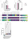
Fig 1.
Fig 1.. Murine macrophage internalization, cytokine production, and neutrophil recruitment differ in responses to different fungal EVs.
A, Percent internalization of Ca EVs, Sc EVs, Cn EVs, and Af EVs by murine macrophages. Significance assessed by an ordinary one-way ANOVA and Dunnet’s multiple comparisons test, ***p=0.0002 vs PBS treated macrophages. n=3. B, Percent endocytosis of Ca EVs and Sc EVs with murine macrophages treated with DMSO or 100μM dynasore. Significance assessed by a two-way ANOVA and Tukey’s multiple comparisons test, ****p= <0.0001 vs respective DMSO controls. n=2. C, Heat maps of induction of TNFα, IFNβ, and MCP-1 (in pg/mL) in WT macrophages stimulated by PBS, 2.5μg cGAMP, Ca, Sc, Cn, and Af EVs. n=2. D, Number of neutrophils that transmigrated towards supernatants from WT macrophage stimulated by PBS, 2.5μg cGAMP, Ca EVs, Sc EVs, Cn EVs, and Af EVs (5 x 10¹⁰ EVs added per stimulation), and 0.1μM fMLP as a positive control. Significance assessed by an ordinary one-way ANOVA and Dunnet’s multiple comparisons test, *p≤0.0203, **p≤0.0073, ****p<0.0001 vs PBS-treated macrophages. n=3.
Fig 2.
Fig 2.. Fungal EVs differentially activate the STING pathway.
A, Representative immunoblots of viperin, phosphorylated IRF3, total IRF3, phosphorylated TBK1, total TBK1, and actin in response to PBS, transfected cGAMP (2.5μg), Ca EVs, Sc EVs, Cn EVs, and Af EVs (5x10¹⁰ EVs/mL added per stimulation) in WT macrophages. B, Immunoblot of viperin in WT macrophages treated with either DMSO (solvent control) or 100μM dynasore and stimulated with Ca EVs, Sc EVs, Cn EVs, and Af EVs (5x10¹⁰ EVs/mL added per stimulation).
Fig 3.
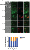
Fig 3.. Fungal EVs induce translocation of cGAS from the nuclear membrane to the cytosol.
A, Representative images of the localization of cGAS (green) when cGAS-GFP expressing macrophages are stimulated for 3h with DiI-labeled (red) PBS, Ca EVs, Sc EVs, Cn EVs, or Af EVs (8x10⁹ EVs added per stimulation). Scale bar=10μm. B, Semi-quantitative analysis of the percentage of cGAS localization that is either nuclear or non-nuclear. cGAS localization analyses were performed on approximately 100 cGAS-GFP expressing macrophages that successfully endocytosed DiI-labeled EVs.
Fig 4.
Fig 4.. Physical and DNA characteristics of Ca EVs, Sc EVs, Cn EVs, and Af EVs.
A, The −log10 values of EV concentration from standard isolation preps. Significance assessed using an ordinary one-way ANOVA and Tukey’s multiple comparisons test with no significance determined. n≥5. B, Quantification of EV concentration according to diameter for Ca EVs, Sc EVs, Cn EVs, and Af EVs. n≥5. C, The median and mode diameters of Ca EVs, Sc EVs, Cn EVs, and Af EVs. n≥5. D, Representative images from TEM of Ca EVs, Sc EVs, Cn EVs, and Af EVs. E, The average DNA concentration (pg per 1x10¹⁰ EVs) of Ca EVs, Sc EVs, Cn EVs, and Af EVs. Significance assessed using an ordinary one-way ANOVA and Tukey’s multiple comparisons test with no significance determined. n≥3. F, Percent GC content of EV DNA reads that mapped to the reference fungal genome as a truncated violin box plot.
Fig 5.
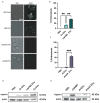
Fig 5.. Outer structural layers on EVs inhibit endocytosis and STING pathway activation.
A, Representative confocal microscopy showing GXM layer present on WT C. neoformans (H99) yeast and EVs but not on cap59Δ yeast and EVs. 1x10⁷ WT yeasts, 1x10⁷ cap59Δ yeasts, and 5x10¹⁰ WT and cap59Δ EVs were added to the respective stimulations. Scale bar=10μm. B, Percent endocytosis of Cn EVs and cap59Δ EVs by WT macrophages. n=2. C, Percent endocytosis of Af EVs and ΔrodA EVs by the total number of WT macrophages. n=2. D, Immunoblot of viperin and actin in WT macrophages stimulated by PBS, Cn EVs, cap59Δ EVs, and the positive control for yeast, Ca EVs (5x10¹⁰ EVs/mL were added per stimulation). E, Immunoblot of viperin and actin in WT macrophages stimulated by PBS, Af EVs, and ΔrodA EVs (5x10¹⁰ EVs/mL were added per stimulation), and positive control for Aspergillus: 2.5μg cGAMP.

References

    1. Fang W, Wu J, Cheng M, Zhu X, Du M, Chen C, et al. Diagnosis of invasive fungal infections: challenges and recent developments. Vol. 30, Journal of Biomedical Science. BioMed Central Ltd; 2023,42. - PMC - PubMed
    1. Vitiello A, Ferrara F, Boccellino M, Ponzo A, Cimmino C, Comberiati E, et al. Antifungal Drug Resistance: An Emergent Health Threat. Vol. 11, Biomedicines. MDPI; 2023. - PMC - PubMed
    1. Pekmezovic M, Dietschmann A, Gresnigt MS. Type I interferons during host fungus interactions: Is antifungal immunity going viral? PLoS Pathog. 2022. Aug 1;18(8). - PMC - PubMed
    1. Pekmezovic M, Hovhannisyan H, Gresnigt MS, Iracane E, Oliveira-Pacheco J, Siscar-Lewin S, et al. Candida pathogens induce protective mitochondria-associated type I interferon signalling and a damage-driven response in vaginal epithelial cells. Nat Microbiol. 2021. May 1;6(5):643–57. - PubMed
    1. Smeekens SP, Ng A, Kumar V, Johnson MD, Plantinga TS, Van Diemen C, et al. Functional genomics identifies type i interferon pathway as central for host defense against Candida albicans. Nat Commun. 2013;4. - PMC - PubMed

Publication types