[Preprint]. 2024 Oct 4:2024.10.03.616528.
doi: 10.1101/2024.10.03.616528.

The C. elegans homolog of Sjögren's Syndrome Nuclear Antigen 1 is required for the structural integrity of the centriole and bipolar mitotic spindle assembly

Affiliations

The C. elegans homolog of Sjögren's Syndrome Nuclear Antigen 1 is required for the structural integrity of the centriole and bipolar mitotic spindle assembly

Jason A Pfister et al. bioRxiv. .

Update in

Abstract

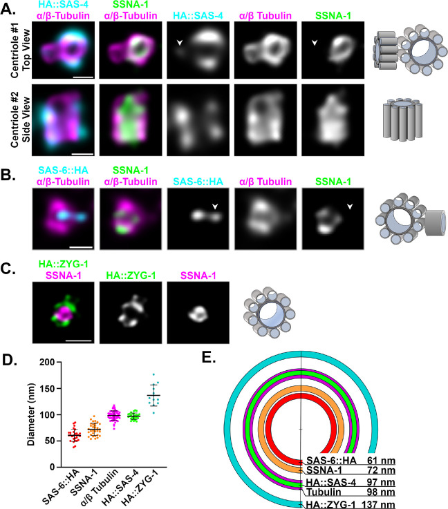
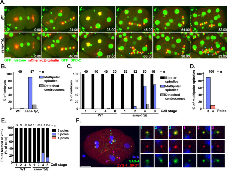
Centrioles play central roles in ciliogenesis and mitotic spindle assembly. Once assembled, centrioles exhibit long-term stability, a property essential for maintaining numerical control. How centriole stability is achieved and how it is lost in certain biological contexts are still not completely understood. In this study we show that SSNA-1, the Caenorhabditis elegans ortholog of Sjogren's Syndrome Nuclear Antigen 1, is a centriole constituent that localizes close to the microtubule outer wall, while also exhibiting a developmentally regulated association with centriole satellite-like structures. A complete deletion of the ssna-1 gene results in an embryonic lethal phenotype marked by the appearance of extra centrioles and spindle poles. We show that SSNA-1 genetically interacts with the centriole stability factor SAS-1 and is required post assembly for centriole structural integrity. In SSNA-1's absence, centrioles assemble but fracture leading to extra spindle poles. However, if the efficiency of cartwheel assembly is reduced, the absence of SSNA-1 results in daughter centriole loss and monopolar spindle formation, indicating that the cartwheel and SSNA-1 cooperate to stabilize the centriole during assembly. Our work thus shows that SSNA-1 contributes to centriole stability during and after assembly, thereby ensuring proper centriole number.


Figures

Fig. 1:
Fig. 1:. C. elegans possesses an SSNA-1 ortholog that plays a critical role in embryogenesis
A. Sequence alignment of various SSNA-1 orthologs. The amino acids are colored as follows: blue and red for negatively and positively charged residues respectively, green for uncharged residues, and gold for hydrophobic residues. B. Table showing percent identity (blue) and percent similarity (red) of SSNA-1 orthologs. C. AlphaFold structural prediction of T07A9.13. D. Embryonic viability of wild-type and ssna-1(Δ) mutant strains at 20°C (left) or 25°C (right). Each datapoint represent progeny of a single hermaphrodite. E. SSNA-1 is maternally required. Embryonic viability among the progeny of wild-type (light blue) or ssna-1(Δ) (dark blue) males and wild-type (light pink) or ssna-1D (dark pink) fem-1(hc17) females. Each datapoint represent progeny of a single female. Note that embryonic lethality correlates with the genotype of the mother and not the father.
Fig. 2:
Fig. 2:. SSNA-1 is a component of the centriole and centriole satellite-like structures.
A. Immunofluorescence images of SSNA-1::SPOT and SAS-4::GFP during the first embryonic division. SSNA-1 is found only at centrioles from meiosis through anaphase of the first cell cycle. During first telophase, additional weaker SSNA-1::SPOT foci can be seen (arrowheads). Scale bar = 10 μm and applies to panels A and B. Insets are 2X magnifications. B. During the second cell cycle SSNA-1::SPOT remains centriolar but beginning around nuclear envelope breakdown (NEBD), SSNA-1::SPOT assembles into satellite-like structures that surround the centriole. Insets are 2X magnifications. C. and D. Immunofluorescence image showing co-localization of SSNA-1::SPOT relative to the PCM marker GFP::SPD-5. C. During anaphase, SSNA-1::SPOT satellite-like structures localize outside of the PCM marked by GFP::SPD-5. Scale bar = 10 μm and applies to panels C and D. D. During telophase when the PCM breaks up into packets, SSNA-1::SPOT satellite-like structures remain distinct from SPD-5 packets. However, a portion of both SSNA-1::SPOT and GFP::SPD-5 co-localize at the centriole. E. Frames from a time-lapse recording of an embryo expressing SSNA-1::wrmScarlet, γ-tubulin::GFP and GFP::Histone H2B. SSNA-1::wrmScarlet reorganizes during division of the ABpl cell (boxed). Time-lapse images reveals that the satellite-like structures disperse over the nucleus during interphase and accumulate at centrosomes as cells enter mitosis. Time is shown as mm:ss. Scale bar = 10 μm.
Fig. 3:
Fig. 3:. Centriolar SSNA-1 localizes between the microtubule-containing outer wall and the cartwheel.
A-C. Ultrastructure Expansion Microscopy (U-ExM) of centrioles from gonad spreads. The schematic shown to the right of each set of images depicts the orientation and configuration of centrioles. A. Co-localization of SSNA-1, HA::SAS-4, and α/β-tubulin from two distinct centrioles showing a top view (centriole #1) and side view (centriole #2). SSNA-1 is localized inside the microtubule outer wall. Arrowhead indicates emergence of a daughter centriole that stains weakly for HA::SAS-4 but not SSNA-1. Scale bars = 100 nm B. Co-localization of SSNA-1, SAS-6::HA and α/β-Tubulin. SSNA-1 is localized outside of the cartwheel defined by SAS-6::HA. Arrowhead indicates emergence of a daughter centriole that stains positive for SAS-6::HA but not SSNA-1. Scale bar = 100 nm C. Co-localization of SSNA-1 and HA::ZYG-1. Scale bar = 200 nm D. The measured diameters of each protein are plotted with each point representing a measurement from a single centriole. The mean and standard deviation are shown. E. Diagram indicating the position of each protein within the centriole. SSNA-1 localizes between SAS-6::HA and α/β-tubulin.
Fig. 4:
Fig. 4:. Deletion of SSNA-1 results in excess centrioles.
A. Time-lapse images of the first four rounds of division from either wild-type or ssna-1(Δ) embryos expressing GFP::histone, mCherry::β-tubulin, and GFP::SPD-2. While wild-type embryos form only bipolar spindles ssna-1(Δ) embryos form multipolar spindles beginning around the four-cell stage. During the third round of division in the ssna-1(Δ) embryo (t=27:00), multiple centrosomes highlighted by cyan, magenta, and white arrowheads appear. These form a multipolar spindle in the EMS cell (t=33:00). Each centrosome is capable of duplication as shown in the subsequent daughter cells E (white arrowheads) and MS (cyan and magenta arrowheads). Scale bar = 10 μm B. Percent of embryos displaying a multipolar spindle or detached centrosome defect. (n= number of embryos scored). C. Percent of embryos at each cell stage displaying a multipolar spindle or detached centrosome defect. (n= number of embryos scored). D. Percentage of tripolar and tetrapolar spindles among all multipolar spindles (n= spindles scored). E. Percentage of cells with a bi-, tri- or tetrapolar spindle at each embryonic cell stage (n= number of cells scored). F. An immunofluorescent image of an ssna-1(Δ) embryo with a multipolar spindle in both the EMS (pink) and ABp (cyan) cells. Note that all spindle poles are positive for both ZYG-1::SPOT and SAS-4 indicating the extra spindle poles contain centrioles. Scale bar = 10 μm.
Fig. 5:
Fig. 5:. SSNA-1 functions during centriole assembly.
A. Embryonic viability of progeny from wild type, zyg-1(it25), ssna-1D, and zyg-1(i25); ssna-1(Δ) strains at 22.5°C. Each data point represents the progeny of a single hermaphrodite. B. Single frames taken from time-lapse recordings of the indicated strains expressing GFP::histone and SPD-2::mCherry. Each image shows a two-cell embryo. A few zyg-1(it25) embryos exhibit monopolar spindles (magenta arrowhead), while ssna-1(Δ) embryos only possess bipolar spindles (white arrowheads). The zyg-1(it25); ssna-1(Δ) double mutant embryos exhibit a mixed phenotype, with monopolar spindle (magenta arrowheads) or multipolar spindles (cyan arrowheads). C. Quantification of spindle defects observed through the first two cell divisions at 22.5°C. The zyg-1(it25); ssna-1D double mutant embryos display twice as many monopolar spindles as zyg-1(it25) embryos. The double mutant also assembles multipolar spindles which are not observed in either single mutant. D. Co-sedimentation assay showing SSNA-1 and ZYG-1 interact. Proteins were incubated alone (left) or in various combinations (right), separated into soluble (S) and pellet (P) fractions by centrifugation, and then detected by immunoblotting. Note that ZYG-1 is found in the soluble fraction when incubated alone or with microtubules but shifts to the pellet fraction upon incubation with SSNA-1. E. Quantitation from three-independent co-sedimentation experiments.
Fig. 6:
Fig. 6:. SSNA-1 is required for the structural integrity of centrioles.
A. Time lapse images from wild-type or ssna-1(Δ) embryos expressing mCherry::histone and GFP::SAS-7. The first and last frames are shown at the top while enlargements show intervening time points with only one spindle pole shown beginning in anaphase (time point 3:00). A. In wild-type embryos two GFP::SAS-7 foci (corresponding to a disengaged centriole pair) first become visible at each spindle pole during telophase (t=9:00, yellow and magenta arrowheads). During the ensuing cell cycle, this cell assembles a bipolar spindle (t=18:00). In ssna-1(Δ) embryos, two GFP::SAS-7 foci first become apparent during prometaphase (t=1.0, yellow and magenta arrowheads). During telophase when centriole separation normally occurs, a third GFP::SAS-7 spot appears (t=8.0, cyan arrowhead). During the ensuring cell cycle this cell assembles a tripolar spindle (t=17:00). Bar = 10 μm. B. Assay to discriminate between centriole fragmentation and premature centriole disengagement. Males whose centrioles are marked with SPOT::SAS-4 (red) are mated to hermaphrodites expressing SAS-4::GFP (green). In wild-type embryos the two paternal centrioles will indelibly be marked red while new centrioles will be marked green. Red mother/green daughter centriole pairs will remain engaged (and appear yellow) until mitotic exit. Centriole instability will be detected as >2 red centrioles per embryo while premature disengagement will be detected as loss of green-red coincidence prior to mitotic exit. C. A wild-type (top), ssna-1(Δ) (middle), and ssna-1(Δ) x WT embryos stained for SPOT::SAS-4 (red), SAS-4::GFP (green), and DNA (blue). The wild-type embryo possesses only two red centrioles indicating centriole stability while the ssna-1(Δ) embryo possesses three red centrioles revealing a centriole fragmentation phenotype. Bar = 10 μm. D. Quantitation of centriole fragmentation in wild-type and ssna-1(Δ) mutant embryos. Note that in early prophase ssna-1(Δ) mutant embryos have two sperm derived centrioles indicating ssna-1(Δ) sperm contain the normal number of centrioles. However, older ssna-1(Δ) embryos frequently contain more than two red foci indicating that the sperm centrioles have fractured. E. ssna-1 and sas-1 genetically interact. The percentage of viable embryos produced by wild-type, ssna-1(Δ), sas-1(t1476, bs272), and sas-1(t1476, bs272); ssna-1(Δ) hermaphrodites.
Fig. 7:
Fig. 7:. SSNA-1 functions to stabilize centrioles during and after assembly.
A model depicting the fate of centrioles possessing or lacking stabilizing elements. Top. Wild-type centrioles, which possess both SSNA-1 (yellow ring) and a fully formed cartwheel, exhibit long-term stability resulting in faithful bipolar spindle assembly. Middle. Centrioles lacking SSNA-1 can be assembled but lack long-term stability leading to centriole fragmentation and multipolar spindle formation. Bottom. Centrioles lacking SSNA-1 and a fully formed cartwheel (due to partial inhibition of zyg-1) experience structural failure during assembly leading to loss of the daughter centriole and monopolar spindle assembly.

Similar articles

References

    1. Breslow D.K., and Holland A.J. (2019). Mechanism and Regulation of Centriole and Cilium Biogenesis. Annu Rev Biochem 88, 691–724. 10.1146/annurev-biochem-013118-111153. - DOI - PMC - PubMed
    1. Izquierdo D., Wang W.J., Uryu K., and Tsou M.F. (2014). Stabilization of cartwheel-less centrioles for duplication requires CEP295-mediated centriole-to-centrosome conversion. Cell Rep 8, 957–965. 10.1016/j.celrep.2014.07.022. - DOI - PMC - PubMed
    1. Le Guennec M., Klena N., Gambarotto D., Laporte M.H., Tassin A.M., van den Hoek H., Erdmann P.S., Schaffer M., Kovacik L., Borgers S., et al. (2020). A helical inner scaffold provides a structural basis for centriole cohesion. Sci Adv 6, eaaz4137. 10.1126/sciadv.aaz4137. - DOI - PMC - PubMed
    1. Hilbert M., Noga A., Frey D., Hamel V., Guichard P., Kraatz S.H., Pfreundschuh M., Hosner S., Fluckiger I., Jaussi R., et al. (2016). SAS-6 engineering reveals interdependence between cartwheel and microtubules in determining centriole architecture. Nat Cell Biol 18, 393–403. 10.1038/ncb3329. - DOI - PubMed
    1. Kitagawa D., Vakonakis I., Olieric N., Hilbert M., Keller D., Olieric V., Bortfeld M., Erat M.C., Fluckiger I., Gonczy P., and Steinmetz M.O. (2011). Structural basis of the 9-fold symmetry of centrioles. Cell 144, 364–375. 10.1016/j.cell.2011.01.008. - DOI - PMC - PubMed

Publication types

LinkOut - more resources