[Preprint]. 2024 Sep 30:2024.09.29.611006.
doi: 10.1101/2024.09.29.611006.

R2DT: A COMPREHENSIVE PLATFORM FOR VISUALISING RNA SECONDARY STRUCTURE

Affiliations

R2DT: A COMPREHENSIVE PLATFORM FOR VISUALISING RNA SECONDARY STRUCTURE

Holly McCann et al. bioRxiv. .

Update in

  • R2DT: a comprehensive platform for visualizing RNA secondary structure.
    McCann H, Meade CD, Williams LD, Petrov AS, Johnson PZ, Simon AE, Hoksza D, Nawrocki EP, Chan PP, Lowe TM, Ribas CE, Sweeney BA, Madeira F, Anyango S, Appasamy SD, Deshpande M, Varadi M, Velankar S, Zirbel CL, Naiden A, Jossinet F, Petrov AI. McCann H, et al. Nucleic Acids Res. 2025 Feb 8;53(4):gkaf032. doi: 10.1093/nar/gkaf032. Nucleic Acids Res. 2025. PMID: 39921562 Free PMC article.

Abstract

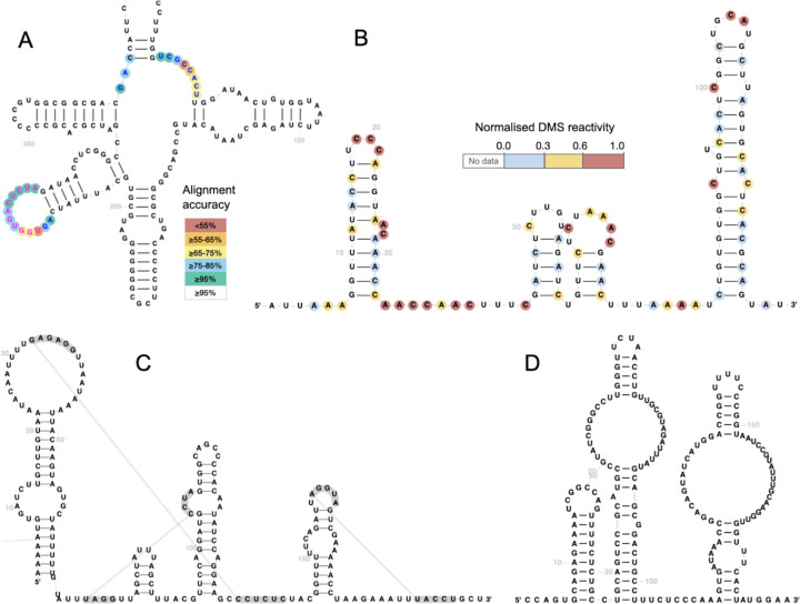
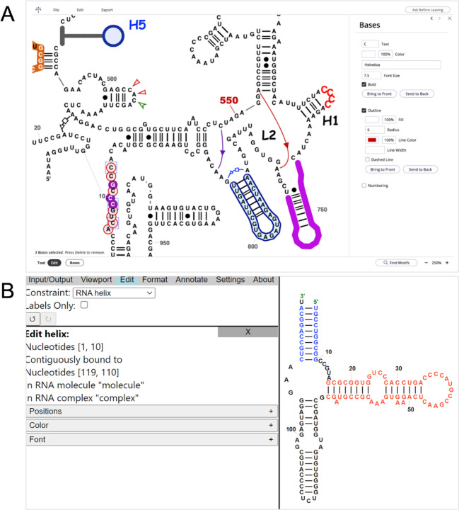
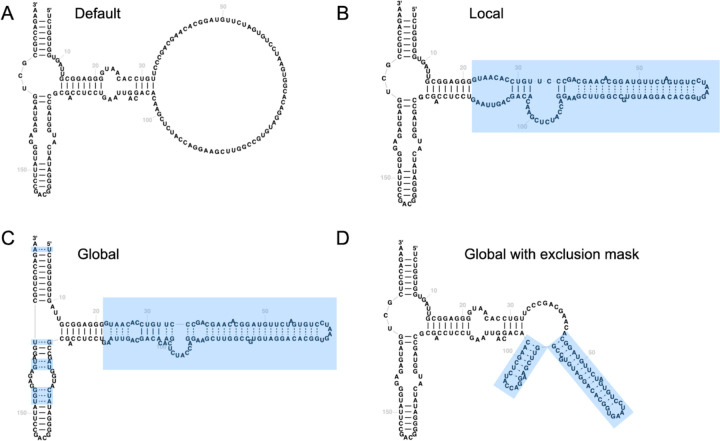
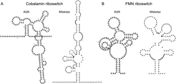
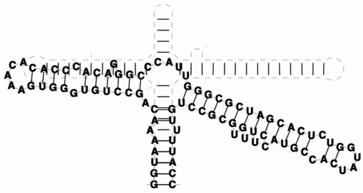
RNA secondary (2D) structure visualisation is an essential tool for understanding RNA function. R2DT is a software package designed to visualise RNA 2D structures in consistent, recognisable, and reproducible layouts. The latest release, R2DT 2.0, introduces multiple significant features, including the ability to display position-specific information, such as single nucleotide polymorphisms (SNPs) or SHAPE reactivities. It also offers a new template-free mode allowing visualisation of RNAs without pre-existing templates, alongside a constrained folding mode and support for animated visualisations. Users can interactively modify R2DT diagrams, either manually or using natural language prompts, to generate new templates or create publication-quality images. Additionally, R2DT features faster performance, an expanded template library, and a growing collection of compatible tools and utilities. Already integrated into multiple biological databases, R2DT has evolved into a comprehensive platform for RNA 2D visualisation, accessible at https://r2dt.bio.


Conflict of interest statement

CONFLICT OF INTEREST None declared.

Figures

Figure 1:
Figure 1:
A) A fragment of a rabbit small ribosomal subunit rRNA sequence visualised using the human template with Infernal posterior probabilities reflecting alignment confidence shown as coloured circles (RNAcentral ID URS000086855E_9986). The highlighted regions help identify species-specific differences between the human and the rabbit rRNAs. B) Normalised DMS reactivities for stem loops SL1-SL4 of SARS-CoV-2 (data from (20)). C) Cripavirus IRES with three pseudoknots (RNAcentral ID URS0000A7638F_12136). D) Bridge RNA from Escherichia coli (INSDC accession CP147105.1/771271–771455) visualised using the new template-free mode (data from (21)).
Figure 2:
Figure 2:
A) Screenshot of the RNAcanvas user interface showing human mitochondrially encoded 12S rRNA (MT-RNR1). The layout and the 2D structure were generated by R2DT, while the fonts, colour, background, and non-canonical interactions were configured in RNAcanvas. B) Screenshot of the XRNA-GT website showing a 5S rRNA.
Figure 3.
Figure 3.
Example diagrams illustrating different constrained folding modes in R2DT. The 5S sequence from Halobacteroides halobius is visualised using a 5S template from Bacillus subtilis (INSDC accession CP003359.1/20186–20360). A) Default R2DT output without constrained folding. B) Local folding mode where RNAfold is used to predict the structure for the insertion relative to the template. C) Global folding mode with additional base pairs predicted throughout the structure. D) An example of using global folding mode with an exclusion mask. In this case all unpaired nucleotides aligned to the template were prevented from forming base pairs. Predicted base pairs are shown as dashed lines. Changes relative to default output are highlighted in blue boxes.
Figure 4.
Figure 4.
Example R2DT templates based on consensus 2D structures of Rfam families visualised using R2R and RNArtist in the left and right subpanels, respectively. A) Cobalamin riboswitch (Rfam ID RF00174). B) FMN riboswitch (Rfam ID RF00050). The R2R layouts contain major overlaps between the structural elements making it difficult to see the nucleotides, while the RNArtist layouts prevent the overlaps and are more legible. For both families, R2DT 2.0 uses the RNArtist layouts by default.
Figure 5.
Figure 5.
A) Screenshot of the 2D and 3D components on the macromolecule page for 23S ribosomal RNA from E. coli, in PDB entry 3CC2. The nucleotide G691 is selected in 3D and also highlighted with orange in 2D. B) Screenshot of the 2D and 3D components on the macromolecule page for 16S ribosomal RNA from E. coli, in PDB entry 5J7L, highlighting a kink-turn RNA 3D module and its non-canonical base pairs annotated using the Leontis- Westhof nomenclature (24).
Video 1.
Video 1.
A still from the animation showing the 2D structure of the reference model from RNApuzzles Puzzle 39 and the 2D structure of a predicted model (Dfold group, model 3). The base pairs were extracted from the PDB files using the RNAView software. The video file is available in Supplementary Data.

References

    1. Cannone J.J., Subramanian S., Schnare M.N., Collett J.R., D’Souza L.M., Du Y., Feng B., Lin N., Madabusi L.V., Müller K.M., et al. (2002) The Comparative RNA Web (CRW) Site: an online database of comparative sequence and structure information for ribosomal, intron, and other RNAs. BMC Bioinformatics, 3, 1–31. - PMC - PubMed
    1. Ponty Y. and Leclerc F. (2015) Drawing and editing the secondary structure(s) of RNA. Methods Mol. Biol., 1269, 63–100. - PubMed
    1. Kerpedjiev P., Hammer S. and Hofacker I.L. (2015) Forna (force-directed RNA): Simple and effective online RNA secondary structure diagrams. Bioinformatics, 31, 3377–3379. - PMC - PubMed
    1. Weinberg Z. and Breaker R.R. (2011) R2R--software to speed the depiction of aesthetic consensus RNA secondary structures. BMC Bioinformatics, 12, 3. - PMC - PubMed
    1. Sieg Jacob P., Forstmeier Peter C., Philip C. Bevilacqua R2easyR.

Publication types

LinkOut - more resources