[Preprint]. 2025 Jan 2:2024.12.31.630961.
doi: 10.1101/2024.12.31.630961.

Autophagy-Mediated Downregulation of AXL and TIM-1 Promotes Sustained Zika Virus Infection

Affiliations

Autophagy-Mediated Downregulation of AXL and TIM-1 Promotes Sustained Zika Virus Infection

Jingyou Yu et al. bioRxiv. .

Update in

Abstract

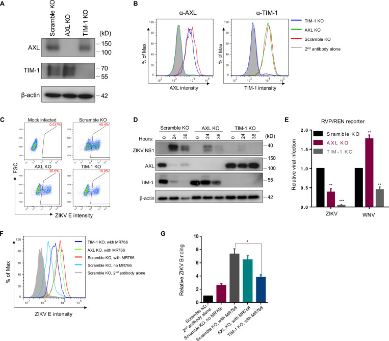
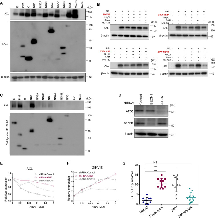
Zika virus (ZIKV) infection can lead to a variety of clinical outcomes, including severe congenital abnormalities. The phosphatidylserine (PS) receptors AXL and TIM-1 are recognized as critical entry factors for ZIKV in vitro. However, it remains unclear if and how ZIKV regulates these receptors during infection. In this study, we investigated AXL and TIM-1 expression in human alveolar basal epithelial A549 cells, glioblastoma U87 cells, and embryonic stem cells-derived trophoblast following ZIKV infection. We found that both the Asian strain FSS13025 and the African strain MR766 of ZIKV downregulate AXL, with a milder effect on TIM-1. We identified several ZIKV proteins, notably envelope (E), NS2A, NS3, and NS4B, that contribute to this downregulation. Notably, treatment with lysosomal inhibitor NH4Cl or the autophagy inhibitor 3-Methyladenine (3-MA) mitigated the AXL/TIM-1 downregulation, indicating autophagy's involvement in the process. Importantly, this downregulation facilitates sustained viral replication and promotes viral spread by preventing superinfection and limiting cell death, which is also associated with impaired innate immune signaling. Our findings uncover a mechanism by which ZIKV downregulates entry factors to enhance prolonged viral replication and spread.

Keywords: AXL; Autophagy; Down-Regulation; TIM-1; ZIKV.


Figures

Figure 1.
Figure 1.. CRISPR knockout of human AXL and TIM-1 differentially modulates ZIKV infection and entry.
AXL and TIM-1 expression levels in stable A549 Scramble KO, AXL KO, and TIM-1 KO cells were examined by immunoblotting (A) or immunostaining (B). (C) A549 Scramble KO, AXL KO, and TIM-1 KO cells were infected with MR766 at MOI=1 for 24 hours. Cells were processed for E protein staining (4G2) and analyzed by flow cytometry. Mock-infected cells served as negative controls. The percents of E protein-positive cells were displayed. (D) The total protein level of ZIKV NS1, AXL and TIM-1 expression following 0, 24 or 36 hours of infection in A549 Scramble KO, AXL KO and TIM-1 KO cells was determined by western blotting. (E) The ZIKV-E or WNV-E pseudotyped one-round RVP/WNV-REN reporter viruses were applied to A549 Scramble KO, AXL KO, and TIM-1 KO cells for 36 hours. Cell lysates were used for measuring the Renilla luciferase activity. Data shown are mean ± SD from three independent experiments. Statistical significance was determined by Student’s two-sided t-test, **p < 0.01, ***p < 0.001. (F-G) Virus binding. A549 Scramble KO, AXL KO, and TIM-1 KO cells (1 × 10⁶ cells) were mixed with 5 × 10⁶ PFU MR766 virus for 2 hours on ice. After three washes with 1 × PBS, cells were stained with 4G2 and FITC-anti-mouse on ice. The binding efficiency of MR766 on A549 cells was analyzed by flow cytometry and plotted as histograms (F). Mock-infected Scramble KO cells served as a negative control, and second antibody staining alone served as a staining control. Geometric means of ZIKV E staining signals are as follows: second antibody alone: 490; no MR766, 1183; Scramble KO with MR766, 3312; AXL KO with MR766, 2962; TIM-1 KO with MR766, 1735. Quantitative analysis of ZIKV binding to three cell lines from three independent experiments; data are shown as mean ± SD. Statistical significance was determined by Student’s two-sided t-test, **p < 0.01, ***p < 0.001.
Figure 2.
Figure 2.. ZIKV infection downregulates AXL in human epithelial, glioblastoma and trophoblast cells.
(A) A549 cells were infected with ZIKV MR766 at MOI=0, 0.05, 0.2, or 1 for 24 hours. ZIKV E protein was intracellularly stained with an anti-flavivirus E protein antibody (4G2). AXL and TIM-1 proteins were stained to examine both cell surface and intracellular expressions using their specific antibodies. CD81 expression upon ZIKV infection was examined by the cell surface staining. Geometric means of intracellular ZIKV E protein staining: second antibody alone, 552; MOI=0, 4380; MOI=0.05, 5904; MOI=0.2, 9219; MOI=1, 12567. Geometric means of the cell surface AXL protein staining: second antibody alone, 552; MOI=0, 37900; MOI=0.05, 34800; MOI=0.2, 9788; MOI=1, 4710. Geometric means of the cell surface TIM-1 protein staining: second antibody alone, 552; MOI=0, 41300; MOI=0.05, 31900; MOI=0.2, 9788; MOI=1, 8412. Bottom left: Geometric means of intracellular AXL: second antibody alone, 952; MOI=0, 38998; MOI=0.05, 36577; MOI=0.2, 27843; MOI=1, 9876. Geometric means of intracellular TIM-1 protein staining: second antibody alone, 952; MOI=0, 47665; MOI=0.05, 45994; MOI=0.2, 43223; MOI=1, 26757. Geometric means of the cell surface CD81 protein staining: second antibody alone, 889; MOI=0, 29891; MOI=1, 30010. (B) Kinetics of AXL and TIM-1 expression on the cell surface upon ZIKV infection within 24 hours. Arrows indicate the approximate time point when downregulation occurred. (C-F) ZIKV infection downregulates AXL in A549 cells (C), U87 cells (D), CHME3 cells (E), and trophoblasts differentiated from stem cells (ESCd) (F), as examined by immunoblotting. ZIKV African strain MR766 (C-D), Asian strain FSS13025 (E), or Dakar strain (F) were applied for 0, 12, 24, and 36 hours. (G) ESCd were infected with ZIKV Dakar strain for 36 hours, and AXL and ZIKV E were detected by immunofluorescence with specific antibodies. Yellow oval indicates area with ZIKV infection. (H) Subcellular localizations of AXL in ZIKV-infected ESCd cells were analyzed by immunofluorescence assay. Yellow and white arrows indicate ZIKV positive and negative cells, respectively. Red, ZIKV E; green, AXL; blue, DAPI. Scale bars: 50 μm.
Figure 3.
Figure 3.. ZIKV infection-induced autophagy mediates AXL downregulation.
(A) A plasmid expressing AXL was co-transfected along with FLAG-tagged ZIKV constructs into HEK293T cells for 36 hours. Immunoblotting was performed to detect the AXL and ZIKV protein expression using AXL-specific or anti-FLAG antibodies. (B) HEK293 cells co-transfected with AXL and ZIKV E, NS2A, NS3, or NS4B were treated with autophagy inhibitor 3-MA, lysosomal inhibitor NH4Cl, or proteosomal inhibitor MG-132 for 8 hours. Cell lysates were harvested and processed for immunoblotting of AXL and β-actin as loading control. (C) A plasmid expressing AXL was co-transfected with FLAG-tagged ZIKV constructs into HEK293T cells, cells were lysed by RIPA buffer, proteins were pulled down with anti-FLAG-conjugated beads and immunoblotted by using anti-AXL or FLAG antibodies. (D) Immunoblotting of Beclin 1 and ATG5 expression in A549 cells expressing shRNA Control, shRNA BECN1, or shRNA ATG5. (E-F) A549 shRNA control, shRNA ATG5, or shRNA BECN1 cells were infected with ZIKV MR766 at MOI=0, 0.01, 0.03, 0.1, 0.3, or 1 for 24 hours, and the cell surface AXL (left) or intracellular ZIKV E (right) expression was examined with specific antibodies. The geometric means of fluorescence signals without ZIKV infection was set to 1.0 for comparisons. Data are representatives of two independent experiments with similar results. (G) A LC3-GFP plasmid was transfected into A549 cells for 24 hours, followed by treatment with DMSO or Rapamycin for 6 hours, or infection with MR766 for 24 hours with or without 3-MA. The number of GFP-LC3 puncta per cell was quantified by counting 10 typical live cells and plotted. Statistical significance was determined by Student’s two-sided t-test, **p < 0.01, ***p < 0.001.
Figure 4.
Figure 4.. ZIKV replication is sustained in AXL knockout cells, correlating with reduced superinfection and cell death.
(A) A549 Scramble KO, AXL KO, and TIM-1 KO cells were infected with ZIKV MR766 or FSS13025 at MOI=0.1 for 24 hours. Cells were imaged after floating cell debris was removed. (B) A549 Scramble KO, AXL KO, and TIM-1 KO cells were infected with ZIKV and at 0, 12, 24, 36, and 48 hours, the viability was quantified by WST-1 assay. Data shown are mean ± SD from three independent experiments. Statistical significance was determined by Student’s two-sided t-test, *p < 0.05, **p < 0.01, ***p < 0.001. (C) A549 cells were treated with DMSO, Rapamycin (Rapa), Stausporin (STS) for 6 hours or infected with MR766 for 24 hours at MOI=0.1 or MOI=1. Cells were lysed and subjected to immunoblotting using anti-Caspase-1, caspase-3, LC3 and ZIKV NS1 specific antibodies; β-actin served as loading control. The intensity of LC3-II and LC-I bands was quantified, and the LC3-II vs. LC3-I ratio was calculated and shown. (D) A549 Scramble KO, AXL KO, and TIM-1 KO cells were infected with ZIKV (MOI=0.1) for 0 or 24 hours, and phosphatidylserine (PS) exposure on the cell surface was measured by flow cytometry using with FITC-Annexin V; dead cells were stained with propidium iodide (PI). (E) Schematic of two-round ZIKV infection procedures. A549 cells were first infected with MR766 for 0, 6, 12, 18, or 24 hours (MOI=1) and subsequently challenged with either ZIKV-REN RVP or rVSV-GFP. The impact of first-round infection on the second round was determined by measuring the Renilla luciferase activity or GFP signal. (F) The relative rate of second-round infection was calculated by setting the infection without the first-round infection to zero. Data shown are mean ± SD from three independent experiments. Statistical significance was determined by Student’s two-sided t-test, ***p < 0.001. (G-H) Long-term replication of MR766 in three A549 cell lines was performed at MOI=0.001 or 0.01. Supernatants containing viruses were collected daily until cell death occurred, and virus titers at each time point were determined by plaque assay.
Figure 5.
Figure 5.. AXL deficiency dampens antiviral type I IFN response and inflammatory signaling, which in turn promotes ZIKV infection.
A549 Scramble KO, AXL KO, and TIM-1 KO cells were infected with ZIKV at an MOI of 5. Eighteen hours after infection, total cellular RNA was extracted and the mRNA levels of ZIKV C (A), IFNB (B), ISG15 (C), and CXCL10 (D) were quantified by qRT-PCR. Alternatively, Poly (I:C) or ZIKV genomic RNA (vRNA) was transfected into A549 Scramble KO, AXL KO, and TIM-1 KO cells, and 18 hours after transfection, mRNA levels of IFNB (E) and CXCL10 (F) were measured by qRT-PCR. Mock-treated cells served as negative controls. (G-H) Conditioned media collected from the transfected A549 cells were added to Vero cells for 24 hours. Subsequently, ZIKV-REN or rVSV-GFP reporter viruses were applied to the treated cells for 24 hours. Cells were then lysed for renilla luciferase activity (G) or GFP (H). In all cases, data are shown as mean ± SD from three independent experiments. Statistical significance was determined by a two-sided Student’s t-test, *p < 0.05, **p < 0.01, ***p < 0.001. (I) Working model for ZIKV infection-induced AXL or TIM-1 downregulation by the autophagy/lysosomal degradation pathway. See text in the Discussion section.

References

    1. Pierson T. C., Diamond M. S., The continued threat of emerging flaviviruses. Nature Microbiology 5, 796–812 (2020). - PMC - PubMed
    1. Honein M. A. et al., Birth Defects Among Fetuses and Infants of US Women With Evidence of Possible Zika Virus Infection During Pregnancy. JAMA 317, 59–68 (2017). - PubMed
    1. Santos T. d. et al., Zika Virus and the Guillain–Barré Syndrome — Case Series from Seven Countries. New England Journal of Medicine 375, 1598–1601 (2016). - PubMed
    1. Pierson T. C., Diamond M. S., The emergence of Zika virus and its new clinical syndromes. Nature 560, 573–581 (2018). - PubMed
    1. Hamel R. et al., Biology of Zika Virus Infection in Human Skin Cells. Journal of Virology 89, 8880–8896 (2015). - PMC - PubMed

Publication types