. 2025 Dec;14(1):2449027.
doi: 10.1080/21623945.2024.2449027. Epub 2025 Jan 13.

Anti-inflammatory effect of Angiotensin 1-7 in white adipose tissue

Affiliations

Anti-inflammatory effect of Angiotensin 1-7 in white adipose tissue

Nozomi Nishida et al. Adipocyte. 2025 Dec.

Abstract

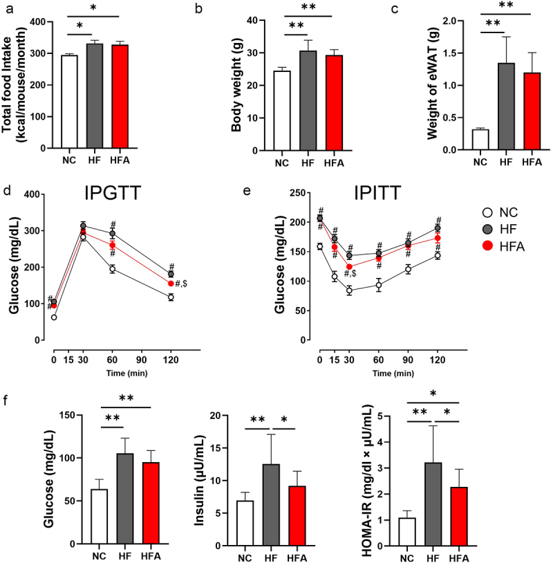
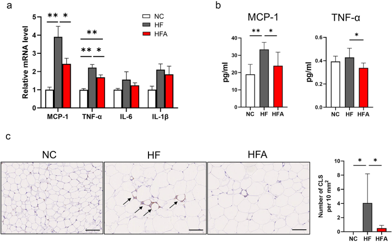
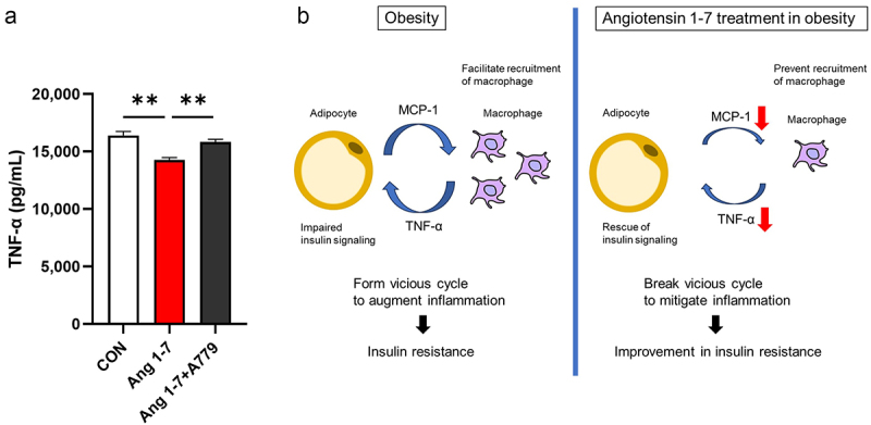
Obesity is a global health concern that promotes chronic low-grade inflammation, leading to insulin resistance, a key factor in many metabolic diseases. Angiotensin 1-7 (Ang 1-7), a component of the renin-angiotensin system (RAS), exhibits anti-inflammatory effects in obesity and related disorders, though its mechanisms remain unclear. In this study, we examined the effect of Ang 1-7 on inflammation of white adipose tissue (WAT) in dietary-induced obese mice. Monocyte chemoattractant protein-1 (MCP-1) produced by white adipocytes and tumour necrosis factor-α (TNF-α) produced by macrophages are pro-inflammatory cytokines and interact to form a pathogenic loop to exacerbate obesity-induced inflammation. We found that Ang 1-7 reduced MCP-1 and TNF-α gene expressions and the number of crown-like structures, which are histological hallmarks of the pro-inflammatory process, in visceral epididymal WAT (eWAT) and reduced circulating MCP-1 and TNF-α levels, accompanied by improvement in insulin resistance, in dietary-induced obese mice. Furthermore, Ang 1-7 reduced MCP-1 and TNF-α secretions in 3T3-L1 white adipocytes and RAW 264.7 macrophages, respectively, which are in vitro experimental models mimicking obesity condition. Our results suggest that Ang 1-7 directly acts on WAT to mitigate obesity-induced inflammation. Thus, this study provides novel insights into the underlying mechanism of anti-obesity effects of Ang 1-7.

Keywords: Angiotensin 1-7; MCP-1; TNF-α; anti-inflammatory effect; obesity.


Conflict of interest statement

No potential conflict of interest was reported by the author(s).

Figures

Angiotensin 1-7 improves insulin resistance and glucose tolerance.
Figure 1.
Angiotensin 1–7 (Ang 1–7) improves insulin resistance and glucose tolerance without affecting body weight, food intake, and epididymal white adipose tissue (eWAT) weight in dietary-induced obese mice. (a) Food intake, n = 4. (b) Body weight, n = 11–16. (c) Weight of eWAT, n = 11–15. (d) Levels of blood glucose during intraperitoneal glucose tolerance test (IPGTT), n = 10–15. (e) Levels of blood glucose during intraperitoneal insulin tolerance test (IPITT), n = 9–15. (f) Levels of blood glucose, serum insulin, and homeostasis model assessment of insulin resistance (HOMA-IR), with sample sizes of n = 11–15, n = 10–14, and n = 10–14, respectively. *p < 0.05, **p < 0.01. #p < 0.05 vs. NC. $p < 0.05 vs. HF. NC, mice fed a normal chow diet; HF, mice fed a high-fat diet alone; HFA, mice fed a high-fat diet and treated with Ang 1–7.
Angiotensin 1–7 reduces inflammation in visceral epididymal white adipose tissue in high-fat diet-induced obese mice.
Figure 2.
Ang 1–7 reduces inflammation in visceral eWAT in HFD-induced obese mice. (a) Relative mRNA level of MCP-1 (n = 6–7), TNF-α (n = 6–7), IL6 (n = 6–8) and IL1-β (n = 6–8) determined using real-time PCR. (b) Serum levels of MCP-1 (n = 6–8) and TNF-α (n = 6–8). (c) Left panel: representative histology of crown-like structures (CLS, indicated by black arrows) in eWAT of NC, HF,and HFA mice. NC, mice fed a normal chow diet alone; HF, mice fed a high-fat diet alone; HFA, mice fed a high-fat diet and treated with Ang 1–7. Scale bars, 100 μm. Right panel: number of CLS in eWAT (n = 5–8). Data are presented as means ± SE. *p < 0.05, **p < 0.01.
Angiotensin 1–7 reduces MCP-1 secretion in an obese model of 3T3-L1 white adipocytes.
Figure 3.
Ang 1–7 reduces MCP-1 secretion in an obese model of 3T3-L1 white adipocytes. (a) MCP-1 concentration in the culture medium of 3T3-L1 adipocytes at 1, 2, and 3 weeks after differentiation, determined by ELISA. CON, control group (vehicle-treated cells), n = 7–8; Ang 1–7, cells treated with Ang 1–7 alone, n = 7–8; Ang 1–7+A779, cells treated with Ang 1–7 and A779, n = 7–8. (b) MCP-1 concentration in the culture medium of TNF-α-treated 3T3-L1 adipocytes, determined by ELISA. CON, control group (vehicle-treated cells), n = 7; TNF-α (cells treated with TNF-α), n = 6; TNF-α+Ang 1–7, cells treated with TNF-α and Ang 1–7, n = 5; TNF-α+Ang 1–7+A779’, cells treated with TNF-α, TNF-α+Ang 1–7+A779, n = 5. Data are presented as means ± SE. *p < 0.05, **p < 0.01.
Angiotensin 1–7 reduces TNF-α secretion in a model of macrophage lipotoxicity.
Figure 4.
Ang 1–7 reduces TNF-α secretion in a model of macrophage lipotoxicity. (a) TNF-α concentrations in the culture medium of LPS- and PA-treated RAW264.7 cells. CON, cells treated with only LPS and PA (control group); Ang 1–7, cells treated with LPS, PA, and Ang 1–7; Ang 1–7+A779, cells treated with LPS, PA, Ang 1–7, and A779. n = 14 for each group. Data are presented as means ± SE. **p < 0.01. (b) Schematic diagram showing the mechanism by which Ang 1–7 reduces inflammation in WAT in obesity. Macrophage-derived TNF-α not only impairs insulin signalling but also stimulates adipocytes to enhance more MCP-1 secretion in obesity. Interaction between adipocyte and macrophage forms a vicious cycle to augment inflammation. Ang 1–7 breaks this vicious cycle by acting on adipocytes and macrophages to reduce MCP-1 and TNF-α secretion, respectively.

References

    1. Ahmed B, Sultana R, Greene MW.. Adipose tissue and insulin resistance in obese. Biomed Pharmacother. 2021;137:111315. doi: 10.1016/j.biopha.2021.111315 - DOI - PubMed
    1. Yaribeygi H, Farrokhi FR, Butler AE, et al. Insulin resistance: review of the underlying molecular mechanisms. J Cell Physiol. 2019;234(6):8152–10. doi: 10.1002/jcp.27603 - DOI - PubMed
    1. Kawai T, Autieri MV, Scalia R. Adipose tissue inflammation and metabolic dysfunction in obesity. Am J Physiol Cell Physiol. 2021;320(3):C375–C391. doi: 10.1152/ajpcell.00379.2020 - DOI - PMC - PubMed
    1. de Kloet AD, Krause EG, Woods SC. The renin angiotensin system and the metabolic syndrome. Physiol Behav. 2010;100(5):525–534. doi: 10.1016/j.physbeh.2010.03.018 - DOI - PMC - PubMed
    1. Santos RAS, Sampaio WO, Alzamora AC, et al. The ACE2/Angiotensin-(1–7)/MAS axis of the renin-angiotensin system: focus on angiotensin-(1–7). Physiol Rev. 2018;98(1):505–553. doi: 10.1152/physrev.00023.2016 - DOI - PMC - PubMed