Vacuolar myopathy with monoclonal gammopathy and stiffness (VAMMGAS)
- PMID: 39804003
- PMCID: PMC11726623
- DOI: 10.1111/ene.70026
Vacuolar myopathy with monoclonal gammopathy and stiffness (VAMMGAS)
Abstract
Background: Monoclonal gammopathy (MG) has been reported in association with numerous neurological disorders but the spectrum of MG-associated myopathies remains poorly described.
Objective: To report a newly acquired myopathy associated with MG.
Methods: Three adult patients with the same phenotype from two French referral centers were prospectively analyzed. Clinical, electrophysiological, muscle biopsy data, and patients' outcomes under treatment are reported.
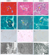
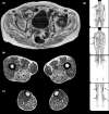
Results: The patients, aged 37, 46, and 56 years, presented progressive weakness with subacute worsening and stiffness, in the context of severe weight loss. The weakness mainly involved the proximal limbs and axial muscles. Creatine kinase levels were 1400-2900 IU/L and electromyography revealed a myopathic pattern with spontaneous complex repetitive discharges. Muscle biopsies showed vacuoles containing glycogen and autophagic material along with the presence of sarcolemmal complement membrane attack complex deposits. There was no evidence of a genetic glycogen metabolic disorder. IgGκ monoclonal gammopathy was identified in all cases, without signs of lymphoplasmocytic proliferation. All patients improved with a treatment combining corticosteroids, intravenous immunoglobulins, and immunosuppressants, and two patients recovered walking ability.
Conclusion and relevance: We report a new muscle disease defined by a vacuolar myopathy characterized by axial and proximal muscle weakness with prominent stiffness and high frequency discharges on electromyography associated with monoclonal gammopathy, defined under the acronym VAMMGAS.
Keywords: elevated CK; monoclonal gammopathy; stiffness; vacuolar myopathy.
© 2025 The Author(s). European Journal of Neurology published by John Wiley & Sons Ltd on behalf of European Academy of Neurology.
Conflict of interest statement
None of the authors have any disclosures relevant to the content of the manuscript.
Figures


Similar articles
-
Treatment-responsive glycogen storage myopathy in a patient with POEMS syndrome: A new monoclonal gammopathy-associated myopathy.Eur J Neurol. 2023 Oct;30(10):3404-3406. doi: 10.1111/ene.16008. Epub 2023 Aug 11. Eur J Neurol. 2023. PMID: 37522432
-
Monoclonal gammopathy with both nemaline myopathy and amyloid myopathy.Neuromuscul Disord. 2017 Oct;27(10):942-946. doi: 10.1016/j.nmd.2017.05.007. Epub 2017 May 11. Neuromuscul Disord. 2017. PMID: 28606401
-
Vacuolar myopathies in adults with myalgias: value of paraspinal muscle investigation.Muscle Nerve. 1997 Oct;20(10):1321-3. doi: 10.1002/(sici)1097-4598(199710)20:10<1321::aid-mus18>3.0.co;2-5. Muscle Nerve. 1997. PMID: 9324092
-
X-linked myopathy with excessive autophagy: a failure of self-eating.Acta Neuropathol. 2015 Mar;129(3):383-90. doi: 10.1007/s00401-015-1393-4. Epub 2015 Feb 3. Acta Neuropathol. 2015. PMID: 25644398 Review.
-
Myopathy and scleromyxedema.J Neurol. 2019 Aug;266(8):2051-2059. doi: 10.1007/s00415-019-09379-w. Epub 2019 May 21. J Neurol. 2019. PMID: 31115676 Review.
References
Publication types
MeSH terms
Supplementary concepts
LinkOut - more resources
Full Text Sources
Medical
Research Materials

