. 2025 Feb 7;24(2):479-490.
doi: 10.1021/acs.jproteome.4c00618. Epub 2025 Jan 13.

Structural Dynamics of the Ubiquitin Specific Protease USP30 in Complex with a Cyanopyrrolidine-Containing Covalent Inhibitor

Affiliations

Structural Dynamics of the Ubiquitin Specific Protease USP30 in Complex with a Cyanopyrrolidine-Containing Covalent Inhibitor

Darragh P O'Brien et al. J Proteome Res. .

Abstract

Inhibition of the mitochondrial deubiquitinating (DUB) enzyme USP30 is neuroprotective and presents therapeutic opportunities for the treatment of idiopathic Parkinson's disease and mitophagy-related disorders. We integrated structural and quantitative proteomics with biochemical assays to decipher the mode of action of covalent USP30 inhibition by a small-molecule containing a cyanopyrrolidine reactive group, USP30-I-1. The inhibitor demonstrated high potency and selectivity for endogenous USP30 in neuroblastoma cells. Enzyme kinetics and hydrogen-deuterium eXchange mass spectrometry indicated that the inhibitor binds tightly to regions surrounding the USP30 catalytic cysteine and positions itself to form a binding pocket along the thumb and palm domains of the protein, thereby interfering its interaction with ubiquitin substrates. A comparison to a noncovalent USP30 inhibitor containing a benzosulfonamide scaffold revealed a slightly different binding mode closer to the active site Cys77, which may provide the molecular basis for improved selectivity toward USP30 against other members of the DUB enzyme family. Our results highlight advantages in developing covalent inhibitors, such as USP30-I-1, for targeting USP30 as treatment of disorders with impaired mitophagy.

Keywords: Hydrogen−Deuterium eXchange-Mass spectrometry; activity-based protein profiling mass spectrometry; cyanopyrrolidine inhibitors; enzyme kinetics; mitophagy; molecular docking; ubiquitin specific protease USP30.


Conflict of interest statement

The authors declare no competing financial interest.

Figures

Figure 1
Figure 1
USP30-I-1 is selective and potent for endogenous USP30. (A) Structure of USP30-I-1. (B) n = 2 Western blot of USP30 with ∼10 kDa mass increase showing HA-Ub-PA bound to USP30 and prevention of HA-Ub-PA binding with increasing USP30-I-1 concentrations. (C) Densitometric quantitation of USP30 HA-Ub-PA labeling in B, fit to Y = 100/(1 + 10 × ((X – Log IC50))) for IC50 value extraction. (D) HA-Ub-PA binding to USP30 and prevention by USP30-I-1 as shown in B. The supernatant and eluate of a HA immunoprecipitation shows efficient pull down of USP30 with HA-Ub-PA labeling, and a reduction in the amount of USP30 immunoprecipitated where USP30-I-1 prevents HA-Ub-PA binding. The anti-HA blot shows efficient immunoprecipitation of HA-Ub-PA-labeled DUBs. (E) LC–MS/MS quantitation of the DUB-ABP complex immunoprecipitation shown in C. DUB activity is the intensity of the DUB in the presence of USP30-I-1 normalized to the positive HA-Ub-PA control. (n = 2, ****p < 0.0001).
Figure 2
Figure 2
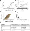
Covalent USP30-I-1 tightly binds to recombinant USP30. (A) Dose-dependent inhibition of USP30 by USP30-I-1. (B) Progress curves recorded on the FLIPR Tetra. Traditional method for determining kinetic constants associated with covalent binders. kobs, determined by fitting the progress curves to eq 2 (methods), is plotted vs [Compound], and fitted to eq 3 (methods) to determine KI and kinact. (C) Krippendorf method used for determining covalent kinetic constants. Time-dependent IC50 curves. Each curve represents inhibition data at an individual incubation time from 3 to 600 s. (D) IC50 values vs incubation time fitted to eq 1 (see the methods section) to obtain KI and kinact. (E) Data table of kinetic constants.
Figure 3
Figure 3
USP30-I-1 binds to the catalytic cysteine of USP30. (A) HDX-MS shows that the USP30 region 70LVNLGNNTCF78 which surrounds the catalytic Cys77 (underlined) is significantly solvent protected in the presence of USP30-I-1. This implies that it is the primary location of compound binding and interaction with the protein. All labeling time points for the two peptides covering this region are shaded in dark green. (B) Modeled structure of human USP30 in complex with USP30-I-1. Structure of human USP30 catalytic domain highlighting the modeled position of USP30-I-1 shown as a stick representation with carbon atoms colored green. The thumb, palm and fingers subdomains of the catalytic domain and the catalytic cysteine, Cys77, are highlighted. Regions identified in the HDX-MS analysis of USP30 in the presence of USP30-I-1 are colored red (mean perturbation over 60 min of 5–20%) and magenta (mean perturbation over 60 min of <60%). (C). Close-up view of the putative USP30-I-1 binding site highlighting flanking residues and key hydrogen-bonding interactions represented as dotted lines. The positions of blocking loop 1 (BL1), blocking loop 2 (BL2), and the switching loop (SL) are highlighted. Figure prepared using PyMOL (The PyMOL Molecular Graphics System, version 2.5.8; Schrödinger, LLC).
Figure 4
Figure 4
Comparison of USP30-I-1 binding to USP30 with Ub and analogous covalent USP30 inhibitors. (A). Superposition of the modeled structure of human USP30 in complex with USP30-I-1 on the X-ray structures of human USP30 in complex with the covalent inhibitors, 552 (PDB code: 8D1T) and 829 (PDB code: 8D0A). The thumb, palm and fingers subdomains of the catalytic domain and the catalytic cysteine, Cys77, are highlighted. The catalytic domain of the modeled structure of human USP30 in complex with USP30-I-1 is colored gray with regions implicated in compound binding from the HDX-MS analysis colored red (mean perturbation over 60 min of 5–20%) and magenta (mean perturbation over 60 min of <60%). USP30-I-1 (carbon atoms in green), 552 (carbon atoms in hot pink), and 829 (carbon atoms in cyan) are shown as stick representations. The catalytic domain of USP30 in complex with 552 and 829 are colored brown and violet, respectively. The modeled pose of USP30-I-1 correlates well with 552 and 829. (B). Close-up view of the superimposed structure of human USP30 in complex with 552 (carbon atoms in hot pink) with the modeled structure of USP30-I-1 (carbon atoms in green). The positions of BL1, BL2, and SL are highlighted. (C). Close-up view of the superimposed structure of human USP30 in complex with 829 (carbon atoms in cyan) with the modeled structure of USP30-I-1 (carbon atoms in green). The positions of BL1, BL2, and SL are highlighted. (D). Superposition of the modeled structure of human USP30 in complex with USP30-I-1 on the X-ray structure of human USP30 in complex with ubiquitin-propargylamide (UbPA; PDB code: 5OHK). The catalytic domain of the modeled structure of human USP30 in complex with USP30-I-1 is colored gray with regions implicated in compound binding from HDX-MS analysis are colored as above. USP30-I-1 is shown with carbon atoms colored green. The catalytic domain of USP30 in complex with UbPA is colored lime with UbPA shown in orange. USP30-I-1 is predicted to bind in the thumb-palm cleft that guides the ubiquitin C-terminus into the active site. (E). Close-up view of the superimposed structures of human USP30 in complex with UbPA and the modeled structure of USP30-I-1 (carbon atoms in green). Catalytic triad residues (C77, H452 and S477) are highlighted and shown as stick representations. Figure prepared using PyMOL (The PyMOL Molecular Graphics System, version 2.5.8; Schrödinger, LLC).
Figure 5
Figure 5
Comparison of covalent and noncovalent inhibitor binding in USP7 and USP30. (A) Superposition of the modeled structure of human USP30 in complex with USP30-I-1 on the X-ray structure of human USP7 in complex with the covalent inhibitor, FT827 (PDB code: 5NGF; r.m.s.d. = 2.5 Å, 249 residues aligned). (B) Close-up view of the superimposed modeled structure of human USP30 in complex with USP30-I-1 (green carbon atoms) on the X-ray structure of human USP7 in complex with the covalent inhibitor, FT827 (orange carbon atoms). The positions of BL1, BL2, SL, and the catalytic cysteines (Cys77 in USP30 and Cys223 in USP7) are highlighted. The Cαs of Cys77 and Cys223 are separated by approximately 5.2 Å, which, combined with differences in the conformations of BL1, BL2, and SL, result in FT827 extending closer toward the fingers subdomain than USP30-I-1. (C) Superposition of the modeled structures of human USP30 in complex with USP30-I-1 and USP30inh. The catalytic domain of the modeled structure of human USP30 in complex with USP30-I-1 is colored gray with regions implicated in compound binding from HDX-MS analysis colored red (mean perturbation over 60 min of 5–20%) and magenta (mean perturbation over 60 min of <60%). USP30-I-1 is shown with carbon atoms colored green. The catalytic domain of the modeled structure of human USP30 in complex with USP30inh is colored blue. USP30inh is shown with carbon atoms colored yellow. (D) Close-up view of the superimposed modeled structures of human USP30 in complex with USP30-I-1 (carbon atoms in green) and USP30inh (yellow carbon atoms). The positions of BL1, BL2, and SL are highlighted. Both inhibitors are predicted to bind within the thumb-palm cleft with USP30inh residing approximately 7.9 Å away from Cys77 and extending out toward the fingers subdomain. (E) HDX-MS residual plot of USP30 in complex with USP30-I-1 and USP30inh. A greater overall solvent protection is observed for USP30 in the presence of the noncovalent inhibitor, as compared to its covalent counterpart. A cutoff of 90% D perturbation was used for ease of comparison, which is the highest value observed for the USP30inh study and common to both experiments. USP30-I-1 primarily induces solvent protection in the region encompassing the catalytic Cys77, whereas the noncovalent USP30inh results in smaller HDX-MS perturbations, albeit, extended to several regions of the protein. Figure prepared using PyMOL (The PyMOL Molecular Graphics System, version 2.5.8; Schrödinger, LLC).

References

    1. Picca A.; Faitg J.; Auwerx J.; Ferrucci L.; D’Amico D. Mitophagy in human health, ageing and disease. Nat. Metab. 2023, 5 (12), 2047–2061. 10.1038/s42255-023-00930-8. - DOI - PubMed
    1. Liu J.; Liu W.; Li R.; Yang H. Mitophagy in Parkinson’s Disease: From Pathogenesis to Treatment. Cells 2019, 8 (7), 712.10.3390/cells8070712. - DOI - PMC - PubMed
    1. Geisler S.; Holmstrom K. M.; Treis A.; Skujat D.; Weber S. S.; Fiesel F. C.; Kahle P. J.; Springer W. The PINK1/Parkin-mediated mitophagy is compromised by PD-associated mutations. Autophagy 2010, 6 (7), 871–878. 10.4161/auto.6.7.13286. - DOI - PubMed
    1. Wang Y.; Long L.; Luo Q.; Huang X.; Zhang Y.; Meng X.; Chen D. Aflatoxin B1 induces ROS-dependent mitophagy by modulating the PINK1/Parkin pathway in HepG2 cells. Basic Clin. Pharmacol. Toxicol. 2024, 135, 195.10.1111/bcpt.14034. - DOI - PubMed
    1. Aaluri G. R.; Choudhary Y.; Kumar S. Mitochondria-Associated MicroRNAs and Parkinson’s Disease. Neurosci. Insights 2024, 19, 26331055241254846.10.1177/26331055241254846. - DOI - PMC - PubMed

LinkOut - more resources