. 2025 Aug 1;60(8):499-507.
doi: 10.1097/RLI.0000000000001152. Epub 2025 Jan 14.

ADAMTS4-Specific MR Peptide Probe for the Assessment of Atherosclerotic Plaque Burden in a Mouse Model

Affiliations

ADAMTS4-Specific MR Peptide Probe for the Assessment of Atherosclerotic Plaque Burden in a Mouse Model

Dilyana B Mangarova et al. Invest Radiol. .

Abstract

Introduction: Atherosclerosis is the underlying cause of multiple cardiovascular pathologies. The present-day clinical imaging modalities do not offer sufficient information on plaque composition or rupture risk. A disintegrin and metalloproteinase with thrombospondin motifs 4 (ADAMTS4) is a strongly upregulated proteoglycan-cleaving enzyme that is specific to cardiovascular diseases, inter alia, atherosclerosis.

Materials and methods: Male apolipoprotein E-deficient mice received a high-fat diet for 2 (n = 11) or 4 months (n = 11). Additionally, a group (n = 11) receiving pravastatin by drinking water for 4 months alongside the high-fat diet was examined. The control group (n = 10) consisted of C57BL/6J mice on standard chow. Molecular magnetic resonance imaging was performed prior to and after administration of the gadolinium (Gd)-based ADAMTS4-specific probe, followed by ex vivo analyses of the aortic arch, brachiocephalic arteries, and carotid arteries. A P value <0.05 was considered to indicate a statistically significant difference.

Results: With advancing atherosclerosis, a significant increase in the contrast-to-noise ratio was measured after intravenous application of the probe (mean precontrast = 2.25; mean postcontrast = 11.47, P < 0.001 in the 4-month group). The pravastatin group presented decreased ADAMTS4 expression. A strong correlation between ADAMTS4 content measured via immunofluorescence staining and an increase in the contrast-to-noise ratio was detected ( R2 = 0.69). Microdissection analysis revealed that ADAMTS4 gene expression in the plaque area was significantly greater than that in the arterial wall of a control mouse ( P < 0.001). Laser ablation-inductively coupled plasma-mass spectrometry confirmed strong colocalization of areas positive for ADAMTS4 and Gd.

Conclusions: Magnetic resonance imaging using an ADAMTS4-specific agent is a promising method for characterizing atherosclerotic plaques and could improve plaque assessment in the diagnosis and treatment of atherosclerosis.

Keywords: ADAMTS4; atherosclerosis; inflammation; molecular imaging; mouse model; plaque; vulnerability.


Conflict of interest statement

Conflicts of interest and sources of funding: The authors declare that they have no competing interests. This work was supported by the Deutsche Forschungsgemeinschaft/German Research Foundation (Project-ID 372486779-SFB 1340, B01, B02).

Figures

FIGURE 1
FIGURE 1
In vivo MRI using an ADAMTS4-specific molecular probe. A, En face lipid oil red O staining showed early lesions in the thoracic portion of the aorta, the aortic arch, carotid arteries, and brachiocephalic arteries in the 2-month group, whereas more extensive, large surface lesions were observed in the 4-month group. B, After in vivo administration of the ADAMTS4-specific probe, a significantly stronger enhancement in the contrast-to-noise ratio between the control group (n = 10), the 2-month group (n = 11) (p[sham, 2 months] < 0.001), the pravastatin group (n = 11) (p[2 months, pravastatin] < 0.001), and the 4 months group (n = 11) (p[pravastatin, 4 months] < 0.001) was measured in MRI. Data are presented as mean values ± SD. C, A group of C57BL/6J mice fed with a standard laboratory chow was used as the control group and showed no pathological changes of the aortic wall in vivo and ex vivo as well as no ADAMTS4 expression and macrophage migration. In the 2-month, 4-month, and pravastatin group, strong signal enhancement in the plaque area (white arrows) was observed on T1-weighted MRI sequences after administration of the ADAMTS4-specific probe. Ex vivo histology confirmed the formation of atherosclerotic plaques in the brachiocephalic artery and a strong expression of ADAMTS4 within the plaque area. The ADAMTS4 expression was colocalized with macrophage migration. ADAMTS4, a disintegrin and metalloproteinase with thrombospondin motifs 4; MRI, magnetic resonance imaging; EvG, Elastica van Gieson staining; HE, hematoxylin-eosin staining; ECM, extracellular matrix; ApoE−/−, apoliproprotein E deficient; * indicates the arterial lumen; # indicates the plaque area; scale bars represent 100 μm.
FIGURE 2
FIGURE 2
Analyses of the selectivity of the ADAMTS4-specific probe. A, By using a cyclic peptide with similar size to the ADAMTS4-specific probe as negative control peptide (Cys*-Phe-His-Pro-Tyr-Cys*-linker (Gd-DOTA)), no significant difference in CNR was measured in vivo in the sham group (n = 3) between precontrast MRI, postapplication of the negative control peptide (P = 1), and the ADAMTS4-specific probe (P = 1). In the 2 months group (n = 3), no significant CNR enhancement was observed between precontrast MRI and the negative control peptide (P = 1), whereas in the same group, the application of the ADAMTS4-specific probe resulted in a strong and significant increase in CNR in the atherosclerotic plaque (P < 0.001). Data are presented as mean values ± SD. B, In the competition experiment (n = 3), a significant decrease in the CNR compared with the first injection of the Gd-labeled ADAMTS4-specific probe alone was observed, confirming the selective binding of the probe (P < 0.001). Data are presented as mean values ± SD. C, In vivo administration of an ADAMTS4-specific fluorescent probe compared with ex vivo immunofluorescence with ADAMTS4-specific antibody yielded a strong overlap of both stainings in the atherosclerotic plaque area. The color of the FLP was changed to green from Cy5 fluorescence for better differentiation in the merged picture. Gd, gadolinium; ADAMTS4, a disintegrin and metalloproteinase with thrombospondin motifs 4; MRI, magnetic resonance imaging; CNR, contrast-to-noise ratio; FLP, fluorescence probe; scale bars represent 100 μm.
FIGURE 3
FIGURE 3
Analyses of ADAMTS4- and CD68-expression within the arterial wall. A, A significant increase in ADAMTS4-expression after 2 months of high fat diet in ApoE−/− mice in immunofluorescence staining was observed. By prolonging the HFD to 4 months, the ADAMTS4-expression increases further. The treatment with pravastatin did not significantly decrease the ADAMTS4 expression. Data are presented as mean values ± SD. B, The ex vivo immunofluorescence measurements of ADAMTS4 were in good correlation with the in vivo contrast-to-noise ratio (n = 7 per group; y = 0.81x + 3.91; R2 = 0.80). C, Immunofluorescence staining for ADAMTS4 and CD68 showed a high correlation for mice after 2 months and 4 months of HFD, as well as 4 months of HFD in combination with pravastatin treatment, whereas for the control group, no relevant expression of ADAMTS4 or CD68 was observed (n = 5 per group; y = 0.86x + 0.27; R2 = 0.90). D, mRNA sequencing by NanoString revealed increasing ADAMTS4 levels (left hand, P < 0.001) as well as increasing levels of the macrophage marker CD68 (right hand, P < 0.001) in the plaque area compared with the control group (n = 7 per group). ADAMTS4, a disintegrin and metalloproteinase with thrombospondin motifs 4; HFD, high fat diet; CNR, contrast-to-noise ratio; ApoE−/−, apolipoprotein E-deficient.
FIGURE 4
FIGURE 4
Histological analyses of human atherosclerotic plaque specimens. Immunostaining of CD68 in a stable human atherosclerotic plaque (erosion-prone plaque; left hand) and a rupture-prone plaque (thin-cap-fibrous atheroma; right hand) showed high colocalization with staining of a parallel section with the ADAMTS4-specific fluorescence probe. Both the macrophage marker CD68 and ADAMTS4 are highly expressed in the human atherosclerotic plaque area, especially in the fibrous cap area. ADAMTS4, a disintegrin and metalloproteinase with thrombospondin motifs 4; FLP, fluorescence probe; scale bars indicate 2.5 mm (upper row) and 0.5 mm (lower row).
FIGURE 5
FIGURE 5
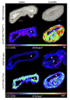
Laser ablation-inductively coupled plasma-mass spectrometry (LA-ICP-MS) analysis for spatial localization of Gd and Ca. First row: Light microscopy image of a cross section of the brachiocephalic artery without atherosclerotic changes (left, control) and with atherosclerotic plaque (right, 2 months group). Second row: Visualization of Gd from the ADAMTS4-specific probe by LA-ICP-MS within the aortic wall and the atherosclerotic plaque (right hand) and of a control section (left hand). Third row: Immunofluorescence of ADAMTS4 of a control section (left hand) and the atherosclerotic section (right hand) yielded areas positive for ADAMTS4 in the atherosclerotic section with a favorable overlay to the Gd location from the LA-ICP-MS. Fourth row: Calcium binding to the elastic layers was observed. Additionally, calcification spots occur in the plaque lesion. * indicates the aortic lumen; # indicates the plaque area. ADAMTS4, a disintegrin and metalloproteinase with thrombospondin motifs 4; LA-ICP-MS, laser ablation–inductively coupled plasma–mass spectrometry; Ca, calcium; Gd, gadolinium. Scale bars indicate 100 μm.

References

    1. Benjamin EJ Blaha MJ Chiuve SE, et al. Heart Disease and Stroke Statistics-2017 update: a report from the American Heart Association. Circulation. 2017;135:e146–e603. - PMC - PubMed
    1. Virmani R Burke AP Farb A, et al. Pathology of the vulnerable plaque. J Am Coll Cardiol. 2006;47(8 Suppl):C13–C18. - PubMed
    1. Cybulsky MI, Gimbrone MA. Endothelial expression of a mononuclear leukocyte adhesion molecule during atherogenesis. Science. 1991;251:788–791. - PubMed
    1. Libby P Buring JE Badimon L, et al. Atherosclerosis. Nat Rev Dis Primers. 2019;5:56. - PubMed
    1. Wight TN. A role for proteoglycans in vascular disease. Matrix Biol J Int Soc Matrix Biol. 2018;71-72:396–420. - PMC - PubMed