. 2025 Jan 13;21(1):e1011355.
doi: 10.1371/journal.pgen.1011355. eCollection 2025 Jan.

Ambivalent partnership of the Drosophila posterior class Hox protein Abdominal-B with Extradenticle and Homothorax

Affiliations

Ambivalent partnership of the Drosophila posterior class Hox protein Abdominal-B with Extradenticle and Homothorax

Jesús R Curt et al. PLoS Genet. .

Abstract

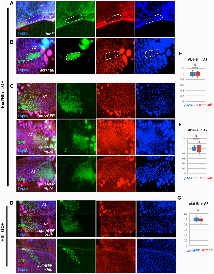
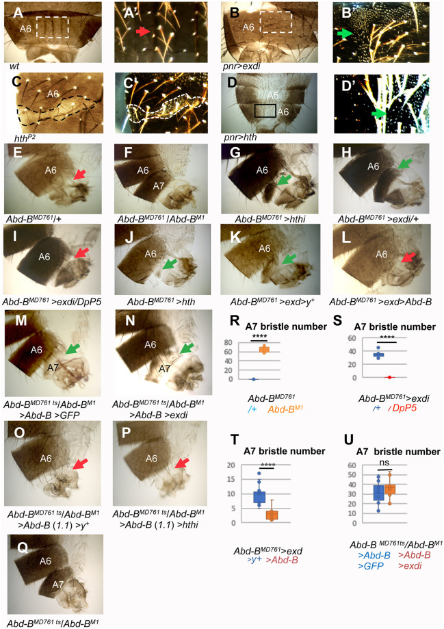
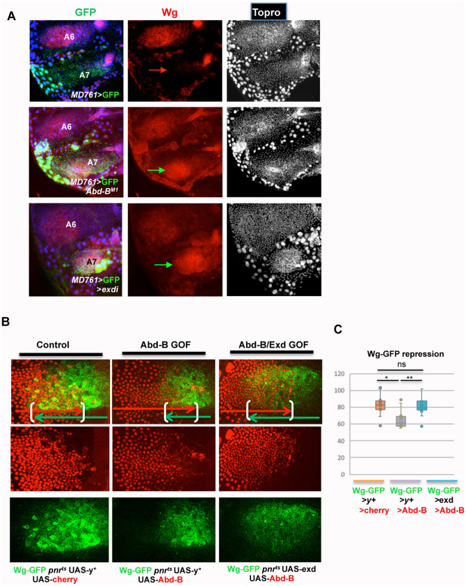
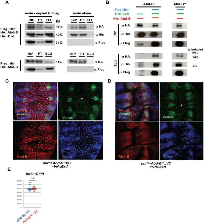
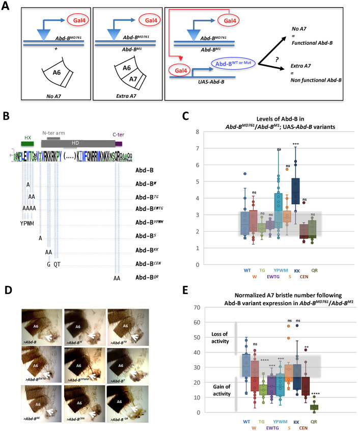
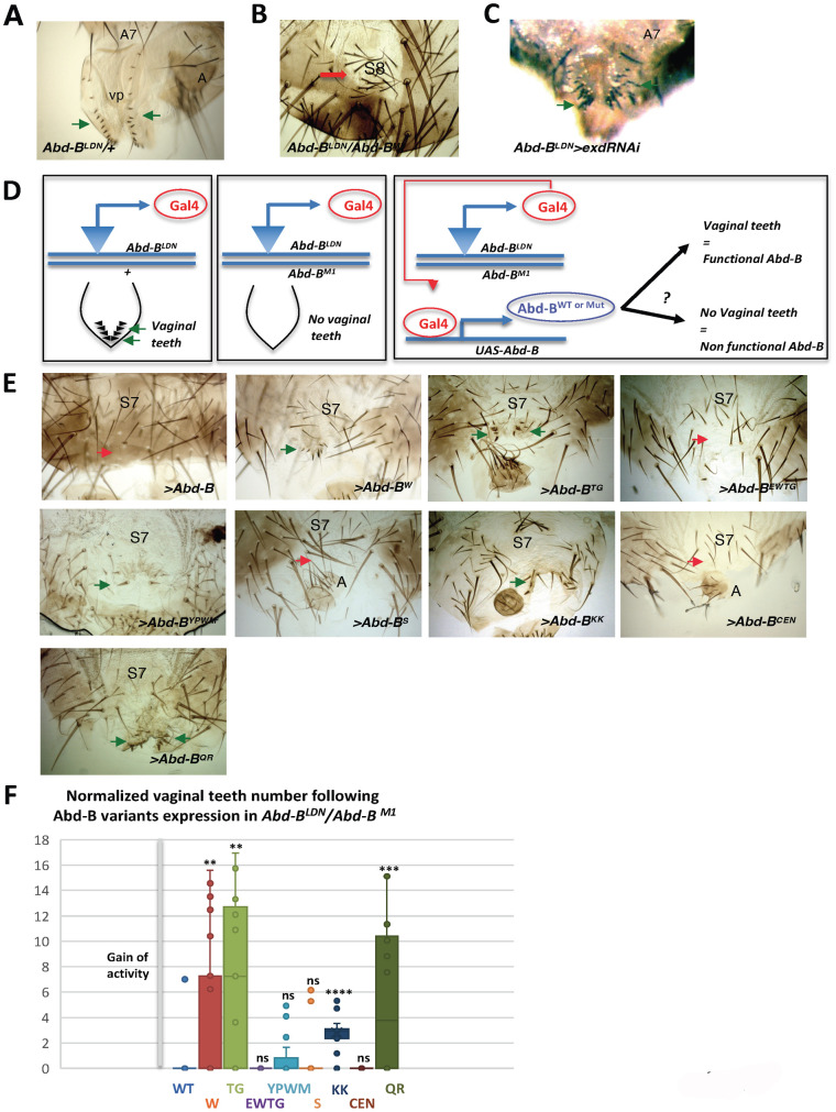
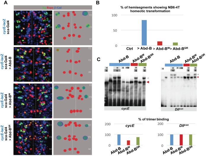
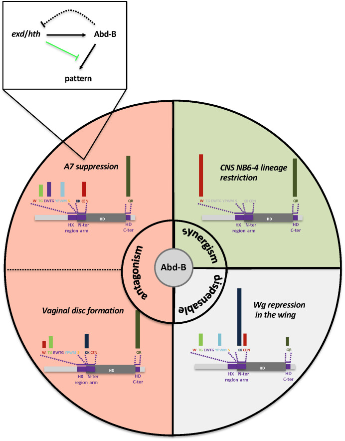
Hox proteins, a sub-group of the homeodomain (HD) transcription factor family, provide positional information for axial patterning in development and evolution. Hox protein functional specificity is reached, at least in part, through interactions with Pbc (Extradenticle (Exd) in Drosophila) and Meis/Prep (Homothorax (Hth) in Drosophila) proteins. Most of our current knowledge of Hox protein specificity stems from the study of anterior and central Hox proteins, identifying the molecular and structural bases for Hox/Pbc/Meis-Prep cooperative action. Posterior Hox class proteins, Abdominal-B (Abd-B) in Drosophila and Hox9-13 in vertebrates, have been comparatively less studied. They strongly diverge from anterior and central class Hox proteins, with a low degree of HD sequence conservation and the absence of a core canonical Pbc interaction motif. Here we explore how Abd-B function interface with that of Exd/Hth using several developmental contexts, studying mutual expression control, functional dependency and intrinsic protein requirements. Results identify cross-regulatory interactions setting relative expression and activity levels required for proper development. They also reveal organ-specific requirement and a binary functional interplay with Exd and Hth, either antagonistic, as previously reported, or synergistic. This highlights context specific use of Exd/Hth, and a similar context specific use of Abd-B intrinsic protein requirements.


Conflict of interest statement

The authors have declared that no competing interests exist.

Figures

Fig 1
Fig 1. Abd-B, exd and hth wildtype expression and regulation of exd and hth by Abd-B.
(A) Male wildtype pupa showing higher levels of Abd-B expression (in green) in the A7 than in the A6. Hth is in red and Topro is in blue. (B) Male A7 segment of a pupa showing the coincident expression of Abd-B (red), Exd (green) and Hth (blue) in histoblasts and Larval Epidermal Cells. (C) Abd-BM5 clone, induced in the pupal male A7 and marked by the absence of GFP, showing there is no major change in Exd (red) or Hth (blue) expression with respect to wildtype cells. (D) In pnr-Gal4 UAS-GFP males, the pnr domain of the A7 segment has similar levels of hth that the pnr- domain of the same segment. (E) The reduction of Abd-B expression in the pnr region of the male A7 (pnr-Gal4 UAS-GFP UAS- Abd-BRNAi) does not change Hth levels of expression with respect to the pnr- domain. (F) The ectopic expression of Abd-B (line 1.1) in the pupal A4 segment, marked with GFP, reduces Exd (red) and Hth (blue) expression with respect to adjacent, wildtype cells. (G) In pnr-Gal4 UAS-GFP pupae, the A4 segment has similar levels of expression of Exd (red) and Hth (blue) in the pnr+ and pnrdomains. In pnr-Gal4 UAS-GFP UAS-Abd-B (line 1.1) pupae, by contrast, the levels of expression of Exd (red) and Hth (blue) in the pnr+ domain are substantially reduced in comparison with those of the pnr- domain. The white arrow indicate LECs that express GFP (and therefore ectopically express Abd-B) but do not reduce Exd or Hth levels as compared with LECs that do not activate Abd-B (yellow arrow). (H, I) Quantification of the ratio of expression in a central (pnr+) with respect to a more lateral (pnr-) domain of Exd (H) or Hth (I) in pnr-Gal4 UAS-GFP/+ and pnr-Gal4 UAS-GFP UAS-Abd-BRNAi male A7 histoblast nests. (J, K) Quantification of the ratio of expression in a central (pnr+) with respect to a more lateral (pnr-) domain of Exd (J) or Hth (K) in pnr-Gal4 UAS-GFP/+ and pnr-Gal4 UAS-GFP UAS-Abd-B male A3-A4 histoblast nests. Pupae in all panels are of about 24-30h APF. Statistical analysis of the data in H-K was done by two-tailed t-tests, with n = 10–12 pupae.
Fig 2
Fig 2. Regulation of Abd-B by exd and hth in the male pupal abdomen.
(A) hthP2 mutant clone in the pupal A7, marked by the absence of GFP, showing reduced expression of Abd-B (red). Topro is in blue. (B) Flip-out clone marked with GFP and expressing an hthRNAi construct (act>stop>Gal4 UAS-GFP UAS-hthRNAi) in the male A7 segment showing reduced expression of Abd-B (red). Topro, marking nuclei, is in blue. (C) In the male A7 segment of pnr-Gal4 UAS-GFP pupae the cells in the pnr domain have similar levels of Abd-B (in red) as those not expressing pnr. Topro is in blue. If either exd or hth expression is reduced in the male A7 pnr domain (pnr-Gal4 UAS-GFP UAS-exdRNAi or pnr-Gal4 UAS-GFP UAS-hthRNAi), the expression of Abd-B is not significantly altered with respect to the domain not expressing pnr. (D) Upper panels: male A7 segment of a pnr-Gal4 UAS-hth pupa showing similar levels of Abd-B expression (red) in the pnr+ and pnr- domains; lower panels: flip-out clone, marked with GFP, induced in the male A7 segment and expressing hth. The levels of Abd-B in the clone (in red) do not change with respect to surrounding cells. (E-G) Quantification of the ratio of expression of Abd-B in a central (pnr+) with respect to a more lateral (pnr-) domain in pnr-Gal4 UAS-GFP/+ and pnr-Gal4 UAS-GFP UAS-exdRNAi (E), pnr-Gal4 UAS-GFP UAS-hthRNAi (F), and pnr-Gal4 UAS-GFP UAS-hth (G) male A7 histoblast nests. Pupae in all panels are of about 24-30h APF except in A, which was of about 34h APF. Statistical analysis of the data in E-G was done by two-tailed t-tests, with n = 10–12 pupae.
Fig 3
Fig 3. Reduction or excess of hth/exd cause anteriorwards transformations in the male abdomen.
(A, A’) Bright (A) and dark field (A’, inset) images of the dorsal A6 segment of a wildtype male, showing no trichomes in the medial region of the segment (red arrow). (B, B’) In pnr-Gal4 UAS-exdRNAi males, the dorsal central domain of the A6 segment, where pnr is expressed, presents many trichomes (B’, dark field image of inset, green arrow), suggesting transformation to a more anterior segment. (C, C’) Bright (C) and dark field (C’) images of a clone mutant for hthP2, marked with yellow and outlined, showing trichomes within the clone, suggesting transformation to a more anterior segment. (D, D’) In the pnr-Gal4 UAS-hth abdomen, the central dorsal region of A5 and A6, where pnr is expressed, present depigmentation and a great number of trichomes (inset of D in D’, dark field, green arrow), again suggesting transformation to a more anterior segment. (E) Abd-BMD761/+ males have no A7 segment, like the wildtype. In this and subsequent panels the red arrows indicate absence of A7 and the green arrows presence of this segment. (F) Abd-BMD761/Abd-BM1 male (mutant background for rescue experiments). The A7 is largely transformed into the A6. (G-I) The reduction of either hth (G) or exd (H) results in the development of a small A7 segment. The formation of this segment depends on the amount of Abd-B, since an extra dose of Abd-B (with DpP5) in an Abd-BMD761 UAS-exdRNAi background (I) reverts the mutant phenotype to the wildtype (compare H with I). (J-L) The increase in the amount of hth (J) or exd (K) also forms an A7 segment, and this development is prevented if Abd-B is concomitantly expressed (L). (M, N) The expression of Abd-B in a tub-Gal80ts/UAS-Abd-B; Abd-BMD761 UAS-GFP/Abd-BM1 male (shift from 18°C to 29°C at third larval stage) partially reverts the phenotype of the Abd-B mutant background (M, compare with F and Q); this phenotype does not significantly change if there is reduction of exd (UAS-exdRNAi/UAS-Abd-B; Abd-BMD761 tub-Gal80ts /Abd-BM1 male with the same temperature shift) (N). (O) UAS-y+/UAS-Abd-B (1.1); Abd-BMD761 tub-Gal80ts /Abd-BM1 male (shift from 18°C to 29°C at the third instar) showing complete suppression of A7 development. (P) The reduction of hth expression in UAS-Abd-B (1.1)/+; Abd-BMD761 tub-Gal80ts /Abd-BM1 UAS-hthRNAi males, with a similar temperature shift, shows also no A7 segment. (Q) Abd-BMD761 tub-Gal80ts/Abd-BM1 male (the mutant background for experiments in O, P), also shifted from 18°C to 29°C in third instar larva, showing transformation of A7 into A6. (R-U) Quantification of the number of bristles in the following genotypes: Abd-BMD761/+ (n = 11) and Abd-BMD761 /Abd-BM1 (n = 14) (R), Abd-BMD761 UAS-exdRNAi, and Abd-BMD761 UAS-exdRNAi DpP5 (n = 10 for both genotypes) (S), Abd-BMD761 UAS-exd UAS-y+ (n = 14) and Abd-BMD761 UAS-exd UAS-Abd-B (n = 10) (T), and tub-Gal80ts/UAS-Abd-B; Abd-BMD761 UAS-GFP/Abd-BM1 (n = 18) and UAS-exdRNAi/UAS-Abd-B; Abd-BMD761 tub-Gal80ts /Abd-BM1 males (n = 14) (U). Statistical analysis of the data in R-U was done by two-tailed t-tests.
Fig 4
Fig 4. Regulation of wg expression by exd/hth and Abd-B.
(A) Expression of Wg in the male A7 of Abd-BMD761 UAS-GFP/+, Abd-BMD761 /Abd-BM1, and Abd-BMD761/UAS-exdRNAi males, showing no Wg expression in the A7 in the first genotype (red arrow) and ectopic Wg expression in this segment of the two latter genotypes (green arrows). (B) Expression of wg-GFP in the A4 segment of male pupae: when Abd-B is overexpressed (wg-GFP/UAS-Abd-B; pnr-Gal4 tub-Gal80ts/UAS-y+; n = 10) there is partial repression of wg-GFP, as compared to the control (wg-GFP/UAS-y+; pnr-Gal4 tub-Gal80ts/UAS-cherry; n = 10). This repression is partially reverted by the concomitant expression of exd (UAS-exd; wg-GFP/UAS-Abd-B; pnr-Gal4 tub-Gal80ts/+; n = 12). Larvae of the three genotypes were shifted from 18°C to 29°C in the third larval stage. (C) Quantification of the extent of overlap of the wg-GFP and cherry, or wg-GFP and Abd-B, signals in the three genotypes. Statistical analysis was done by One-way ANOVA.
Fig 5
Fig 5. Co-immunoprecipitation and BiFC experiments related to Abd-B and Exd interaction.
(A) Abd-B, Exd and Hth co-immunoprecipitations (co-IPs). INP, FT, and Elu stand, respectively, for input (the material loaded on the resin), flow through (the material not captured by the resin), and elution (the material bound to the resin and boiled eluted). Protein present in the INP, FT and ELU is detected by the fused tags: Flag for Hth, which also serves for capturing on the anti-Flag resin, HA for Abd-B and His for Exd. The resin not coupled to anti-Flag serves as a control (three right columns). Above is the co-IP in the presence of Hth, Abd-B and Exd, and below the co-IP in the presence of Hth and Abd-B. Abd-B co-precipitates with Hth in the presence and absence (to a lesser extent) of Exd. % next to the panels indicates the ratio Elu/INP (x100). (B) Comparative Abd-B and Abd-BW co-IPs. Protein present in the co-IPs is indicated above the gels. There is a significant decrease in the amount of the Abd-BW protein captured on the resin when compared to the Abd-B wildtype protein (24%), while Exd remains almost unchanged (2%). (C, D) Dorsal views of pupae of approximately 24-28h APF of the genotypes pnr-Gal4 tub-Gal80ts UAS-Abd-B::VC UAS-VN::Exd (C) and pnr-Gal4 tub-Gal80ts UAS-Abd-BW::VC UAS-VN::Exd (D) showing Venus signal (in green), Abd-B expression (in red) and Topro, marking nuclei (in blue). See that there is Venus signal, indicating complementation between the Venus fragments of the Abd-B and Exd proteins in histoblasts but not (or just a few) in the polytene LECs (asterisks). The preparation in D has a reduced number of LECs due to tearing of the tissue during mounting. (E) Quantification of BiFC signals. There is similar Venus signal in the experiments performed with the Abd-B and Abd-BW constructs (n = 12 for the two genotypes). Statistical analysis was done by two-tailed t-test.
Fig 6
Fig 6. Activity of the wildtype and Abd-BW mutant proteins.
(A) Scheme of the genetic experiments performed to ascertain the rescue of the male Abd-BMD761/Abd-BM1 mutant phenotype (A7 transformed into A6) by the expression of wildtype Abd-B or different Abd-B variants. (B) Scheme of Abd-B protein mutations. The sequence of the Abd-B protein is indicated, as well as its most conserved domains (in arthropods and human), the Homeodomain (HD), the Hexapeptide (HX), and sequences immediately C-terminal to the HD. The N-terminal arm of the HD is highlighted. Web logo was obtained using sequences for the following Abd-B sequences (Drosophila (AAA84402), Tribolium (AAF36721.1), Anophela (XM311628), Sacculine (AAQ49317.1), Folsomia (AAK52499.1) and human (BCO10023). (C) Levels of Abd-B protein expression in the rescue experiment. Levels of the wild type (WT) and different Abd-B variants (referred to according to the labeling in B) measured as amount of protein in a defined area of A7/amount of protein in the same area of A6 dorsal histoblast nests in 24-28h APF pupae; n = 20 (Abd-B), 19 (W), 11 (TG), 11 (EWTG), 19 (YPWM), 15 (S), 12 (KK), 11 (CEN) and 15 (QR). (D) Examples of the posterior abdomens of males expressing wild type or Abd-B variants (UAS constructs represented as > Abd-B [x]) in the Abd-BMD761/Abd-BM1 common genetic background. Arrows point to the A7. (E) Quantification (measured as number of bristles) of the rescue of the A7 mutant phenotype observed in Abd-BMD761/Abd-BM1 animals by the different Abd-B protein variants. Bristle numbers were normalized relative to the level of Abd-B and Abd-B protein variant expression (counts x ratio Abd-B variants/ Abd-B); n = 18 (Abd-B), 26 (W), 15 (TG), 16 (EWTG), 21 (YPWM), 11 (S), 16 (KK), 14 (CEN) and 12 (QR). Statistical analysis on normalized bristle number values was performed using the Wilcoxon test.
Fig 7
Fig 7. Rescue of the female genitalia mutant phenotype by different Abd-B proteins.
(A) Abd-BLDN/+ female genitalia, showing the two rows of vaginal teeth (arrows). Vp, vaginal plates. (B) In Abd-BLDN/Abd-BM1 females the genitalia are eliminated and frequently replaced by an eighth sternite (S8). (C) Reduction of exd produces more, bigger and disorganized vaginal teeth (arrows). (D) Scheme of the genetic experiments performed to ascertain the rescue of the Abd-B mutant phenotype of Abd-BLDN/Abd-BM1 females (elimination of genitalia, including vaginal teeth) by the expression of wildtype or different Abd-B variants. (E) Examples of female genitalia phenotypes of Abd-BLDN/Abd-BM1 (common genetic background) expressing wild type or Abd-B variants (UAS constructs represented as > Abd-B [x]), showing no rescue of the mutant phenotype (red arrows) or a small rescue, with presence of some vaginal teeth (green arrows). S7, seventh sternite; A, analia. (F) Quantification (measured as number of vaginal teeth) of the rescue of the absence of genitalia phenotype observed in Abd-BLDN /Abd-BM1 animals expressing the Abd-B wild type or the Abd-B variants; n = 24 (Abd-B), 28 (W), 11 (TG), 12 (EWTG), 19 (YPWM), 17 (S), 12 (KK), 13 (CEN) and 20 (QR). Vaginal teeth numbers were normalized relative to the level of Abd-B and Abd-B protein variant expression (counts / ratio Abd-B variants/ Abd-B).
Fig 8
Fig 8. Functional synergy of Abd-B and Exd/Hth in the embryonic CNS.
(A) Impact of thoracic expression of Abd-B, Abd-BW or Abd-BQR on the NB6-4 CNS lineage. Thoracic expression was achieved using the scabrous (sca) Gal-4 driver, active from early stages in all neuroblasts, including thoracic NB6-4. Eagle (Eg; in green) was used as a marker to identify the NB6-4 lineage (neuron + glia) and Repo (in red) as a general marker of glial cells. The 1.9 cycE enhancer, shown to recapitulate the Hox-dependent cycE expression in NB6-4, was monitored to assess the impact of Abd-B W and QR mutations on cycE expression and NB6-4 abdominal lineage specification. Abd-B promotes a thoracic to abdominal transformation of NB6-4 evidenced by the lack of NB6-4/CycE positive cells in the thorax, a transformation that requires the integrity of the W and QR residues. T2 and T3 indicate second and third thoracic segments, and A1 the first abdominal one. ML, midline. (B) Quantification of the Abd-B, Abd-BW and Abd-BQR abdominal homeotic transformation, assessed by the percentage of hemisegments showing NB6-4 homeotic transformation. (C) EMSA experiments assessing the impact of the W and QR mutation on the formation of an Abd-B/Exd/Hth/DNA complex, using the cycE and Dllcon targets. While the W and QR mutations do not affect Abd-B/Exd/Hth/DNA complex formation on the Dllcon sequence, they do so on the cycE DNA target. E and H refer to Exd and Hth, respectively, and HM to the N-ter domain of Hth that mediate contact with Exd (10).
Fig 9
Fig 9. Summary of Abd-B partnership with Exd/Hth and intrinsic protein requirements.
Within the circle, functional antagonism is highlighted in red, synergism in green, and dispensability in grey. Protein intrinsic requirement are summarized for each of the four developmental contexts illustrated. Bars above the scheme of Abd-B protein are a qualitative representation of protein sequence requirement. Non-assessed protein sequences are in grey. A functional relationship between Exd/Hth and Abd-B for A7 suppression is indicated.

Similar articles

References

    1. Lambert SA, Jolma A, Campitelli LF, Das PK, Yin Y, Albu M, Chen X, Taipale J, Hughes TR, Weirauch MT. The Human Transcription Factors. Cell. 2018; 175(2):598–599. doi: 10.1016/j.cell.2018.09.045 - DOI - PubMed
    1. Gehring WJ, Affolter M, Bürglin T. Homeodomain proteins. Annu Rev Biochem. 1994; 63:487–526. doi: 10.1146/annurev.bi.63.070194.002415 - DOI - PubMed
    1. Bobola N, Merabet S. Homeodomain proteins in action: similar DNA binding preferences, highly variable connectivity. Curr Opin Genet Dev. 2017; 43:1–8. doi: 10.1016/j.gde.2016.09.008 - DOI - PubMed
    1. Noyes MB, Christensen RG, Wakabayashi A, Stormo GD, Brodsky MH, Wolfe SA. Analysis of homeodomain specificities allows the family-wide prediction of preferred recognition sites. Cell. 2008; 133(7):1277–89. doi: 10.1016/j.cell.2008.05.023 - DOI - PMC - PubMed
    1. Berger MF, Badis G, Gehrke AR, Talukder S, Philippakis AA, Peña-Castillo L et al.. Variation in homeodomain DNA binding revealed by high-resolution analysis of sequence preferences. Cell. 2008;133(7):1266–76. doi: 10.1016/j.cell.2008.05.024 - DOI - PMC - PubMed

MeSH terms

LinkOut - more resources