. 2025 Jan 13;21(1):e1012861.
doi: 10.1371/journal.ppat.1012861. eCollection 2025 Jan.

Casein Kinases 2-dependent phosphorylation of the placental ligand VAR2CSA regulates Plasmodium falciparum-infected erythrocytes cytoadhesion

Affiliations

Casein Kinases 2-dependent phosphorylation of the placental ligand VAR2CSA regulates Plasmodium falciparum-infected erythrocytes cytoadhesion

Dominique Dorin-Semblat et al. PLoS Pathog. .

Abstract

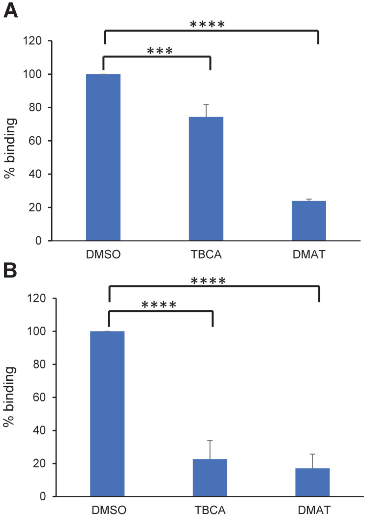
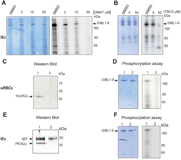
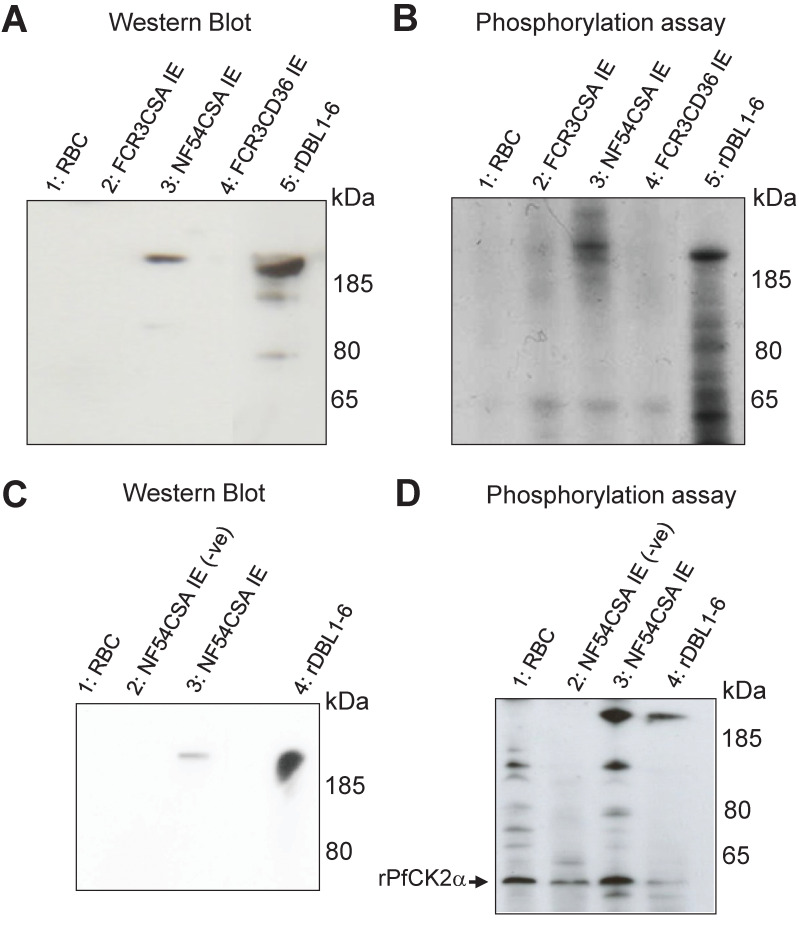
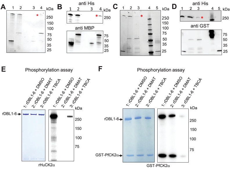
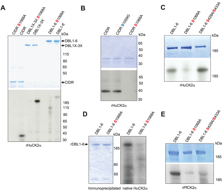
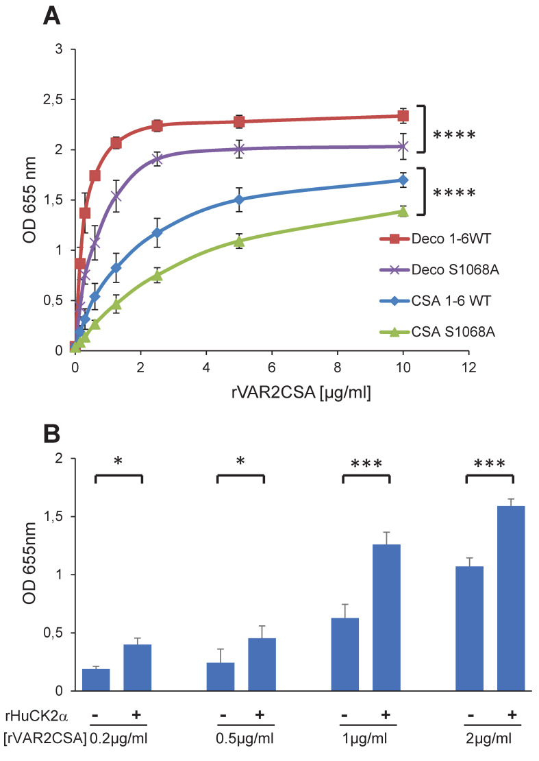
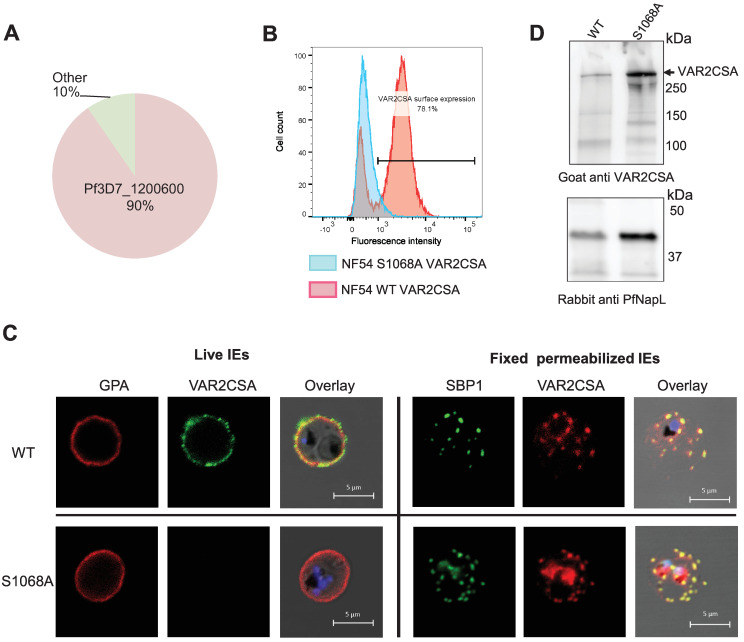
Placental malaria is characterized by the massive accumulation and sequestration of infected erythrocytes in the placental intervillous blood spaces, causing severe birth outcomes. The variant surface antigen VAR2CSA is associated with Plasmodium falciparum sequestration in the placenta via its capacity to adhere to chondroitin sulfate A. We have previously shown that the extracellular region of VAR2CSA is phosphorylated on several residues and that the phosphorylation enhances the adhesive properties of CSA-binding infected erythrocytes. Here, we aimed to identify the kinases mediating this phosphorylation. We report that Human and Plasmodium falciparum Casein Kinase 2α are involved in the phosphorylation of the extracellular region of VAR2CSA. We notably show that both CK2α can phosphorylate the extracellular region of recombinant and immunoprecipitated VAR2CSA. Mass spectrometry analysis of recombinant VAR2CSA phosphorylated by recombinant Human and P. falciparum CK2α combined with site-directed mutagenesis led to the identification of residue S1068 in VAR2CSA, which is phosphorylated by both enzymes and is associated with CSA binding. Furthermore, using CRISPR/Cas9 we generated a parasite line in which phosphoresidue S1068 was changed to alanine. This mutation strongly impairs infected erythrocytes adhesion by abolishing VAR2CSA translocation to the surface of infected erythrocytes. We also report that two specific CK2 inhibitors reduce infected erythrocytes adhesion to CSA and decrease the phosphorylation of the recombinant extracellular region of VAR2CSA using either infected erythrocytes lysates as a source of kinases or recombinant Human and P. falciparum casein kinase 2. Taken together, these results undoubtedly demonstrate that host and P. falciparum CK2α phosphorylate the extracellular region of VAR2CSA and that this post-translational modification is important for VAR2CSA trafficking and for infected erythrocytes adhesion to CSA.


Conflict of interest statement

The authors have declared that no competing interests exist.

Figures

Fig 1
Fig 1. CK2 inhibitors affect cytoadhesion but not VAR2CSA expression or trafficking.
(A) Parasitized RBCs at trophozoite stage and (B) ring stage were treated for 1 h or 16 h, respectively, with 50μM of TBCA or DMAT CK2 inhibitors prior to performing static binding assays on CSA and compared to the DMSO control. Bound mature stages IEs were counted in five random fields in 3 independent experiments. Results were expressed as a percentage of treated culture binding to CSA compared to DMSO-treated culture. Mean and Standard deviation are indicated. Statistics (Paired t test; ****p = 0.0001; ***p = 0.0007).
Fig 2
Fig 2. Total IEs extracts, red blood cell HuCK2α and P. falciparum CK2α phosphorylate rVAR2CSA DBL1-6.
In vitro radioactive [γ—32P] ATP phosphorylation assays of recombinant His tagged VAR2CSA DBL1-6 protein (1μg in all assays) were performed in the presence of total IEs lysates and increasing concentrations of DMAT (A) or TBCA (B). (A) rDBL1-6 + DMSO; rDBL1-6 + DMAT 2μM; rDBL1-6 DMAT 10μM; rDBL1-6 + DMAT 50μM; (B) rDBL1-6 + DMSO; rDBL1-6 + TBCA 5μM; rDBL1-6 + TBCA 50μM. (C) Western blot analysis using a goat anti-Human CK2α; mouse anti-Human CK2α was used to immunoprecipitate the native HuCK2α from the membrane of uninfected erythrocytes. Control was performed with a non-specific mouse IgG isotype. Lane 1: immunoprecipitation with mouse anti-Human CK2α; lane 2: immunoprecipitation with mouse IgG control; (D) In vitro phosphorylation of rDBL1-6 by native HuCK2α. Immunoprecipitates were used in a standard [γ—32P] ATP phosphorylation kinase assay with 1μg of rDBL1-6. The reactions were loaded on a gel and stained with Coomassie (left panel) prior to being exposed for autoradiography (right panel). rDBL1-6 is indicated with an arrow. Lane 1: immunoprecipitation with mouse anti-HuCK2α; lane 2: immunoprecipitation with mouse control IgG isotype. (E) Western Blot anti PfCK2α. Rabbit anti-PfCK2α was used to immunoprecipitate the native kinase from total parasite lysates of IEs. Control was performed with the corresponding rabbit pre-immune immunopurified antibody. Samples were loaded on a SDS PAGE prior to transfer and detection with the same anti-PfCK2α. lane 1: immunoprecipitation with anti-PfCK2α; lane 2: immunoprecipitation with pre-immune immunopurified rabbit IgG. (F) In vitro phosphorylation of rDBL1-6 by endogenous PfCK2α. Immunoprecipitates were used in a standard [γ—32P] ATP phosphorylation kinase assay with 1μg of rDBL1-6. The reactions were loaded on a gel and stained with Coomassie (left panel) prior to being exposed for autoradiography (right panel). Lane 1: immunoprecipitation with immunopurified anti-PfCK2α; lane 2: immunoprecipitation with a pre-immune immunopurified antibody.
Fig 3
Fig 3. Recombinant Human and P. falciparum CK2α phosphorylate immunoprecipitated VAR2CSA.
(A) A rabbit polyclonal anti-VAR2CSA was used to immunoprecipitate VAR2CSA from NF54CSA and FCR3CSA IEs membrane extracts. Membrane extracts from uninfected RBC and FCR3CD36 IEs were also processed using the same protocol. A fraction of immunoprecipitated material was loaded on a gel for western blot probed with a mouse monoclonal anti-VARCSA antibody. Lane 1: uRBC membrane extracts immunoprecipitation; lane 2: IEs FCR3CSA membrane extracts immunoprecipitation; lane 3: IEs NF54CSA membrane extracts immunoprecipitation; lane 4: IEs FCR3CD36 membrane extracts immunoprecipitation; lane 5: rDBL1-6 control. (B) In vitro phosphorylation of endogenous immunoprecipitated VAR2CSA by rHuCK2α. The immunoprecipitated materials were used as substrates in a standard [γ-32P] ATP phosphorylation kinase assay using 250ng of rHuCK2α. Phosphorylation of rDBL1-6 was performed as a control. Lane 1: immunoprecipitated uRBC membrane extracts; lane 2: immunoprecipitated FCR3CSA IEs membrane extracts; lane 3: immunoprecipitated NF54CSA IEs membrane extracts; lane 4: immunoprecipitated FCR3CD36 IEs membrane extracts; lane 5: rDBL1-6 control. (C) Immunoprecipitation with a rabbit polyclonal anti-VARCSA from uninfected and NF54CSA IEs membrane lysates. A rabbit IgG isotype was used as a negative control with NF54CSA IEs lysates. Western blot analysis of the immunoprecipitated material with a mouse monoclonal anti-VAR2CSA antibody; lane 1: uRBC membrane extracts immunoprecipitated material using anti-VAR2CSA polyclonal antibodies; lane 2: NF54CSA IEs lysates immunoprecipitated material with rabbit IgG isotype (-ve); lane 3: NF54CSA IEs lysates immunoprecipitated material using anti-VAR2CSA polyclonal antibodies; lane 4: rDBL1-6 control. (D) In vitro phosphorylation of endogenous immunoprecipitated VAR2CSA by rPfCK2α. The immunoprecipitated materials were used as substrates in a standard [γ-32P] ATP phosphorylation kinase assay using 250ng of rPfCK2α. Phosphorylation of rDBL1-6 was performed as a control. lane 1: Immunoprecipitated uRBC membrane material; lane 2: Immunoprecipitated material with rabbit IgG isotype from NF54CSA IEs membrane extracts (-ve); lane3: Immunoprecipitated material with anti VAR2CSA antibody from NF54CSA IEs membrane extracts. Lane 4: rDBL1-6 control.
Fig 4
Fig 4. Recombinant Human and P. falciparum CK2α interact and phosphorylate rDBL1-6.
(A) MBP-tagged HuCK2α or MBP alone were incubated with His-tagged rDBL1-6. Complexes containing the MBP-tagged proteins were then purified using amylose resin beads, and any bound His-tagged proteins were detected by stain-free SDS gel prior Western blot. (B) Western blot analysis using anti-His HRP (upper panel) or anti-MBP (lower panel). Lane 1: MBP + His DBL1-6 (input); lane2: MBPHuCK2α + HisDBL1-6 (input); lane 3: MBP + HisDBL1-6 (bound fraction); lane 4: MBPHuCK2α + HisDBL1-6 (bound fraction). (C) GST-tagged PfCK2α or GST alone were incubated with HisDBL1-6. Complexes containing the GST-tagged proteins were then purified using glutathione agarose beads, and any bound His-tagged proteins were detected by stain-free SDS gel prior Western blot. (D) Western blot analysis using anti-His HRP (upper panel) or anti-GST (lower panel). Lane 1: GST + His DBL1-6 (input); lane2: GST-PfCK2α + HisDBL1-6 (input); lane 3: GST-PfCK2α + HisDBL1-6 (bound fraction); lane 4: MW: lane 5: GST + His DBL1-6 (bound fraction). (E, F) rDBL1-6 was used as a substrate for in vitro phosphorylation assays in the presence of [γ—32P] ATP with rHuCK2α (E) or rPfCK2α (F) in the presence or not of the CK2 inhibitors DMAT and TBCA at 50μM. Lane 1: rDBL1-6 + DMSO; lane2: rDBL1-6 + DMAT; lane 3: rDBL1-6 + TBCA.
Fig 5
Fig 5. Identification of targeted VAR2CSA domains and phosphosites.
(A, B) Mapping of phosphorylated VAR2CSA extracellular domains. Single and multidomains of recombinant VAR2CSA DBL1-6 were used as substrates for in vitro phoshorylation assays with rHuCK2α and rPfCK2α. Autoradiograms are shown on the lower panels and the corresponding Coomassie-stained gels are in the top panel. (A) Phosphorylation assays in the presence of [γ—32P] ATP and rHuCK2α. (B) Phosphorylation assays in the presence of [γ—32P] ATP and rPfCK2α. Lane 1: DBL1X; lane 2: DBL2X; lane 3: DBL3X; lane 4: DBL1X-DBL2X; lane 5: INT1-CIDR; lane 6: CIDR; lane 7: DBL1X-3X; lane 8: DBL1X-6ε; lane 9: DBL4ε-6ε. rDBL1-6 were used as substrates for in vitro cold phosphorylation assays with rHuCK2α and rPfCK2α. (C) Schematic view of rDBL1-6 identified phosphorylation sites by rPfCK2α and by rHuCK2α; TM: transmembrane domain; ATS: Acidic Terminal Segment corresponds to the cytoplasmic region of PfEMP1s proteins.
Fig 6
Fig 6. Validation and effect of phosphosites mutation on VAR2CSA phosphorylation and CSA adhesion.
(A, B, C). The wild type and mutated VAR2CSA recombinant CIDR, DBL1X-3X and DBL1X-6 proteins indicated in the figure were tested for phosphorylation in a standard in vitro phosphorylation assays in the presence of [γ-32P] ATP with HuCK2α. (D) Wild type and S1068A DBL1X-6 recombinant proteins were tested for phosphorylation in standard in vitro phosphorylation assays in the presence of [γ-32P] ATP with endogenous immunoprecipitated HuCK2α. (E) The wild type and mutated rDBL1X-6, DBL1X-6 S1068A and DBL1X-6 S429A/S433A proteins indicated in the figure were tested for phosphorylation in a standard in vitro phosphorylation assays in the presence of [γ-32P] ATP with the wild type recombinant PfCK2α or the catalytically inactive mutant K72M recombinant PfCK2α. In all panels, similar amounts of wild-type and mutated proteins were loaded on SDS-PAGE, as shown by Coomassie blue staining.
Fig 7
Fig 7. rDBL1-6 S1068A mutation impairs CSA-binding while its phosphorylation by rHuCK2α enhances CSA binding.
(A) Wild-type DBL1X-6 (1-6WT) and mutated DBL1X-6 S1068A (S1068A) recombinant proteins were assayed by ELISA at different protein concentrations for in vitro binding to CSA or decorin coated ELISA plates. Increasing concentrations of recombinant DBL1X-6ε proteins at serial dilutions of 0.156 to 10 μg/mL were added to wells previously coated with CSA, or decorin. Error bars correspond to SD between 3 independent experiments. Each experiment was performed in triplicate. P values were determined by running an unpaired t test on the two area-under-the-curve (AUC) according to the method described in the following web link: https://www.graphpad.com/support/faqid/2031/. (B) Human casein kinase 2 plus ATP increases binding of VAR2CSA to CSA. 0.2, 0.5, 1 and 2 μg/mL of VAR2CSA recombinant proteins were preincubated 30 min at 30°C in kinase buffer supplemented with 10μM ATP with 300 ng or without Human CK2α kinase and added to wells previously coated with CSA. Error bars correspond to SD between 3 independent experiments. Statistics (paired t test; *p = 0.018; *p = 0.03; ***p = 0.0008 and ***p = 0.0002 for respectively 0.2, 0.5, 1 and 2μg/ml rDBL1-6). CSA, chondroitin sulfate A; Deco, decorin; ELISA, enzyme-linked immunosorbent assay; OD, optical density at 655nm; SD standard deviation.
Fig 8
Fig 8. Phenotype of the transgenic NF54CSA S1068A line.
(A) Var2csa transcriptional profile of S1068A shown by qPCR. Transcriptional levels of each var genes were normalized with the housekeeping gene, seryl-tRNAtransferase. (B) Flow cytometry analysis of wild-type and S1068A NF54CSA IEs. IEs were labelled with rabbit anti-VAR2CSA antibodies. Geometric means of fluorescence intensities and percentage of IEs expressing VAR2CSA are indicated. (C) VAR2CSA immunofluorescence assays (IFA). IFA staining was performed on live cells and fixed and permeabilized smears on wild-type NF54CSA IEs and transgenic NF54 S1068A IEs with rabbit anti-VAR2CSA, anti-GPA and anti-SBP1 antibodies. (D) Western blot analysis with a goat anti-VAR2CSA antibody was performed on total lysates of wild-type NF54CSA and NF54 S1068A IEs (Upper panel). An anti-PfNapL (PfNucleosome Assembly protein L) serum was used as a loading control for both lysates (Lower panel).

Similar articles

References

    1. WHO. World Malaria Report 2022. ISBN 978-92-4-006490-4. (World Health Organization, Geneva, 2022). 2022.
    1. Craig AG, Khairul MFM, Patil PR. Cytoadherence and severe malaria. The Malaysian journal of medical sciences: MJMS. 2012;19(2):5–18. - PMC - PubMed
    1. Smith JD, Rowe JA, Higgins MK, Lavstsen T. Malaria’s deadly grip: cytoadhesion of Plasmodium falciparum-infected erythrocytes. Cell Microbiol. 2013;15(12):1976–83. Epub 2013/08/21. doi: 10.1111/cmi.12183 . - DOI - PMC - PubMed
    1. Storm J, Jespersen JS, Seydel KB, Szestak T, Mbewe M, Chisala NV, et al.. Cerebral malaria is associated with differential cytoadherence to brain endothelial cells. EMBO molecular medicine. 2019;11(2):e9164. doi: 10.15252/emmm.201809164 - DOI - PMC - PubMed
    1. Ganguly AK, Ranjan P, Kumar A, Bhavesh NS. Dynamic association of PfEMP1 and KAHRP in knobs mediates cytoadherence during Plasmodium invasion. Sci Rep. 2015;5:8617. Epub 2015/03/03. doi: 10.1038/srep08617 . - DOI - PMC - PubMed

Substances

LinkOut - more resources