. 2025 Jan 13;21(1):e1012434.
doi: 10.1371/journal.ppat.1012434. eCollection 2025 Jan.

Identification of novel PfEMP1 variants containing domain cassettes 11, 15 and 8 that mediate the Plasmodium falciparum virulence-associated rosetting phenotype

Affiliations

Identification of novel PfEMP1 variants containing domain cassettes 11, 15 and 8 that mediate the Plasmodium falciparum virulence-associated rosetting phenotype

Florence E McLean et al. PLoS Pathog. .

Abstract

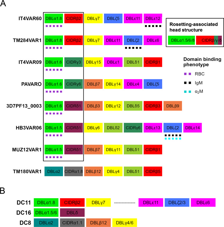
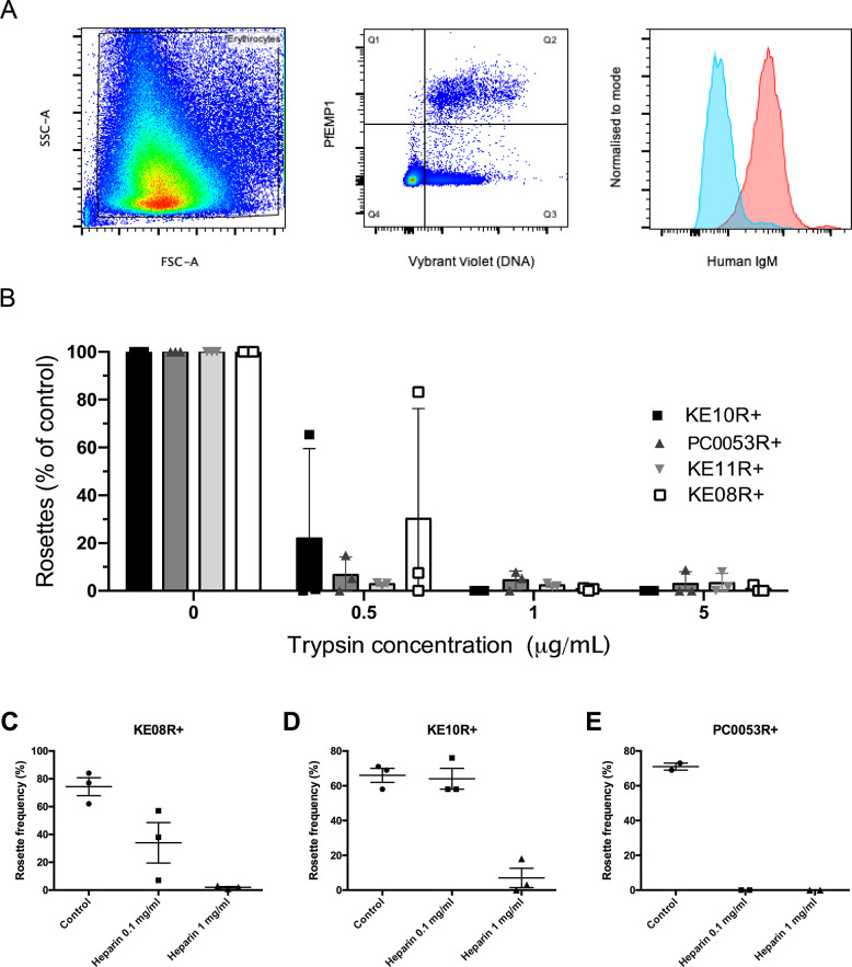
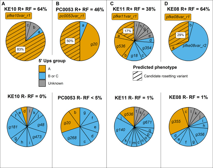
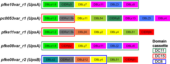
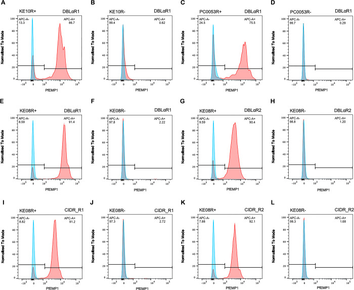
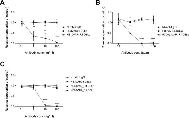
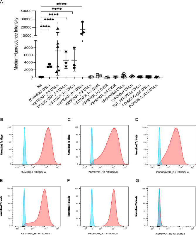
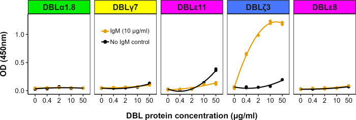
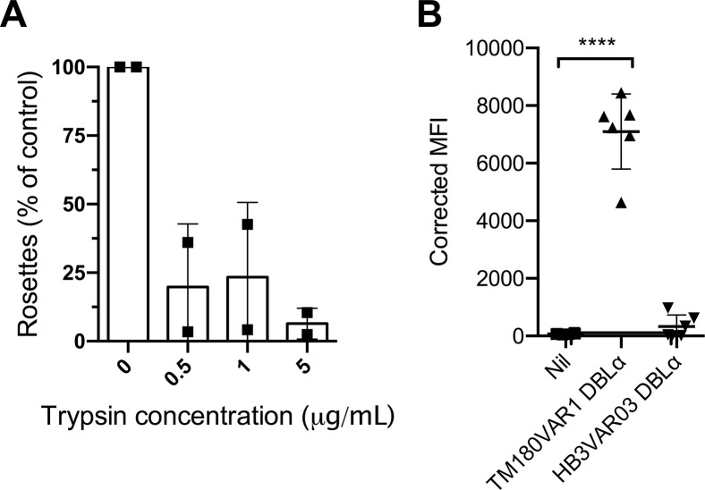
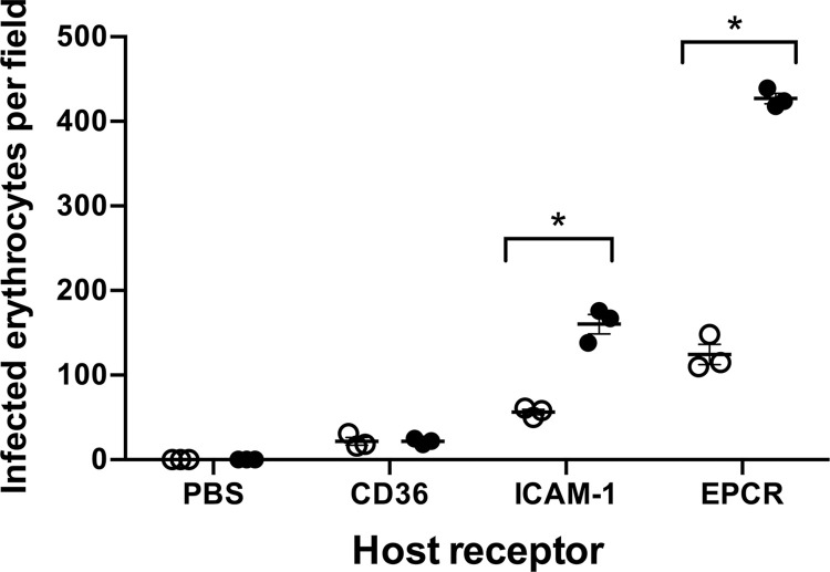
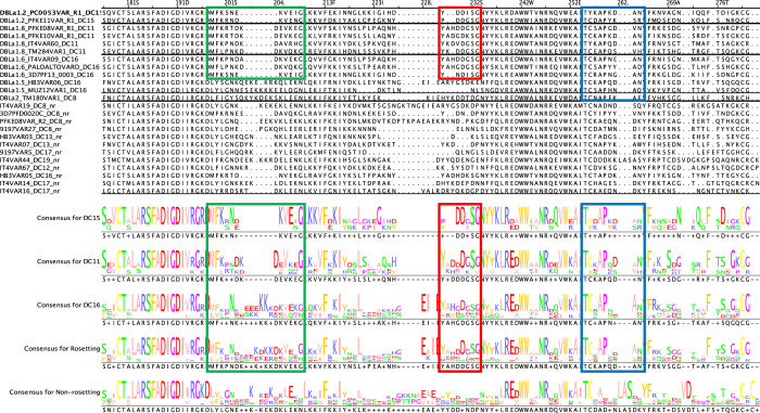
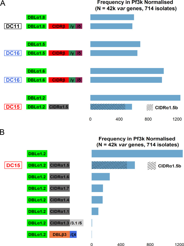
Plasmodium falciparum erythrocyte membrane protein 1 (PfEMP1) is a diverse family of variant surface antigens, encoded by var genes, that mediates binding of infected erythrocytes to human cells and plays a key role in parasite immune evasion and malaria pathology. The increased availability of parasite genome sequence data has revolutionised the study of PfEMP1 diversity across multiple P. falciparum isolates. However, making functional sense of genomic data relies on the ability to infer binding phenotype from var gene sequence. For P. falciparum rosetting, the binding of infected erythrocytes to uninfected erythrocytes, the analysis of var gene/PfEMP1 sequences encoding the phenotype is limited, with only eight rosette-mediating PfEMP1 variants described to date. These known rosetting PfEMP1 variants fall into two types, characterised by N-terminal domains known as "domain cassette" 11 (DC11) and DC16. Here we test the hypothesis that DC11 and DC16 are the only PfEMP1 types in the P. falciparum genome that mediate rosetting, by examining a set of thirteen recent culture-adapted Kenyan parasite lines. We first analysed the var gene/PfEMP1 repertoires of the Kenyan lines and identified an average of three DC11 or DC16 PfEMP1 variants per genotype. In vitro rosette selection of the parasite lines yielded four with a high rosette frequency, and analysis of their var gene transcription, infected erythrocyte PfEMP1 surface expression, rosette disruption and erythrocyte binding function identified four novel rosette-mediating PfEMP1 variants. Two of these were of the predicted DC11 type (one showing the dual rosetting/IgM-Fc-binding phenotype), whereas two contained DC15 (DBLα1.2-CIDRα1.5b) a PfEMP1 type not previously associated with rosetting. We also showed that a Thai parasite line expressing a DC8-like PfEMP1 binds to erythrocytes to form rosettes. Hence, these data expand current knowledge of rosetting mechanisms and emphasize that the PfEMP1 types mediating rosetting are more diverse than previously recognised.


Conflict of interest statement

The authors have declared that no competing interests exist.

Figures

Fig 1
Fig 1. Known rosette-mediating PfEMP1 variants and associated domain cassettes.
A) Diagram of the PfEMP1 domain architecture of known rosette-mediating variants [–17]. The rosetting-associated head structure, DBLα1.5/6/8-CIDRβ/γ/δ, is boxed. Domains that bind erythrocytes (RBC) or serum proteins (IgM and alpha2Macroglobulin, α2M) are indicated. B) Diagram of domain cassettes (DCs) [as described by [27]] which are relevant to the rosetting PfEMP1 variants characterised previously.
Fig 2
Fig 2. Properties of the new Kenyan rosetting lines.
A) IgM binding by KE10R+ infected erythrocytes detected by flow cytometry. Forward and side scatter were used to gate on erythrocytes and exclude debris (left panel) and PfEMP1-expressing infected erythrocytes were detected by staining with 20μg/ml of polyclonal rabbit IgG against NTS-DBLα of KE10VAR_R1 followed by 1/1000 dilution of Alexa Fluor 647-conjugated goat anti-rabbit IgG secondary antibody and 1/2500 dilution of Vybrant DyeCycle Violet (middle panel Q2). IgM staining of the Q2 cell population was detected with 1/1000 dilution of an Alexa Fluor 488-conjugated goat anti-human IgM heavy chain antibody (red). The negative control (blue) was parasites grown in IgM-depleted medium and stained with the same antibodies. Results are representative of two independent experiments. B) Effect of low-dose trypsinisation on rosetting. Purified infected erythrocytes were treated with 0.5, 1 or 5 μg/ml of trypsin for 5 mins and the rosette frequency relative to a control with no added enzyme was calculated. The mean and standard deviation from three independent experiments per parasite line is shown. The rosette frequency of the untreated control was between 46%-81% (KE10R+), 45%-89% (PC0053R+), 16%-30% (KE11R+) and 61%-91% (KE08R+). C-E) Effect of heparin on rosetting. The mean and standard error from two or three independent experiments per parasite line is shown.
Fig 3
Fig 3. DBLα expression tag profiling of the new Kenyan rosetting lines.
DBLα tag expression profiles showing the proportion each gene tag contributed to the overall expressed tag profile for each parasite population. var gene tags encoding candidate rosetting variants (hatched; percentage of all tags given in white box) are identified as predominant genes expressed in R+ parasites but not/rarely in isogenic R- parasites. Other large pie slices are identified by gene (g) number for each parasite line, and small slices are given letters, with the full information for each gene given in S1 Table. var gene groups are identified by colour. Slices labelled as “unknown” are from sequence tags that did not map back to an assembled var gene in that parasite’s repertoire [35]. The rosette frequency (RF) in the cycle in which the RNA was collected is shown for each parasite line. Results are representative of two independent experiments.
Fig 4
Fig 4. Domain architecture of the candidate rosette-mediating PfEMP1 variants.
Domain cassettes are as described by Rask et al [27]. The alternative names for these genes in the Pf3k database [35] are as follows: pfke10var_r1/PX0203.g54; pc0053var_r1/PC0053-C.g687; pfke11var_r1/ PFKE11.g448; pfke08var_r1/ PFKE08.g502; pfke08var_r2/ PFKE08.g501.
Fig 5
Fig 5. Staining of the surface of live infected erythrocytes with PfEMP1 antibodies.
Fluorescence intensity histograms of rosetting (R+) and non-rosetting (R-) parasite lines stained with rabbit IgG against rosetting PfEMP1 variants (red) or negative control (rabbit IgG against the NTS-DBLα domain of an irrelevant PfEMP1 variant, HB3VAR03, blue). A) parasite line KE10R+ with antibodies to KE10VAR_R1 NTS-DBLα. B) parasite line KE10R- with antibodies to KE10VAR_R1 NTS-DBLα. C) parasite line PC0053R+ with antibodies to PC0053VAR_R1 NTS-DBLα. D) parasite line PC0053R- with antibodies to PC0053VAR_R1 NTS-DBLα. E) parasite line KE08R+ with antibodies to KE08VAR_R1 NTS-DBLα. F) parasite line KE08R- with antibodies to KE08VAR_R1 NTS-DBLα. G) parasite line KE08R+ with antibodies to KE08VAR_R2 NTS-DBLα. H) parasite line KE08R- with antibodies to KE08VAR_R2 NTS-DBLα. I) parasite line KE08R+ with antibodies to KE08VAR_R1 CIDR. J) parasite line KE08R- with antibodies to KE08VAR_R1 CIDR. K) parasite line KE08R+ with antibodies to KE08VAR_R2 CIDR. L) parasite line KE08R- with antibodies to KE08VAR_R2 CIDR. PfEMP1 was detected with 20μg/ml rabbit IgG against PfEMP1 and 1/1000 dilution of Alexa Fluor 647-conjugated goat anti-rabbit IgG secondary antibody. Gates shows the percentage of mature infected erythrocytes positive for the variant. The rosette frequency at the time of staining was 70–90% for the R+ lines and <2% for the R- lines, and the percentage of positive staining infected erythrocytes (APC-A+, top right corner of each histogram) closely matched the rosette frequency in each parasite line. Results are representative of at least two experiments for each parasite line.
Fig 6
Fig 6. Rosette disruption with rabbit IgG raised against candidate rosetting PfEMP1 variants.
Antibodies to the candidate rosette-mediating PfEMP1 variant NTS-DBLα domains were tested in rosette-disruption assays over a range of concentrations from 0.1–100 μg/mL. The rosette frequency in the presence of antibody is shown as the proportion of the control value with no added antibody. IgG from a non-immunised (NI) rabbit and from a rabbit immunised with the NTS-DBLα domain of a non-rosetting PfEMP1 variant HB3VAR03 were used as negative controls. Data represent three independent experiments with the mean and standard deviation of the three experiments shown. A) parasite line KE10R+; the rosette frequency of the no antibody control ranged from 71%-87%. B) Parasite line PC0053R+; the rosette frequency of the no antibody control ranged from 49%-60%. C) Parasite line KE08R+; the rosette frequency of the no antibody control ranged from 69%-92%. Data were analyzed by two-tailed paired t tests corrected for multiple comparisons with the Holm-Sidak method ** P<0.01, *** P<0.001, **** P<0.0001.
Fig 7
Fig 7. Binding of PfEMP1 recombinant proteins to erythrocytes.
A) Recombinant proteins were incubated with uninfected erythrocytes and bound protein detected with specific antibodies to each domain. The positive control was the NTS-DBLα domain of the well-characterised rosetting variant IT4VAR60 [15,17] and negative controls were the NTS-DBLα domains from human brain endothelial cell binding PfEMP1 variants HB3VAR03 (PFHB3_130080100), IT4VAR07 (PFIT_060036700), 3D7_PFD0020c (PF3D7_0400400), PC0053-C.g96 and PC0053-C.g410 that are known to be non-rosetting [50]. The mean and standard deviation of the Alexa Fluor 488 median fluorescence intensity from at least three independent experiments per protein is indicated. Data were log transformed and analysed by one way ANOVA with Dunnett’s multiple comparisons test compared to the “Nil” (no added protein) control. **** P<0.0001. B-G) Example Alexa Fluor 488 fluorescence intensity histograms of recombinant PfEMP1 domains bound to erythrocytes and detected by indirect immunofluorescence (red) compared to a non-rosetting NTS-DBLα domain negative control (blue). The domain tested is indicated below each histogram.
Fig 8
Fig 8. The DBLζ3 domain of PFKE10VAR_R1 binds human IgM.
IgM was coated onto the wells of an ELISA plate and individual PfEMP1 recombinant protein domains were added at four different 5-fold dilutions. Wells with no IgM were included as negative controls. Binding was detected using an HRP-conjugated anti-His tag antibody with TMB substrate and the absorbance read at 450nm. The domains are shown from N- to C-terminal and the panels are colour-coded as previously described [27]. One representative experiment is shown out of at least two performed for each domain.
Fig 9
Fig 9. The NTS-DBLα domain of TM180VAR1 mediates rosetting.
A) Effect of low dose trypsinisation on TM180R+. Infected erythrocytes were treated with 0.5, 1 or 5 μg/ml of trypsin for 5 mins in each experiment and the rosette frequency relative to a control sample with no added enzyme was calculated. Data represent two independent experiments with the mean indicated by bar height and the standard deviation shown. The rosette frequency in the untreated controls were 72% and 61% respectively. B) Corrected Median Fluorescence Intensities (MFI) of uninfected erythrocytes in binding experiments with recombinant TM180VAR1 NTS-DBLα and the negative control HB3VAR03 NTS-DBLα compared to “Nil” no added protein control. Data are from six independent experiments and the mean and standard deviation are shown. Significant differences by one-way ANOVA with Dunnett’s multiple comparisons test compared to the “Nil” negative control are indicated. ****P<0.0001.
Fig 10
Fig 10. Spot binding assay of KE08R+ to endothelial cell receptor proteins.
Adhesion of KE08R+ infected erythrocytes to recombinant host receptor proteins was determined with (black circles) and without (white circles) prior rosette disruption with antibodies to KE08VAR_R1 NTS-DBLα. The number of infected erythrocytes bound per field was determined using an inverted microscope with a 40X objective and six fields were counted per spot in each experiment. The difference in mean binding values compared with the same receptor in the presence and absence of rosette from n = 3 independent experiments was analyzed by two-tailed paired t tests corrected for multiple comparisons with the Holm-Sidak method. *P< 0.05.
Fig 11
Fig 11. Rosetting motifs.
The NTS-DBLα amino acid sequences from known rosetting (upper) and non-rosetting (lower) sequences were aligned in Jalview and amino acid motifs over-represented in the rosetting variants are shown in green, red and blue boxes. The amino acid sequences are given in S1 Text. nr: non-rosetting.
Fig 12
Fig 12. Frequency of rosetting-associated domains in the global P. falciparum population.
A) A bar plot showing the frequency of rosetting-associated domains and their respective domain cassettes in the Pf3k normalised varDB [35]. The domains are colour-coded as defined by Rask et al [27]. B) A bar plot showing the frequency of different domains linked to DBLα1.2 in the Pf3k normalised varDB.

Similar articles

References

    1. Lee WC, Russell B, Renia L. Evolving perspectives on rosetting in malaria. Trends Parasitol. 2022;38(10):882–9. doi: 10.1016/j.pt.2022.08.001 - DOI - PubMed
    1. Rowe A, Obeiro J, Newbold CI, Marsh K. Plasmodium falciparum rosetting is associated with malaria severity in Kenya. Infect Immun. 1995;63(6):2323–6. - PMC - PubMed
    1. Carlson J, Helmby H, Hill AV, Brewster D, Greenwood BM, Wahlgren M. Human cerebral malaria: association with erythrocyte rosetting and lack of anti-rosetting antibodies. Lancet. 1990;336(8729):1457–60. doi: 10.1016/0140-6736(90)93174-n - DOI - PubMed
    1. Normark J, Nilsson D, Ribacke U, Winter G, Moll K, Wheelock CE, et al.. PfEMP1-DBL1alpha amino acid motifs in severe disease states of Plasmodium falciparum malaria. Proc Natl Acad Sci U S A. 2007;104(40):15835–40. - PMC - PubMed
    1. Doumbo OK, Thera MA, Kone AK, Raza A, Tempest LJ, Lyke KE, et al.. High levels of Plasmodium falciparum rosetting in all clinical forms of severe malaria in African children. Am J Trop Med Hyg. 2009;81(6):987–93. - PMC - PubMed

MeSH terms

Substances

LinkOut - more resources