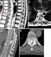
Resection of a ventrally located ossified thoracic spinal meningioma: illustrative case
- PMID: 39805109
- PMCID: PMC11734618
- DOI: 10.3171/CASE24603
Resection of a ventrally located ossified thoracic spinal meningioma: illustrative case
Abstract
Background: Resection of calcified meningiomas in the ventral thoracic spinal canal remains a formidable surgical challenge despite advances in technology and refined microsurgical techniques. These tumors, which account for a small percentage of spinal meningiomas, are characterized by their hardness, complicating safe resection and often resulting in worse outcomes than their noncalcified counterparts.
Observations: The authors present the case of a 68-year-old woman with a ventrally located ossified meningioma at the T9-10 level, successfully treated via a posterolateral transpedicular approach. Additionally, they conducted a systematic review of the literature following Preferred Reporting Items for Systematic Reviews and Meta-Analyses guidelines, focusing on the origins, imaging findings, surgical strategies, and outcomes of calcified meningiomas in the ventral thoracic spinal canal. Data were extracted and analyzed from 15 articles encompassing 18 cases.
Lessons: Calcified meningiomas in the ventral thoracic spinal canal require meticulous preoperative planning and a tailored surgical approach to optimize outcomes. The posterolateral transpedicular approach offers a balance between adequate exposure and minimizing spinal cord manipulation, making it a viable option for resecting these challenging tumors. A single facetectomy and pediculotomy do not compromise long-term spinal stability. Technological adjuncts, including ultrasonic aspiration, neuromonitoring, and endoscopic assistance, can further enhance surgical safety and effectiveness. https://thejns.org/doi/10.3171/CASE24603.
Keywords: intraoperative monitoring; ossified spinal meningioma; spinal tumor; surgical technique; ventral thoracic spinal canal.
Figures


References
-
- Cohen-Gadol AA, Zikel OM, Koch CA, Scheithauer BW, Krauss WE. Spinal meningiomas in patients younger than 50 years of age: a 21-year experience. J Neurosurg. 2003;98(3 suppl):258-263. - PubMed
-
- Doita M, Harada T, Nishida K, Marui T, Kurosaka M, Yoshiya S. Recurrent calcified spinal meningioma detected by plain radiograph. Spine. 2001;26(11):E249-E252. - PubMed
-
- Nakayama N, Isu T, Asaoka K, et al. Two cases of ossified spinal meningioma. Article in Japanese. No Shinkei Geka Neurol Surg. 1996;24(4):351-355. - PubMed
-
- Alafaci C, Grasso G, Granata F, Salpietro FM, Tomasello F. Ossified spinal meningiomas: clinical and surgical features. Clin Neurol Neurosurg. 2016;142:93-97. - PubMed
LinkOut - more resources
Full Text Sources

