. 2025 Apr;31(4):1303-1311.
doi: 10.1038/s41591-025-03496-y. Epub 2025 Jan 13.

Oral obeldesivir provides postexposure protection against Marburg virus in nonhuman primates

Affiliations

Oral obeldesivir provides postexposure protection against Marburg virus in nonhuman primates

Robert W Cross et al. Nat Med. 2025 Apr.

Abstract

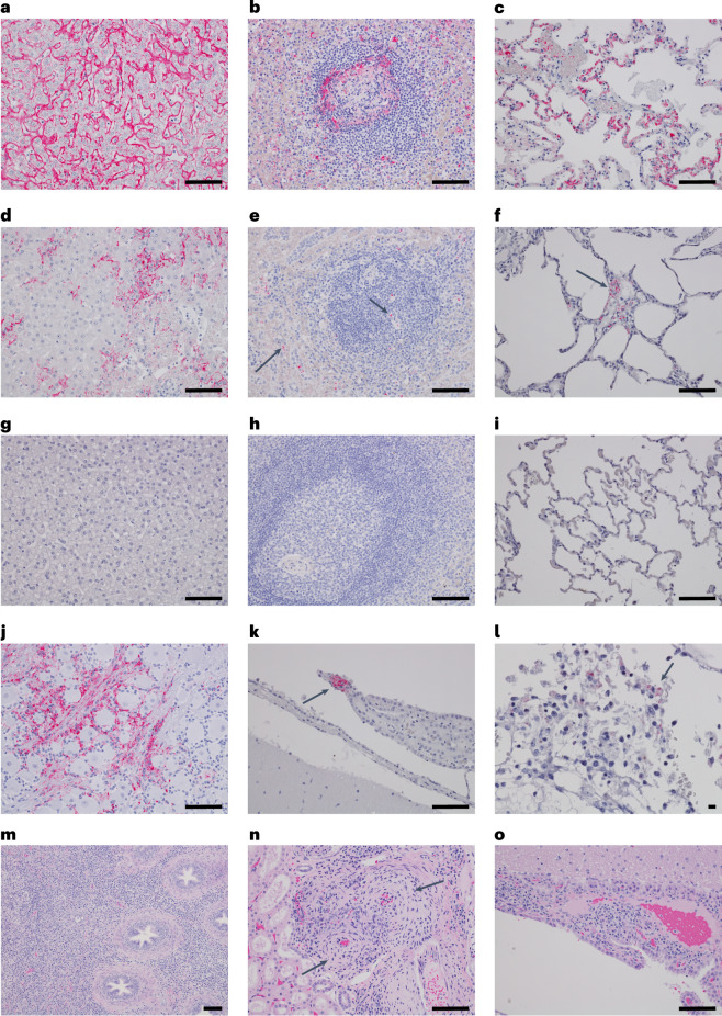
The recent outbreak of Marburg virus (MARV) in Rwanda underscores the need for effective countermeasures against this highly fatal pathogen, with case fatality rates reaching 90%. Currently, no vaccines or approved treatments exist for MARV infection, distinguishing it from related viruses such as Ebola. Our study demonstrates that the oral drug obeldesivir (ODV), a nucleoside analog prodrug, shows promising antiviral activity against filoviruses in vitro and offers significant protection in animal models. Here with cynomolgus macaques (n = 6), a 10 day regimen of once-daily ODV, initiated 24 h after exposure, provided 80% protection against a thousandfold lethal MARV challenge, delaying viral replication and disease onset. Transcriptome analysis revealed that early adaptive responses correlated with successful outcomes. Compared with intravenous options, oral antivirals such as ODV offer logistical advantages in outbreak settings, enabling easier administration and broader contact coverage. Our findings support the potential of ODV as a broad-spectrum, oral postexposure prophylaxis for filoviruses.


Conflict of interest statement

Competing interests: T.C., A.-Q.N., D.B., R.B., M.S.V. and V.C.C. are paid employees of Gilead Sciences, Inc. and may own company stock. The other authors declare no competing interests.

Figures

Fig. 1
Fig. 1. Survival analysis, clinical scoring and virus replication kinetics in cynomolgus macaques challenged with MARV and treated with ODV.
a, Kaplan–Meier survival curves for MARV-challenged cynomolgus macaques. The in-study control is plotted separately; however, for statistical comparison, the in-study control was grouped with historical controls (HC) from previous studies. Differences in curves were tested by the Mantel–Cox log–rank test. b, Clinical scores were assigned based on daily cage-side observations of behavior and apparent physical health. The horizontal dashed line indicates the minimum clinical score by which euthanasia criteria were met. c,d, Viral load was determined by RT-qPCR of RNA from whole blood (c) or plaque titration of plasma (d) collected at predetermined sampling points or at euthanasia. e, Viral load in selected tissues collected at necropsy as determined by RT-qPCR detection of vRNA. For ce, individual data points represent the mean of two technical replicates. For HC, the geometric mean ± geometric s.d. for the cohort is plotted. To fit on a log-scale axis, zero values (below LOQ) are plotted as ‘1’ (100). Dashed horizontal lines indicate the LLOQ for the assay (1,000 GEq ml−1 of blood or GEq g−1 of tissue for RT-qPCR; 25 PFU ml−1 for plaque titration). To fit on a log-scale axis, zero values (below LLOQ) are plotted as ‘1’ (100). ALN, axillary lymph node; ILN, inguinal lymph node; Liv, liver; Spl, spleen; Kid, kidney; Adr, adrenal gland; BrFr, brain frontal cortex; BrSt, brain stem; CSC, cervical spinal cord; Pan, pancreas; Uri, urinary bladder; Gon, gonad; Ut/Pro, uterus or prostate; NaMu, nasal mucosa; Conj, conjunctiva. Source data
Fig. 2
Fig. 2. Survival analysis and comparisons of circulating MARV vRNA and infectious virus in cynomolgus and rhesus macaques.
a, Kaplan–Meier survival curves for MARV-challenged HC cynomolgus and rhesus macaques. Survival data from the in-study control cynomolgus macaque (C-1) is included. Differences in curves were tested by the Mantel–Cox log–rank test. b, Comparison of the MTD of unprotected control cynomolgus macaques and that of rhesus macaques following infection with MARV. Statistical significance was tested by Welch’s t-test. c, Comparison of circulating MARV vRNA in cynomolgus and rhesus macaques at 3 DPI as measured by RT-qPCR. d, Comparison of circulating infectious MARV in cynomolgus and rhesus macaques at 3 DPI as measured by plaque titration. For b, statistical comparison was made using Welch’s t-test. For c and d, the horizontal dashed lines indicate the LLOQ for the assay (1,000 GEq ml−1 for RT-qPCR; 25 PFU ml−1 for plaque titration). Individual data points represent the mean of two technical replicates. The bars indicate the geometric mean ± s.d. For statistical comparisons, undetectable values are plotted as values below the LLOQ (999 GEq ml−1 for RT-qPCR; 24 PFU ml−1 for plaque titration). Statistical comparisons were made using the Mann–Whitney U-test. All reported P values are two tailed. NS, not significant.
Fig. 3
Fig. 3. Serum MARV-neutralizing antibody titers in cynomolgus macaques exposed to MARV and treated with ODV.
Neutralizing antibody titers were determined by PRNT with a lower threshold of 50% (PRNT50). a, MARV-neutralizing antibody titers on the day of challenge (0 DPI) and at 14 DPI. The asterisk indicates that data from animals Tx-4 and C-1, which succumbed at 13 and 7 DPI, respectively, are plotted with 14 DPI data. The bars indicate the mean percentage neutralization ± s.d. at each timepoint after the challenge. b, Neutralization curves from surviving ODV-treated macaques at the study endpoint (35 DPI). The horizontal dashed line indicates 50% neutralization. Source data
Fig. 4
Fig. 4. Representative hematoxylin and eosin-stained and IHC images of anti-MARV NP antibody in NHPs.
ao, Images taken from the in-study positive control, C-1 (ac); ODV-treated nonsurvivor at 13 DPI, Tx-4 (df,jl); and ODV-treated survivors at 35 DPI, Tx-5 (gi), TX-2 (m) and Tx-1 (n,o). The images were captured with ×20 (ak,n,o), ×40 (l) and ×10 (m) objectives (×200, ×400 and ×100 total magnification, respectively). Liver with widespread, diffuse immunolabeling (red) of hepatic sinusoidal lining cells (a), multifocal to coalescing immunolabeling of hepatic sinusoidal lining cells (d) and no appreciable immunolabeling in the liver (g). Spleen with immunolabeling of individual to clusters of mononuclear cells within the white and red pulp (b), scattered individual immunolabeling of mononuclear cells within the red and white pulp (arrows) (e) and no appreciable immunolabeling in the spleen (h). Lung with locally extensive immunolabeling of the alveolar septum and rare alveolar macrophages (c), immunolabeling of rare clusters of mononuclear cells within the alveolar septum (arrow) (f) and no appreciable immunolabeling in the lung (i). Positive immunolabeling was also noted in satellite cells, glial cells and axons of the trigeminal ganglion (j), ependymal cells and mononuclear cells of the choroid plexus (arrow) (k) and mononuclear cells clustered within the meninges (arrow) (l). Chronic inflammatory infiltrates were noted in various organs in surviving NHPs. Lymphohistiocytic and eosinophilic epididymitis (m), lymphohistiocytic arteritis of the kidney (arrows) (n) and lymphohistiocytic choroid plexitis (o). For l, scale bar = 10 µm. For all other panels, scale bars = 100 µm.
Fig. 5
Fig. 5. Transcriptional profiling of ODV-treated macaques exposed to MARV.
Blood RNA samples from all subjects (n = 4 ODV-treated survivors; n = 1 ODV-treated fatal; n = 5 control subjects). a, Pathway analysis of differentially expressed transcripts (Benjamini–Hochberg-adjusted P < 0.01, two tailed) comparing transcriptomic changes in ODV-treated survivors versus controls across each disease progression stage (1–2 DPI, 4–5 DPI, 7 DPI and 10 DPI). Pathways are sorted by the topmost positive (top) or negative (bottom) z-scores for the ODV survivor group at 10 DPI. Red indicates increased expression; blue indicates decreased expression; white indicates no change in expression. b, Volcano plot depicting −log10(P value, two-tailed) and log2(fold change) values of differentially expressed transcripts between ODV-treated survivors (n = 4) and the treated fatal case (n = 1) across all timepoints. Key to horizontal lines: solid, P < 0.01; dashed, P < 0.05; dotted, P < 0.10; dotted-dashed, P < 0.50. c, Heat map showing top differentially expressed genes (Benjamini–Hochberg-adjusted P < 0.05, two tailed) in ODV-treated survivors compared with the treated fatal at each timepoint (sorted by the ODV survivor group at 1–2 DPI). Any differentially expressed transcripts with a Benjamini–Hochberg false discovery rate-corrected P value less than 0.05 were deemed significant. d, Immune cell transcriptional profiling of controls (n = 5), the treated fatal (n = 1) and ODV-treated survivors across all timepoints. Higher cell-type scores indicate a higher abundance of transcripts mapping to the specific cell subset. Source data
Extended Data Fig. 1
Extended Data Fig. 1. Differential Pathway Activation in ODV-Treated Survivors.
Volcano plots depicting -log10 p-values and log2 fold-change values of mRNA in ODV-treated survivors compared to a pre-challenge baseline (0 DPI) across sequential disease stages: post-challenge (1-2 DPI), early (4 DPI), mid (7 DPI), and late (10 DPI). Functional enrichment was performed on differentially expressed transcripts (Benjamini-Hochberg adjusted p-value < 0.1, two-tailed); pathways are sorted by the topmost positive (red) or negative (blue) z-scores. DPI, days post infection; ODV, obeldesivir. Source data

References

    1. Feldmann, H., Sanchez, A. & Geisbert, T. W. in Fields Virology Vol. 1 (eds Knipe, D. M. & Howley, P. M.) Ch. 32, 923–956 (Lippincott Williams & Wilkins, 2013).
    1. Feldmann, H., Sprecher, A. & Geisbert, T. W. Ebola. N. Engl. J. Med.382, 1832–1842 (2020). - PubMed
    1. Bausch, D. G. et al. Marburg hemorrhagic fever associated with multiple genetic lineages of virus. N. Engl. J. Med.355, 909–919 (2006). - PubMed
    1. Towner, J. S. et al. Marburgvirus genomics and association with a large hemorrhagic fever outbreak in Angola. J. Virol.80, 6497–6516 (2006). - PMC - PubMed
    1. World Health Organization. Marburg virus disease—Rwanda https://www.who.int/emergencies/disease-outbreak-news/item/2024-DON537 (2024).

Substances

LinkOut - more resources