. 2025 Jan 13;16(1):619.
doi: 10.1038/s41467-024-55439-9.

Conformational diversity in class C GPCR positive allosteric modulation

Affiliations

Conformational diversity in class C GPCR positive allosteric modulation

Giuseppe Cannone et al. Nat Commun. .

Abstract

The metabotropic glutamate receptors (mGlus) are class C G protein-coupled receptors (GPCR) that form obligate dimers activated by the major excitatory neurotransmitter L-glutamate. The architecture of mGlu receptor comprises an extracellular Venus-Fly Trap domain (VFT) connected to the transmembrane domain (7TM) through a Cysteine-Rich Domain (CRD). The binding of L-glutamate in the VFTs and subsequent conformational change results in the signal being transmitted to the 7TM inducing G protein binding and activation. The mGlu receptors signal transduction can be allosterically potentiated by positive allosteric modulators (PAMs) binding to the 7TMs, which are of therapeutic interest in various neurological disorders. Here, we report the cryoEM structures of metabotropic glutamate receptor 5 (mGlu5) purified with three chemically and pharmacologically distinct PAMs. We find that the PAMs modulate the receptor equilibrium through their different binding modes, revealing how their interactions in the 7TMs impact the mGlu5 receptor conformational landscape and function. In addition, we identified a PAM-free but agonist-bound intermediate state that also reveals interactions mediated by intracellular loop 2. The activation of mGlu5 receptor is a multi-step process in which the binding of the PAMs in the 7TM modulates the equilibrium towards the active state.


Conflict of interest statement

Competing interests: The authors declare no competing interests.

Figures
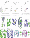
Fig. 1
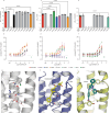
Fig. 1. Pharmacology of mGlu5 receptor with different PAMs and the cryoEM structure of orthosteric agonist quisqualate and VU0424465 ago-PAM-bound conformation.
a Evaluation of VU0424465, PAM VU29 and VU0409551 intrinsic agonist activity using cell-based assay. Dose-dependent potentiation of quisqualate agonist-induced response of mGlu5 by VU0424465, VU29 and VU0409551 were determined by the measurement of IP1 accumulation in HEK cells transiently transfected with mGlu5-Δ856. Data are normalised to the response measured for quisqualate alone and are mean ± SEM of three independent experiments (n = 3) performed in technical duplicates. Parameters from the dose-response curves are listed in Supplementary Table 1. Source data are provided as a Source Data file. b mGlu5 structure displays binding of the ago-PAM VU0424465 (yellow sphere) to both 7TMs and both VFTs display bound quisqualate (blue spheres). c CryoEM map (magenta) sharpened with B = −95 Å2 at 8 σ showing density for the ago-PAM VU0424465 in stick representation (3 Å carving around the atoms; carbon atoms in yellow) surrounded by key side chain residues shown (carbon atoms in blue and green). d Superposition of the two VU0424465-bound 7TMs from the same mGlu5 homodimer. VU0424465 induces a shift of TM6 along its helical axis, towards the extracellular side of the receptor. Resolved density for ICL2 displays an extended helix 3 that stretches out of the detergent micelles. e The active state is stabilised by a limited number of residues localised at the top of TM6. Molecular contact between N796 (ECL3) and D722, P724, Y730 (ECL2) observed only in one protomer. f Ligand binding site of VU0424465 in each protomer. VU0424465 make polar interaction with S805 and with the W785 carbonyl. It is stabilised by hydrophobic contact involving M802, V806, S809 in TM7; W785, Y793, F788, L789 in TM6 pushing out F788. g, h PAMs and NAMs occupy a common binding site. Panels show the superposition of the X-ray structure of thermostabilized mGlu5 7TM bound to NAM alloswitch-1 (g; Cyan, PDB code 7P2L) and M-MPEP (h; Purple, PDB code 6FFI) with 7TM of the mGlu5-5M bound to ago-PAM VU0424465 (blue and green) from this work. Figure panels (bh) were generated using PyMOL (The PyMOL Molecular Graphics System, Schrödinger, LLC.).
Fig. 2
Fig. 2. Conformational diversity of positive allosteric modulation of mGlu5 receptor dimer.
a CryoEM structure of the PAM VU29-bound mGlu5 with only one PAM modelled and the binding mode of VU29 (teal blue). bf Structure comparison of mGlu5 receptor dimer bound to ago-PAM VU0424465, PAMs VU29 and VU0409551 and VU0424465-bound to mGlu5 W785A mutant. The 3D alignment or superposition was performed with residues 25-191 and 331-461 of both lobe-1 of the PAM1_d1 data set (one protomer in green and the other in blue) (VU0424465-bound mGlu5 receptor) and those of VU29 (PAM2, yellow); VU0409551 (PAM3, light purple); the different conformations of VU0424465 including PAM1_d2_c1 (salmon) and PAM1_d2_c2 (cyan) and VU0424465-bound mGlu5 W785A mutant (PAM1_W785, olive). g PAM diversity of mGlu5 recpetor interface at TM6. Extracellular view of the 7TM dimer interface with P790 in stick representation show the difference in interface as illustrated by the movement of TM6 and P790. The panel represents the superimposition of VU042264 (blue and green), VU29 (yellow) and VU0409551 (purple). Figures were generated using PyMOL.
Fig. 3
Fig. 3. Functional role of PAM binding site residues on mGlu5 activity.
a, b VU0424465, c, d VU29, e, f VU0409551-dependent agonist-induced activity of different mutants in the PAM binding site. Single residues were mutated into alanine and functional consequences were determined by measuring the production of inositol monophosphate (IP1) in HEK293 cells transiently expressing the mGlu5-Δ856 and mutated mGlu5-Δ856 receptors. Experiments were performed in the presence of a fixed dose of the orthosteric agonist quisqualate (10 nM) and different doses of PAMs. Bar plots (a, c and e) are pEC50 average values of at least 3 independent experiments +/- SEM. Dose response curve (b, d, and f) are presented for mutants displaying statistically significant effect as compared to the WT type receptor. pEC50 value are presented in Supplementary Table 3. Each independent experiments were performed in technical duplicates (p value are indicated above each bar when required). For VU04244465, pEC50 value were determined for mGlu5-Δ856 (n = 11), for all mutants (n = 4) except for Y659A, F788A and A813L (n = 3). For VU29 pEC50, mGlu5-Δ856 (n = 10), for all mutants (n = 3) except for T781A, W785A, S805A and S809A (n = 4). In case of VU0409551, pEC50 value were determined for mGlu5-Δ856 (n = 12), for mutants T781A, W785A, F788A, A813L (n = 3), for mutants S658A, Y659A, Y792A and F793A (n = 4), and for S809A (n = 5). The Source data are provided as a Source Data file. gi Network of polar interaction between Y659, S809 and W785 is reframed by PAM binding. mGlu5 inactive state relies on W785, Y659, S809 forming polar contact with the NAM alloswitch-1 ligand (g; PDB 7P2L). Ago-PAM VU0424465 binding destabilises polar interactions between Y659, S809 and W785 (h). VU29 pushes away Y659 and W785 but is stabilised by F788 (i). VU0424465 and VU29 stabilise different 7TM conformations. Figures were generated using PyMOL.
Fig. 4
Fig. 4. CryoEM structure of quisqualate bound but PAM free intermediate state (Rcc) of mGlu5.
a Two views of cryoEM map of the Rcc conformation illustrating molecular interaction between the ICL2 loops. b 3D model of the Rcc quisqualate-bound state show closed conformation of the VFTs induced by quisqualate binding and the close proximity of ICL2s. c Comparsion of the monomers of Rcc model with antagonist-bound Roo state (PDB 7FD9) and the agonist and PAM-bound Acc state (PAM1_d1; green) conformation. d CryoEM density for quisqualate (blue mesh) standard binding pose (monomer A), as observed in VU0409551-bound mGlu5 structure, and its alternate binding mode (monomer B) in intermediate state (Rcc). The sharpened map with a B-factor of -70 Å2 at σ = 11 and carve of 2 Å was used to generate the maps with Pymol. e Quisqualate (yellow stick representation) binding mode observed in VU0409551-bound mGlu5 structure (green/magenta)) and its alternate pose in intermediate Rcc state. f Intracellular view of the mGlu5 receptor structures superposition of intermediate Rcc and active Acc states. The ICL2s meet in the Rcc intermediate state during the transition between the Roo (not illustrated in the figure here) and the Acc state. ICL2 movement is illustrated by the I680 localised in ICL2. Figures were generated using PyMOL and Chimera.
Fig. 5
Fig. 5. Diversity of metabotropic glutamate receptor VFT conformations.
a Superimposition of VU0424465-bound mGlu5 structure (PAM1_d1; green) and quisqualate-bound intermediate state (Rcc) (PAM3_c2; purple) and b Superimposition of the quisqualate-bound intermediate state (Rcc) (PAM3_c2; purple) and inactive state Roo (Orange; 7FD9) illustrate the close conformation of the VFTs in the intermediate Rcc state. c Superimposition of the quisqualate-bound intermediate state (Rcc) (PAM3_c2; purple) and intermediate 1a state (Cyan; 8T7H) show a similar conformation. d Superimposition of the quisqualate-bound intermediate state (Rcc) (PAM3_c2; purple) and Rco state of mGlu2/3 heterodimer (Blue; 8JD1) show the difference of open and close conformation for one VFTs. Figures were generated using PyMOL.
Fig. 6
Fig. 6. Comparison of PAM binding mode of mGlu5, mGlu2 and CaSR.
ac superposition of VU0424465-bound mGlu5 receptor (salmon) structure to PAM bound mGlu2 + Gi receptor and PAM-bound CaSR+Gi (grey). Superposition shows only one PAM bound to the G protein-coupled subunit of mGlu2, whereas CaSR displays one PAM bound to each subunit. The PAM VU0424465 is shown in yellow stick representation and the other PAMs of mGlu2 and CaSR are in magenta. The helix of Gi that interacts with the receptor is shown in green colour and the ICL2 of the receptor that interacts with G protein is highlighted in blue. dh mGlu2 and mGlu5 NAM and PAM binds in similar allosteric binding site in the 7TMs but adopting different poses. Figures were generated using PyMOL.
Fig. 7
Fig. 7. Conformational diversity of allosteric binding mode at the 7TMs.
a Superposition of PAM-free 7TM conformation (magenta) in the intermediate Rcc and Alloswitch-1 X-ray structure (grey), b and VU0424465-bound mGlu5 (blue), c and VU29-bound mGlu5 (yellow) and d ADX55164-bound mGlu2 receptor (green; 7MTR). Figures were generated using PyMOL.

References

    1. Sladeczek, F., Pin, J. P., Récasens, M., Bockaert, J. & Weiss, S. Glutamate stimulates inositol phosphate formation in striatal neurones. Nature317, 717–719 (1985). - PubMed
    1. Gregory, K. J. & Goudet, C. International union of basic and clinical pharmacology. cxi. pharmacology, signaling, and physiology of metabotropic glutamate receptors. Pharm. Rev.73, 521–569 (2021). - PubMed
    1. Pin, J.-P. & Bettler, B. Organization and functions of mGlu and GABAB receptor complexes. Nature540, 60–68 (2016). - PubMed
    1. Wang, X. et al. Structural insights into dimerization and activation of the mGlu2-mGlu3 and mGlu2-mGlu4 heterodimers. Cell Res33, 762–774 (2023). - PMC - PubMed
    1. Seven, A. B. et al. G-protein activation by a metabotropic glutamate receptor. Nature595, 450–454 (2021). - PMC - PubMed

Substances

LinkOut - more resources