. 2025 Jan 13;21(1):16.
doi: 10.1186/s12917-024-04456-3.

Uterine histomorphological and immunohistochemical investigation during the follicular phase of estrous cycle in Saidi sheep

Affiliations

Uterine histomorphological and immunohistochemical investigation during the follicular phase of estrous cycle in Saidi sheep

Mahmoud Abd-Elkareem et al. BMC Vet Res. .

Abstract

Background: Saidi sheep are one of the most important farm animals in Upper Egypt, particularly in the Assiut governorate. Since they can provide meat, milk, fiber, and skins from low-quality roughages, sheep are among the most economically valuable animals bred for food in Egypt. Regarding breeding, relatively little is known about the Saidi breed. In mammals, the uterus is a crucial reproductive organ. Therefore, the purpose of this work was to provide further details on the histological, histochemical, and immunohistochemical analyses of superoxide dismutase 2 (SOD2), glutathione reductase (GR), and progesterone receptor alpha (PRA) as well as terminal deoxynucleotidyl transferase (TdT) dUTP nick-end labeling assay (TUNEL) of the uterus during the follicular phase of estrous cycle in Saidi sheep. Thus, 11 healthy Saidi ewes (38.5 ± 2.03 kg weight) ranging in age from 2 to 5 years were used to examine the histological changes in the uterus.

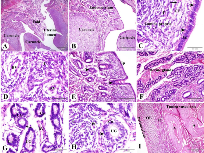
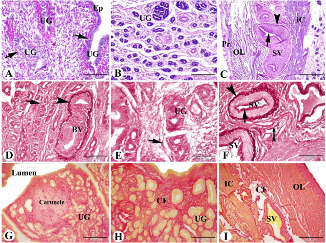
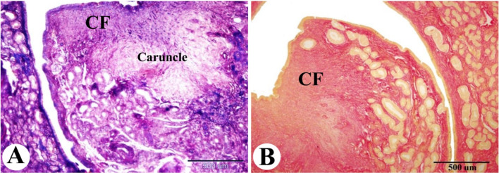
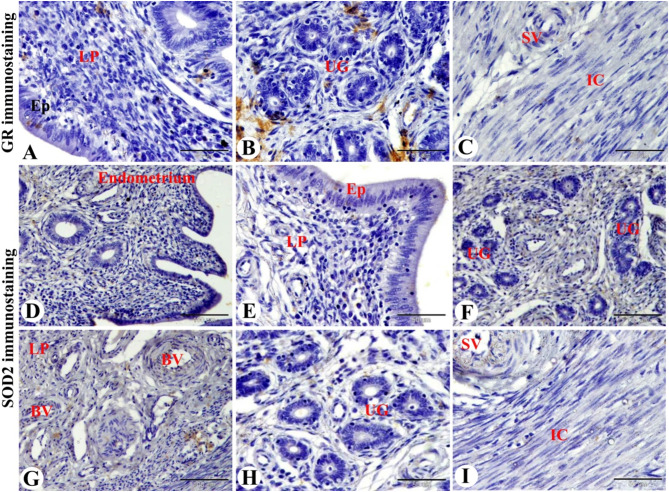
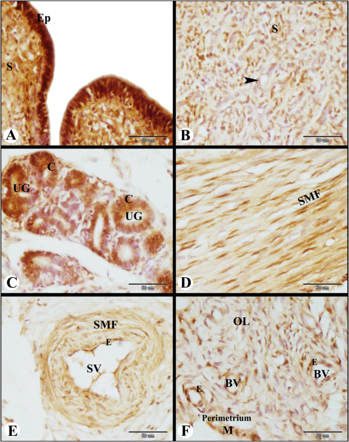
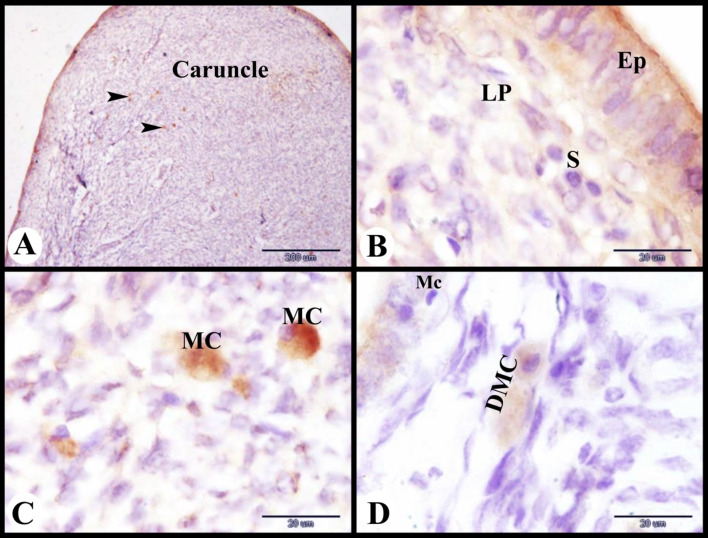
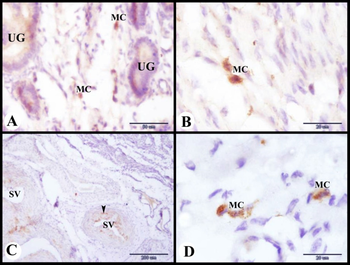
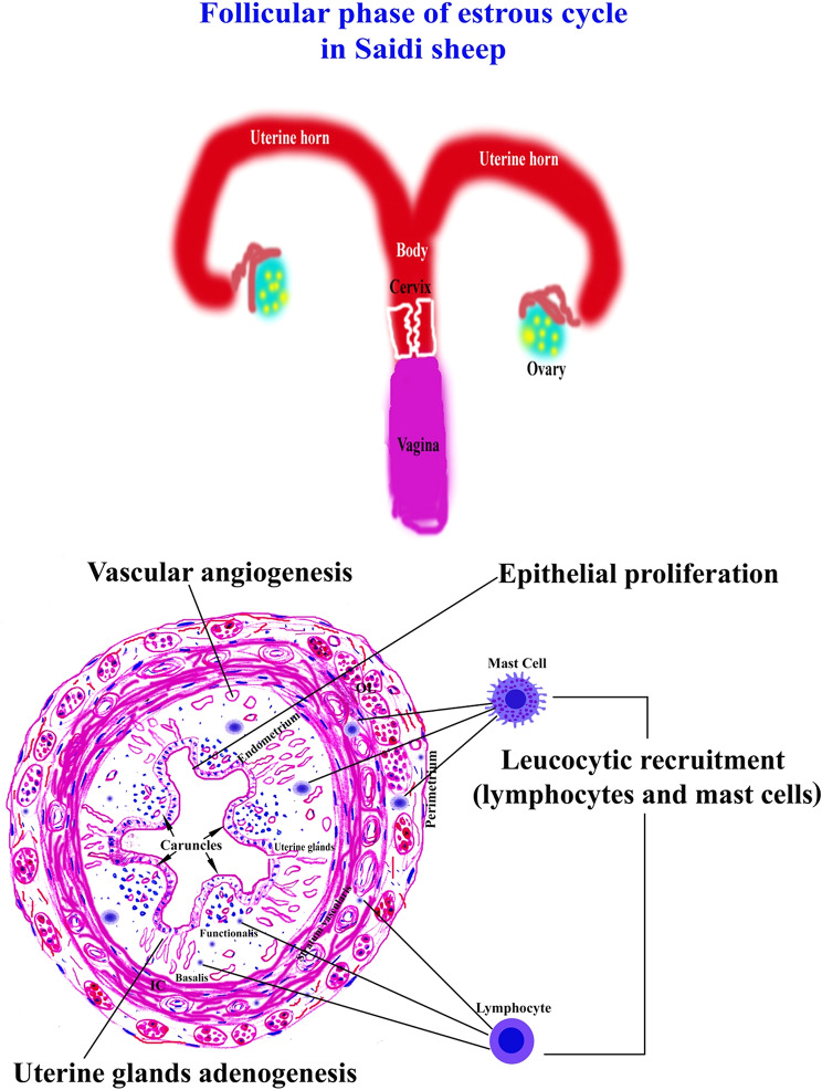
Results: In Saidi sheep, the uterine histological and immunological picture during the follicular phase of the estrous cycle was characterized by epithelial and stromal proliferation and apoptosis. Leucocytic recruitment (lymphocytes, plasma, and mast cells) was also observed. Uterine gland adenogenesis, vascular angiogenesis, oxidative marker expression, and PRA expression in the muscles, stroma, and epithelium were the most noticeable features of the follicular phase.

Conclusion: This study provides new evidence of the role of PRA, SOD2, GR, and mast cells in controlling uterine epithelial proliferation and apoptosis in the Saidi sheep during the follicular phase of the estrus cycle. These findings have growing significance in understanding the key mechanisms that characterize successful reproduction and enhancing the fertility and reproductive efficiency in Saidi Sheep.

Keywords: Endometrium; Estrous cycle; Mast cells; Oxidative stress; Progesterone receptors; Saidi sheep; Uterus.


Conflict of interest statement

Declarations. Ethics approval and consent to participate: The experimental protocol was approved by the Local Ethical Committee and by the Institutional Review Board of Molecular Biology Research and studies Institute, Assiut University (22-2023-0028) and was carried out in accordance with relevant guidelines and regulations. This research was done in compliance with the ARRIVE guidelines and regulations ( https://arriveguidelines.org ). All national and institutional guidelines for animal care and use have been followed throughout the study procedures. Consent for publication: Not applicable. Competing interests: The authors declare no competing interests.

Figures

Fig. 1
Fig. 1
Photomicrograph of the sheep uterus during the follicular phase of the estrous cycle. (A & B): Showing the endometrium with mucosal folds, caruncles, and narrow uterine lumen. (C): Showing the endometrium formed of lamina epithelialis (EP) of pseudostratified columnar epithelium with intraepithelial lymphocytes (arrowheads) and connective tissue lamina propria contained fibroblasts (F), collagen fibers (CF) and lymphocytes (arrow). (D): lamina propria contained many blood vessels (BV) and leucocytic infiltrations (L). (E): Showing many epithelial (EP) invaginations (arrowheads) which form the uterine glands (arrowhead). (F & G): Showing lamina propria with blood vessels (BV) and highly branched and highly coiled uterine glands (UG) which formed of columnar epithelium (Co) and surrounded by myoepithelial cells (arrow). (H): Showing intraepithelial lymphocytes (arrowheads) between the columnar cells (Co) of the uterine glands (UG), plasma cells (PC) and leucocytic infiltration (L) in lamina propria. (I): Showing well vascularized tunica vascularis with many branches of the uterine artery (A) in the outer most layer of inner circular myometrium (IC). Note the outer longitudinal (OL) muscle fibers of myometrium and perimetrium. Original magnification; A: X12.5, scale bar = 1 mm, B & I: X40, scale bar = 500 μm, C, D, G & H: X400, scale bar = 50 μm, E & F: X100, scale bar = 200 μm, haematoxylin and eosin stain
Fig. 2
Fig. 2
Photomicrograph of the sheep uterus during the follicular phase of the estrous cycle. (A & B): Showing the endometrium with PAS positive epithelial basement membrane (arrows) of lamina epithelialis (Ep) and uterine glands (UG). Note no PAS positive secretion in the uterine glands. (C): Showing well-vascularized tunica vascularis with PAS positive internal (arrow) and external (arrowhead) elastic lamina in the outer most layer of inner circular myometrium (IC). Note the outer longitudinal (OL) muscle fibers of the myometrium and the perimetrium (Pr). (D & E): Showing the endometrium in the subepithelial and between the uterine glands (UG) with few elastic fibers (arrow) except for blood vessels; external elastic lamina (arrowhead) of endometrial blood vessels (BV). (F): Showing the myometrium with few elastic fibers between the smooth muscle fibers (forked tail arrow) except for blood vessels of stratum vascularis; in the internal (arrow) and external (arrowhead) elastic lamina. (G & H): Showing the endometrium with a moderate amount of collagen fibers (red color, CF) between the uterine glands (UG) and a few amounts in the caruncle. I: Showing the myometrium with few amount of collagen fibers (red color) in between the smooth muscle fibers of the inner circular (IC) and outer longitudinal (OL) layers. Note the moderate amount of collagen fibers (CF) around blood vessels in the stratum vascularis (SV). Original magnification; A: X200, scale bar = 100 μm, B: X100, scale bar = 200 μm, C: X40, scale bar = 500 μm, PAS & Hx. D: X200, scale bar = 100 μm, E: X100, scale bar = 200 μm, F: X200, scale bar = 100 μm, Orcien stain. G: X40, scale bar = 500 μm, H: X100, scale bar = 200 μm, I: X40, scale bar = 500 μm, Picro-Sirius red technique
Fig. 3
Fig. 3
Photomicrograph illustrating the collagen fiber distributions in the sheep uterus during the follicular phase of the estrous cycle. (A & B): Showing the lamina propria of the endometrium with a moderate amount of collagen fibers (blue color with Masson’s trichrome technique and red color with Picro-Sirius red technique, CF) between the uterine glands (UG) and few amounts in the caruncle. Original magnification; (A & B): X40, scale bar = 500 μm, (A): Masson’s trichrome technique, (B): Picro-Sirius red technique
Fig. 4
Fig. 4
Photomicrograph of GR immunostaining in the sheep uterus during the follicular phase of the estrous cycle. (A & B): Showing slight GR immunostaining in the lamina epithelialis (Ep), in the stroma cells of lamina propria (LP), and negative GR immunostaining in the uterine glands (UG). (C): Showing slight GR immunostaining in the smooth muscle fibers of the blood vessels of the stratum vascularis (SV) and negative GR immunostaining in the smooth muscle fibers of the inner circular layer (IC) of the myometrium. (D-H): Showing slight SOD2 immunostaining in the lamina epithelialis (Ep) and in the stroma cells and the smooth muscle fibers of blood vessels (BV) of the lamina propria (LP) and negative SOD2 immunostaining in the uterine glands (UG). (I): Showing slight SOD2 immunostaining in the smooth muscle fibers of blood vessels of stratum vascularis (SV) and negative SOD2 immunostaining in the smooth muscle fibers of the inner circular layer (IC) of the myometrium. Original magnification; (A, B, C, E, H & I): X400, scale bar = 50 μm, D, F & G: X200, scale bar = 100 μm
Fig. 5
Fig. 5
Photomicrograph of TUNEL assay immunofluorescence in the sheep uterus during the follicular phase of estrous cycle. (A): Showing apoptotic luminal (arrow) and glandular epithelial cells (arrowheads). (B): Showing apoptotic endometrial stromal cells (arrowheads). Note the inner circular layer (IC) of the myometrium with few apoptotic cells (arrow). (C): Showing apoptotic endometrial stromal cells (arrowheads) in the caruncle. (D): Showing apoptotic smooth muscle fibers (arrowheads) of the blood vessels of the stratum vascularis (SV) and some apoptotic smooth muscle fibers of the myometrium (arrow). Original magnification; (A-D): X400, scale bar = 50 μm
Fig. 6
Fig. 6
Photomicrograph of PRA immunolocalization in the sheep uterus during the follicular phase of the estrous cycle. (A): Showing strong PRA immunolocalization in the lamina epithelialis (Ep) and strong to moderate PRA immunolocalization in the subepithelial stroma cells (S). (B): Showing moderate PRA immunolocalization in the stromal cells and endothelial cells (arrowhead) of blood vessels in the caruncles. (C): Showing strong to moderate PRA immunolocalization in the columnar epithelial cells (C) of the uterine glands (UG). (D): Showing moderate PRA immunolocalization in the smooth muscle fibers (SMF) of the inner circular layer of the myometrium. (E): Showing moderate PRA immunolocalization in the endothelium (E) and smooth muscle fibers (SMF) of the blood vessels of the stratum vascularis (SV). (F): Showing moderate PRA immunolocalization in the smooth muscle fibers and endothelial cells (E) of blood vessels (BV) in the outer longitudinal layer (OL) of the myometrium and moderate PRA immunolocalization in the mesothelial cells (M) and in the endothelial cells (E) of blood vessels (BV) of the perimetrium. Original magnification; A-F: X400, scale bar = 50 μm
Fig. 7
Fig. 7
Photomicrograph of mast cells immunolocalization in the sheep uterus during the follicular phase of the estrous cycle. (A): Showing tryptase positive immunostaining mast cells (arrowheads) in the deep lamina propria of the caruncles. (B): Showing the superficial lamina propria (LP) under the lamina epithelialis (Ep) with stroma cells (S) and no mast cells. (C): Showing mast cells (MC) filled with tryptase positive immunostaining granules. (D): Showing degranulated mast cells (DMC) near the macrophage (Mc) in the endometrial lamina propria. Original magnification; A: X100, scale bar = 200 μm, B-D: X1000, scale bar = 20 μm
Fig. 8
Fig. 8
Photomicrograph of mast cells immunolocalization in the sheep uterus during the follicular phase of the estrous cycle. (A): Showing tryptase positive immunostaining mast cells (MC) in-between uterine glands (UG). (B): Showing tryptase positive immunostaining mast cells (MC) in the inner circular smooth muscle layer of the myometrium. (C): Showing tryptase positive immunostaining mast cells (arrowheads) around the blood vessels of the stratum vascularis (SV). (D): Showing tryptase positive immunostaining mast cells (MC) in the outer longitudinal smooth muscle layer of the myometrium. Original magnification; A: X400, scale bar = 50 μm, B & D: X1000, Scale bar = 20 μm, C: X100, scale bar = 200 μm
Fig. 9
Fig. 9
The main uterine characteristics of the follicular phase of the estrous cycle in Saidi sheep

Similar articles

References

    1. Wang H, Feng X, Muhatai G, Wang L. Expression profile analysis of sheep ovary after superovulation and estrus synchronisation treatment. Vet Med Sci. 2022;8:1276–87. - PMC - PubMed
    1. Germot A, Khodary MG, Othman OE-M, Petit D. Shedding light on the Origin of Egyptian Sheep breeds by Evolutionary comparison of mitochondrial D-Loop. Animals. 2022;12:2738. - PMC - PubMed
    1. El-Homosi FF, Abd El-Hafiz GA, REPRODUCTIVE PERPORMANCE OF OSSIMI, AND SAIDI SHEEP UNDER TWO PRE PUBERTAL PLANES OF NUTRITION. Assiut Vet Med J. 1982;101:59–66.
    1. Teleb D, Ahmed N, Tag E, –Din H, Abou El Soud S, Hassan OM, STUDY ON LEVELS OF SOME BLOOD HORMONAL AND BIOCHEMICAL CONSTITUENTS DURING DIFFERENT REPRODUCTIVE STATUS IN SAIDI EWES. Egypt J Sheep Goats Sci. 2019;9:1–10.
    1. Abd-Elkareem M, Khormi MA, Mohamed RH, Ali F, Hassan MS. Histological, immunohistochemical and serological investigations of the ovary during follicular phase of estrous cycle in Saidi sheep. BMC Vet Res. 2024;20:98. - PMC - PubMed

LinkOut - more resources