. 2025 Jan 13;22(1):5.
doi: 10.1186/s12987-024-00608-7.

Venous compression causes chronic cerebral ischaemia in normal pressure hydrocephalus patients

Affiliations

Venous compression causes chronic cerebral ischaemia in normal pressure hydrocephalus patients

Tomohisa Ohmura et al. Fluids Barriers CNS. .

Abstract

Background: Cerebral autoregulation is a robust regulatory mechanism that stabilizes cerebral blood flow in response to reduced blood pressure, thereby preventing cerebral ischaemia. Scientists have long believed that cerebral autoregulation also stabilizes cerebral blood flow against increases in intracranial pressure, which is another component that determines cerebral perfusion pressure. However, this idea was inconsistent with the complex pathogenesis of normal pressure hydrocephalus, which includes components of chronic cerebral ischaemia due to mild increases in intracranial pressure.

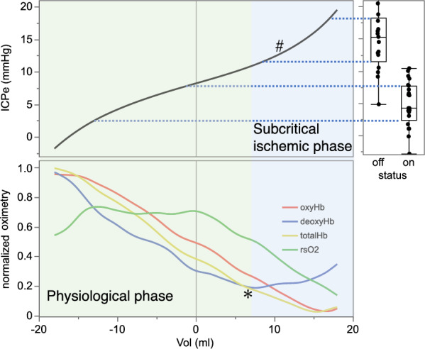
Methods: Twenty-one patients who underwent ventriculoperitoneal shunt surgery for normal pressure hydrocephalus were included in this study. To determine the pressure setting of the Codman-Hakim programmable valve, intracranial pressure was measured after shunt surgery by puncturing the Ommaya reservoir, which formed a closed circuit with the needle and the syringe. Then, intracranial pressure was continuously measured with intermittent infusion of cerebrospinal fluid from the same closed circuit. We also continuously measured oximetry data, such as regional cerebral oxygen saturation derived from near-infrared spectroscopy monitoring. These data were digitized, recorded, and used for calculating intracranial compliance and the relationship between cerebrospinal fluid volume loading and intracranial pressure.

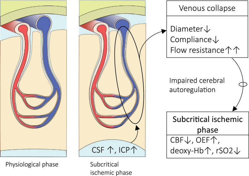
Results: This study demonstrates that in patients with normal pressure hydrocephalus, cerebral venous vascular bed compression induces mild cerebral ischaemia when intracranial pressure is slightly higher than physiological venous pressure. Cerebral venous compression impairs cerebral blood flow by quadratically increasing circulatory resistance according to Poiseuille's law. Furthermore, chronic cerebral ischaemia occurred even at low or normal intracranial pressures when deep and subcortical white matter hyperintensities (DSWMHs) were severe.

Conclusion: The fact that cerebral blood flow impairment begins at very low intracranial pressures indicates that cerebral autoregulation to compensate for reduced venous blood flow is not functioning adequately in NPH. These processes provide a link between impaired cerebrospinal fluid circulation, cerebral autoregulation, and neurological dysfunction, which has been missing in patients with NPH involving small vessel arteriosclerosis. These findings may provide insight into similar conditions, such as normal-tension glaucoma and chronic kidney disease, in which a mild increase in local compartment pressure leads to chronic ischemia in organs protected by autoregulatory mechanisms.


Conflict of interest statement

Declarations. Consent for publication: I confirm the corresponding author has read the journal policies and submit this manuscript in accordance with those policies. Competing interests: The authors declare no competing interests.

Figures

Fig. 1
Fig. 1
Relationship between ICPvein, oximetry data by NIRS, and compliance: Compliance and oximetry curves were derived from the mean values of all patients measured. As ICPvein increased, oxyHb and totalHb decreased. On the other hand, deoxyHb decreased and then increased after 2.5 mmHg. The asterisk (*) in this figure indicates the trough where deoxyHb transitions from decreasing to increasing with increasing ICP. In general, the increase in deoxyHb is due to an increase in cerebral oxygen extraction fraction (OEF). Furthermore, the regional cerebral oxygen saturation (rSO2) also decreased, indicating a subcritical ischemic phase (blue background region in this figure). Whereas the distribution of ICPvein during the valve-on period (-3.3 ± 4.6 mmHg, n = 21) in shunted NPH patients is in a physiological phase, the distribution of ICPvein during the valve-off period is in a subcritical ischemic phase (7.1 ± 7.4 mmHg, n = 17). 3 of the 17 participants were in the physiological phase (green background), while the remaining 14 were in the subcritical ischaemic phase (blue background) dunring the valve-off period. (Note: Only 14 of the 17 patients are shown in the graph with valves off, as two were scaled to a range higher than 15 mmHg and one was a clear outlier at −7.44 mmHg). This indicates that the intracranial pressure of untreated NPH patients is in the subcritical ischemic phase, despite being in the normal range, and that shunt surgery allows them to move into the physiological phase. The ICPvein-compliance curve exhibited a symmetrical bell-shaped curve with a peak at 0 mmHg
Fig. 2
Fig. 2
Relationship between CSF volume loading and ICPeac: Relationship between CSF volume loading and ICPeac was simulated from the ICPvein-compliance curve. CSF volume loading increased ICPeac quadratically above approximately 10 ml (#). Surprisingly, the ICPeac that moved into the subcritical ischemic phase was only 10 mmHg (*). The right-hand graph illustrates that the distribution of ICPeac during the valve-on period (4.9 ± 3.6 mmHg) in shunted NPH patients is in the physiological phase, whereas the distribution of ICPeac during the valve-off period (15.4 ± 5.8 mmHg) is in the subcritical ischemic phase
Fig. 3
Fig. 3
Patients with iNPH are exposed to a subcritical ischemic phase from lower intracranial pressure than patients with sNPH: ICPvein values at the beginning of the transition from the physiological phase to the subcritical ischaemic phase, indicated by the timing of deoxy-Hb turning from the previous decrease to an increase, are lower in patients with iNPH (left top) than in those with sNPH (left bottom). Statistical analysis revealed that ICPvein at the beginning of the transition from the physiological phase to the subcritical ischemic phase was 2.2 ± 1.6 mmHg for iNPH and 4.9 ± 2.5 mmHg for sNPH, which was significantly lower in iNPH (right top). Compliance tended to be higher in iNPH than in sNPH (2.7 ± 1.4 ml/mmHg, 1.3 ± 0.4 ml/mmHg, iNPH, sNPH, respectively) (right bottom)
Fig. 4
Fig. 4
Impact of severity of DSWMH: ICPvein at the beginning of the transition from the physiological phase to the subcritical ischemic phase was 2.4 ± 1.8 mmHg in the DSWMH grade High group and 5.6 ± 2.2 mmHg in the Low group, significantly lower in the High group. Compliance tended to be higher in the DSWMH grade High group than in the Low group (2.5 ± 1.3 ml/mmHg, 1.1 ± 0.2 ml/mmHg, DSWMH grade High and Low, respectively)
Fig. 5
Fig. 5
The pathophysiology of NPH: Patients with NPH present with mild intracranial hypertension due to CSF accumulation caused by impaired CSF circulation. In addition, there is reduced compliance due to venous collapses, which, combined with ageing, arteriosclerosis, and brain atrophy, impairs CA, resulting in a subcritical ischemic phase with decreased CBF, increased OEF, increased deoxy-Hb, and decreased rSO2
Fig. 6
Fig. 6
General course of each disease: As people get older, it becomes progressively more difficult to maintain an active life. In addition, the reserve capacity of motor and cognitive functions declines. In the case of Binswanger disease, which develops after the mid-seventies due to chronic cerebral ischaemia in addition to the usual age-related functional decline, the symptoms of vascular dementia slowly worsen and the modified Rankin Scale (mRS) also worsens. Unlike pure Binswanger disease, iNPH is complicated by impaired CSF circulation. iNPH is a triad of additive effects of cerebral ischaemia due to chronic cerebral ischaemic damage from small artery arteriosclerosis and venous compression, in addition to low motor and cognitive reserve due to old age at onset. Patients are thus already in a subcritical ischemic phase at the onset of the disease. After onset, the mRS declines, but partial improvement is observed with shunt surgery (blue & purple line). However, the subsequent course of the disease is in deficit, with the mRS gradually worsening with age. sNPH is commonly associated with SAH, for example, and the onset of sNPH is usually at a younger age than iNPH. Unlike iNPH, sNPH is less likely to involve ischaemia derived from cerebral arteriosclerosis and more likely to involve purely CSF circulatory disturbance. Due to chronic cerebral ischaemia caused by impaired CSF circulation, the patient is already in a subcritical ischemic phase at the onset of the disease. With onset, the mRS deteriorates to a greater extent than in iNPH, but considerable improvement can be achieved by shunt surgery (blue & purple line). The course of the disease then follows a deficit course, with the mRS gradually worsening with age at the same rate as normal age-related functional decline

Comment in

Similar articles

Cited by

References

    1. Ruland S, Aiyagari V. Cerebral autoregulation and blood pressure lowering. Hypertension. 2007;49:977–8. - PubMed
    1. Lassen NA. Cerebral blood flow and oxygen consumption in man. Physiol Rev. 1959;39:183–238. - PubMed
    1. Faraci FM, Heistad DD. Regulation of large cerebral arteries and cerebral microsvascular pressure. Circ Res. 1990;66:8–17. - PubMed
    1. Cipolla MJ, Smith J, Kohlmeyer MM, Godfrey JA. SK Ca and IK Ca channels myogenic tone, and vasodilator responses in middle cerebral arteries and parenchymal arterioles. Stroke. 2009;40:1451–7. - PMC - PubMed
    1. Greenfield JC, Tindall GT. Effect of acute increase in intracranial pressure on blood flow in the internal carotid artery of man. J Clin Invest. 1965;44:1343–51. - PMC - PubMed

LinkOut - more resources