Review
. 2025 Jan 13;18(1):6.
doi: 10.1186/s13045-024-01634-6.

Harnessing the tumor microenvironment: targeted cancer therapies through modulation of epithelial-mesenchymal transition

Affiliations
Review

Harnessing the tumor microenvironment: targeted cancer therapies through modulation of epithelial-mesenchymal transition

Antonino Glaviano et al. J Hematol Oncol. .

Abstract

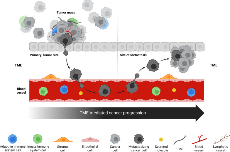
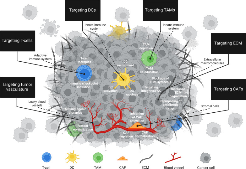
The tumor microenvironment (TME) is integral to cancer progression, impacting metastasis and treatment response. It consists of diverse cell types, extracellular matrix components, and signaling molecules that interact to promote tumor growth and therapeutic resistance. Elucidating the intricate interactions between cancer cells and the TME is crucial in understanding cancer progression and therapeutic challenges. A critical process induced by TME signaling is the epithelial-mesenchymal transition (EMT), wherein epithelial cells acquire mesenchymal traits, which enhance their motility and invasiveness and promote metastasis and cancer progression. By targeting various components of the TME, novel investigational strategies aim to disrupt the TME's contribution to the EMT, thereby improving treatment efficacy, addressing therapeutic resistance, and offering a nuanced approach to cancer therapy. This review scrutinizes the key players in the TME and the TME's contribution to the EMT, emphasizing avenues to therapeutically disrupt the interactions between the various TME components. Moreover, the article discusses the TME's implications for resistance mechanisms and highlights the current therapeutic strategies toward TME modulation along with potential caveats.

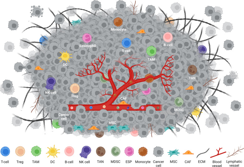
Keywords: Cancer; Cancer-associated fibroblasts (CAFs); Chimeric antigen-receptor (CAR) T-cell therapy; Dendritic cells (DCs); Epithelial-mesenchymal transition (EMT); Extracellular matrix (ECM); Metastasis; Myeloid-derived suppressor cells (MDSCs); Natural killer (NK) cells; T-cell receptor (TCR) therapy; T-cells, B-cells, tumor-associated macrophages (TAMs); Theranostics; Tumor microenvironment (TME); Tumor-associated neutrophils (TANs).


Conflict of interest statement

Declarations. Ethics approval and consent to participate: Not Applicable. Consent for publication: We confirm that the manuscript has been read and approved by all named authors and that there are no other persons who satisfied the criteria for authorship but are not listed. We further confirm that the order of authors listed in the manuscript has been approved by all the authors. All figures are original and were drawn using BioRender by the authors for this review. Competing interests: The authors declare that they have no known competing financial interests or personal relationships that could have appeared to influence the work in this paper. M.H.A. is founder, shareholder and scientific advisor of IO Biotech ApS. LMC is a consultant for Evergreen Theragnostics. S.K. has consulted for Telix Pharmaceuticals Ltd., acknowledges support for investigator services from RayzeBio and holds the following patent: PCT/US2021/039418 (THOR cell (tumor homing radio-emitting cell).

Figures

Fig. 1
Fig. 1
Metastasis is responsible for the diffusion of tumor cells to distant regions of the body leading to increased drug resistance, therapy failure, and mortality. The plasticity of EMT suggests that metastasis is regulated by extracellular signals from the TME, a complex multicellular and unique tumor-surrounding ecosystem, which denotes the non-malignant cells and their released molecules present in the tumor via epigenetic modifications in cancer cells. The TME comprises various cell types such as immune cells from the adaptive immune system, immune cells from the innate immune system, and stromal cells, as well as blood/lymphatic vascular network, ECM, and secreted molecules; all of which communicate to signals modulating EMT. The constant interdependent interaction between cancer cells and their TME, as well as the heterogeneity of TME, represent the major contributors toward metastasis, cancer progression, and reduced therapeutic response. TME: Tumor microenvironment; ECM: Extracellular matrix. This figure has been created with BioRender.com
Fig. 2
Fig. 2
TME is composed of different cell types, cell structures, and secreted factors. The TME is populated by heterogeneous cancer cells and various cell types including immune cells such as T-cells, B-cells, NK cells, DCs, TAMs, MDSCs, neutrophils, monocytes, and eosinophils; stromal cells including CAFs and MSCs; as well as blood and lymphatic vascular networks. CAF: Cancer-associated fibroblast; DC: Dendritic cell; ECM: Extracellular matrix; ESP: Eosinophil; MDSC: Myeloid-derived suppressor cells; MSC: Mesenchymal stromal cells; NK cell: Natural killer cell; TAM: Tumor-associated macrophages; TAN: Tumor-associated neutrophil; Treg: Regulatory T-cells. These cells secrete ECM components, cytokines, growth factors, and ECVs important for signaling among different cell types in the TME. This figure has been created with BioRender.com
Fig. 3
Fig. 3
Each cell type present in the TME can contribute to the regulation of cancer progression and therapeutic response individually and thus several TME-directed therapies have been developed. The major strategies, which either have been FDA-approved or are currently being under clinical investigation, principally focus on the targeting of T-cells, DCs, TAMs, CAFs, ECM, and tumor vasculature; and thus, are indicated in the figure (black boxes) and referenced in the review. Targeting T-cells includes immune checkpoint inhibition, and T-cell therapies; targeting DCs comprises DC activation, DC recruitment, and DC vaccines; targeting TAMs consists of TAM depletion, and TAM re-education; targeting CAFs includes CAF depletion, inhibition of CAF activation, and CAF normalization; targeting ECM comprises increased ECM degradation, blockage of ECM synthesis, repurposing of drugs with antifibrotic properties, and targeting integrins or the downstream effector FAK; and targeting tumor vasculature consists of antiangiogenic therapies, and vessel normalization. DCs: Dendritic cells; TAMs: Tumor-associated macrophages; CAFs: Cancer-associated fibroblasts; ECM: Extracellular matrix; FAK: Focal adhesion kinase. This figure has been created with BioRender.com
Fig. 4
Fig. 4
Timeline of FDA approvals for ICIs targeting T-cells, B-cells, TAMs, CAFs, and tumor vasculature. Timeline of FDA approvals for ICIs targeting T-cells (blue rectangles), B-cells (red rectangles), TAMs (orange rectangle), CAFs (yellow rectangles), and tumor vasculature (green rectangles) is shown. Black arrows below the ICIs: years in which the first FDA approval occurred. Grey squares at the top-left of each inhibitor: cancer type/s related to the first FDA approval of the corresponding inhibitor. ASTs: Advanced solid tumors; BCC: Basal cell carcinoma; CLL: Chronic lymphocytic leukemia; CRC: Colorectal cancer; GC: Gastric cancer; GISTs: Gastrointestinal stromal tumors; HCC: Hepatocellular carcinoma; HL: Hodgkin lymphoma; MCL: Mantle cell lymphoma; MEL: Melanoma; MZL: Marginal zone lymphoma; NSCLC: Non-small cell lung cancer; PC: Pancreatic cancer; RCC: Renal cell carcinoma; SLL: Acute lymphocytic leukemia; STS: Soft tissue sarcoma; TC: Thyroid cancer; TGCTs: Tenosynovial giant cell tumors; UC: Urothelial cancer; WM: Waldenström macroglobulinemia. Asterisk (*) beside grey-squared ASTs: year in which the first (of a series of) AST FDA approval occurred for a particular ICI. Circled capital letters at the top-right of each inhibitor: biopharmaceutical companies related to the first FDA approval of inhibitors. Ⓐ: AstraZeneca; Ⓑ: Bayer AG; Ⓓ: Daiichi Sankyo Company; Ⓔ: Eli Lilly and Company; Ⓖ: GlaxoSmithKline (GSK); Ⓙ: Janssen Pharmaceuticals, Inc; Ⓜ: Merck Inc; Ⓝ: Novartis; Ⓟ: Pfizer Inc; Ⓡ: Bristol-Myers Squibb Company; Ⓢ: Sanofi SA; Ⓣ: Genentech Inc; Ⓥ: AbbVie Inc
Fig. 5
Fig. 5
Targeting of different TME components (adaptive immune system, innate immune system, CAFs, tumor vasculature, and ECM), for cancer therapy. Diverse agents are being/have been used to target different TME components such as adaptive immune system (T-cells and B-cells), innate immune system (TAMs, MDSCs, and DCs), CAFs, tumor vasculature, and ECM, for cancer therapy used in clinical trials or approved by the FDA. Targeted molecules (ANG2-TIE2, BCR, CCL2, CCR2, CD40, CD47, Collagen, CSF1R, CTGF, CTLA4, CXCR4, FAK, FAP, FGFR, FLT3L, GM-CSF, HA, Hedgehog, ICOS, LAG-3, LOXL2, OX40, PD-1, PDE5, PDGFR, PD-L1, PI3K, ROCK, RTK, SIRPα, TGF-β, TIGIT, TIM-3, TREM2, uPAR, VEGF, VEGFR, VISTA, Vitamin A, Vitamin D) are written (along the lines) in red. ANG2-TIE2: Angiopoietin-2-TIE2; BCR: B-cell receptor; CCL2: CC-motif chemokine ligand 2; CCR2: CC-chemokine receptor 2; CD40: Cluster of differentiation 40; CD47: Cluster of differentiation 47 or integrin associated protein (IAP); CSF1R: Colony-stimulating factor-1 receptor; CTGF: Connective tissue growth factor; CTLA4: Cytotoxic T lymphocyte-associated protein-4; CXCR4: C-X-C chemokine receptor type 4; FAK: Focal adhesion kinase; FAP: Fibroblast activation protein; FGFR: Fibroblast growth factor receptor; FLT3L: Fms-related tyrosine kinase 3 ligand; GM-CSF: Granulocyte–macrophage colony-stimulating factor; HA: Hyaluronan; HIF1α: Hypoxia-inducible factor 1α; ICOS: Inducible T-cell co-stimulatory; LAG-3: Lymphocyte activation gene-3; LOXL2: Lysyl oxidase like-2; OX40: OX40 receptor or tumor necrosis factor receptor superfamily, member 4 (TNFRSF4); PD-1: Programmed cell death protein 1; PDE5: Phosphodiesterase 5; PDGFR: platelet-derived growth factor receptor; PD-L1: Programmed death-ligand 1; PI3K: Phosphoinositide 3-kinase; ROCK: Rho-associated protein kinase; RTK: Receptor tyrosine kinase; SIRPα: Signal regulatory protein α; TGF-β: Transforming growth factor-β; TIGIT: T-cell immunoreceptor with Ig and ITIM domains; TIM-3: T-cell immunoglobulin and mucin-domain containing-3; TREM2: Triggering receptor expressed on myeloid cells 2; uPAR: urokinase-type plasminogen activator receptor; VEGF: Vascular endothelial growth factor; VEGFR: Vascular endothelial growth factor receptor; VISTA: V-domain Ig suppressor of T-cell activation. TM: Targeted molecule; Abs: Antibodies; SMIs: Small-molecule inhibitors; Rec. cytokines: Recombinant cytokines; Others: Recombinant fragment fusion proteins, Vitamin A metabolite, and PEGylated enzyme. Drugs (written in bold): FDA-approved drugs
Fig. 6
Fig. 6
Therapeutic targeting of T-cells to augment anti-cancer activity. T-cell antitumor activity can be increased through 1) inhibition of several immune checkpoint molecules, or 2) adoptive transfer of CAR T-cells, TCR T-cells, or tumor-infiltrating lymphocytes (TILs). The scheme on the bottom left of the figure displays five major protein/ligand interactions (e.g. PD-1/PD-L1). Dotted-lines select the square enlargement including protein/ligand interaction between a T-cell and a DC. The scheme on the bottom right of the figure shows the five main steps of CAR T-cell and TCR T-cell therapy: 1) T-cell collection from blood patients; 2) viral vector-mediated introduction of a CAR gene or a TCR gene; 3) generation of CAR T-cells or TCR T-cells; 4) ex-vivo expansion of engineered T-cells (CAR T-cells or TCR T-cells); 5) infusion of CAR T-cells or TCR T-cells back into patients. This figure has been created with BioRender.com
Fig. 7
Fig. 7
Therapeutic targeting of TAMs to enhance anti-cancer activity. Several strategies have been and are being developed to determine TAM depletion and/or reprogramming to increase anti-cancer immune activity. The major approaches currently used or being evaluated and indicated in the black boxes and comprise: 1) enhancing TAM-induced phagocytosis of tumor cells by inhibiting the “do not eat me” CD47/SIRPα pathway; 2) reprogramming TAMs by augmenting their antigen presentation to T-cells via CD40 agonists, or by endorsing their re-education to anti-cancer phenotypes by inhibition of TREM2 or PI3Kγ; 3) inhibiting TAM recruitment to the TME through suppression of CCL2 or CCR2; 4) depleting or re-educating TAMs through inhibition of CSF1R signaling. This figure has been created with BioRender.com
Fig. 8
Fig. 8
Therapeutic targeting of DCs to increase anti-cancer activity. Different targeting strategies have been established to enhance DC-promoted T-cell priming, such as 1) GM-CSF administration-mediated endorsement of DC survival, proliferation, and differentiation; 2) FLT3L administration-induced DC expansion and maturation; and 3) ex vivo DC manipulation and administration in the form of a DC vaccine. The DC vaccines manipulate DCs ex vivo to augment their presentation capacity for specific TAAs in vivo, and are created through the following steps: 1) CD14+ monocytes or CD34+ hematopoietic stem and progenitor cells (HSPCs) are isolated from the blood of a cancer patient; 2) CD14+ monocytes or CD34+ HSPCs are differentiated into immature monocyte-derived dendritic cells (moDCs); 3) immature moDCs are subjected to TAA loading normally attained from tumor lysates; 4) DCs are genetically engineered to increment their cell-intrinsic features, e.g. cross-presentation, cytokine production, and lymph node migration, thereby augmenting their anticancer functions, and complete maturation of DC is accomplished by diverse maturation cocktails; finally, 5) TAA-loaded matured DCs are then injected back into the cancer patient, either subcutaneously or intradermally, resulting in the increase and improvement of cancer-specific immune responses. The types of DC vaccines can differ depending on cells used for ex vivo manipulation, strategy for TAA delivery, or activation status of DCs infused into the cancer patient. Ag: antigen. This figure has been created with BioRender.com
Fig. 9
Fig. 9
Therapeutic targeting of CAFs to augment anti-cancer activity. CAFs can be targeted using various strategies, such as interfering with CAF activation using TGFβ and FBFR inhibitors, CAF signaling using TGFβ, CXCR4, FAP, ROCK signaling and Hedgehog signaling inhibitors, or CAF normalization using vitamin A metabolites and vitamin D analogues, which are either FDA-approved or currently being evaluated in clinical trials. CAFs: Cancer-associated fibroblasts; CXCR4: C-X-C chemokine receptor type 4; FAK: Focal adhesion kinase; FAP: Fibroblast activation protein; FGFR: Fibroblast growth factor receptor; FSP1: Fibroblast-specific protein 1 (FSP1); Hh: Hedgehog; ITGA11: Integrin alpha 11; ITGB1: Integrin β-1; LOX: Lysyl oxidases; PDGFR: Platelet-derived growth factor receptor; RTK: Receptor tyrosine kinase; TGF-β: Transforming growth factor-β. This figure has been created with BioRender.com
Fig. 10
Fig. 10
Therapeutic targeting of tumor vasculature to enhance anticancer activity. Inhibition of VEGF and/or VEGFR is the most used antiangiogenic strategy accomplished with several FDA-approved agents, such as anti-VEGF and VEGF-TRAP (VEGF decoy receptors), and/or VEGFR-specific antibodies and tyrosine kinase inhibitors (RTK inhibitors), respectively. Alternatively, ANG2-TIE2 inhibitors currently being tested in the clinic can also be used to promote antiangiogenesis. The drugs targeting tumor vasculature, either FDA-approved or being-evaluated at different stages of clinical development drugs, are referenced in the text. VEGF: Vascular endothelial growth factor; VEGFR: Vascular endothelial growth factor receptor; ANG2: Angiopoietin-2; TIE2: TEK receptor tyrosine kinase; TKIs: Tyrosine kinase inhibitors. TME: Tumor microenvironment. This figure has been created with BioRender.com
Fig. 11
Fig. 11
Therapeutic targeting of ECM to increase anti-cancer activity. The secreted ECM can be targeted with different strategies, such as interfering with integrin signaling using FAK inhibitors, destabilizing collagen network through inhibition of LOX enzymes using LOXL2 antibodies, degrading hyaluronan using hyaluronidases, and enhancing antifibrotic properties by reducing collagen synthesis and production using collagen inhibitors. CAFs: Cancer-associated fibroblasts; CXCR4: C-X-C chemokine receptor type 4; CTGF: Connective tissue growth factor; FAK: Focal adhesion kinase; FAP: Fibroblast activation protein; FGF: Fibroblast growth factor; LOX: Lysyl oxidases; TGF-β: transforming growth factor-β. This figure has been created with BioRender.com.
Fig. 12
Fig. 12
Metabolic interactions in the TME. The TME is represented in the center by a group of cancer cells coated with activated fibroblasts and surrounded by CAFs and immune cells. At the top: In hypoxic condition, TME promotes the production of angiogenic factors (VEGF, TGF-β, FGF and PDGF) to induce rapid angiogenesis, resulting in the formation of aberrant blood vessels with reduced pericyte coverage, low levels of leukocyte adhesion molecules, and low levels of T-cell recruiting cytokines, therefore impeding the recruitment of anti-tumor immune cells. On the left: under certain stimuli, CAFs are activated and acquire a pro-inflammatory signature with the expression of immunomodulatory molecules (TGF-β and PDL-1) and lead to ECM remodeling into a rigid fibrotic matrix. They also form a stromal matrix surrounding the tumor core through the desmoplastic reaction. On the right: Cancer cells drain energy from the surrounding immune cells by competing for nutrients and amino acids, stealing their mitochondria through nanotubes, and hiding from them using protective stromal matrix formed by CAFs which limits cytotoxic cell infiltration. CAF: CAFs: Cancer-associated fibroblasts; VEGF: Vascular endothelial growth factor; TGF-β: Transforming growth factor-β; FGF: Fibroblast growth factor; PDGF: Platelet-derived growth factor; PDL-1: Programmed death-ligand 1. This figure has been created with BioRender.com

References

    1. Collaborators GCRF. The global burden of cancer attributable to risk factors, 2010–19: a systematic analysis for the global burden of disease study 2019. Lancet. 2022;400(10352):563–91. - PMC - PubMed
    1. Ganesh K. Uncoupling Metastasis from Tumorigenesis. N Engl J Med. 2023;388(7):657–9. - PMC - PubMed
    1. Li R, Liu X, Huang X, Zhang D, Chen Z, Zhang J, et al. Single-cell transcriptomic analysis deciphers heterogenous cancer stem-like cells in colorectal cancer and their organ-specific metastasis. Gut. 2024;73(3):470–84. - PMC - PubMed
    1. Chiang AC, Massagué J. Molecular basis of metastasis. N Engl J Med. 2008;359(26):2814–23. - PMC - PubMed
    1. Yu Y, Lyu C, Li X, Yang L, Wang J, Li H, et al. Remodeling of tumor microenvironment by extracellular matrix protein 1a differentially regulates ovarian cancer metastasis. Cancer Lett. 2024;596: 217022. - PubMed

LinkOut - more resources