. 2024 Oct 29;3(12):102396.
doi: 10.1016/j.jscai.2024.102396. eCollection 2024 Dec.

Can Contrast Injections Cause or Propagate Coronary Injuries? Insights From Vessel and Guiding Catheter Hemodynamics

Affiliations

Can Contrast Injections Cause or Propagate Coronary Injuries? Insights From Vessel and Guiding Catheter Hemodynamics

Daniel Chamié et al. J Soc Cardiovasc Angiogr Interv. .

Abstract

Background: The mechanistic association between the hydraulic forces generated during contrast injection and the risk of coronary injury is poorly understood. In this study, we sought to evaluate whether contrast injections increase intracoronary pressures beyond resting levels and estimate the risk of hydraulic propagation of coronary dissections.

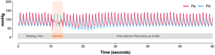
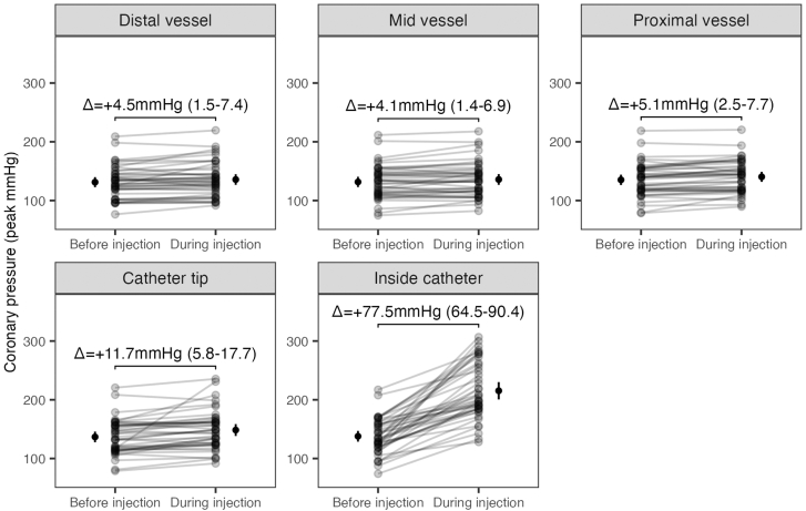
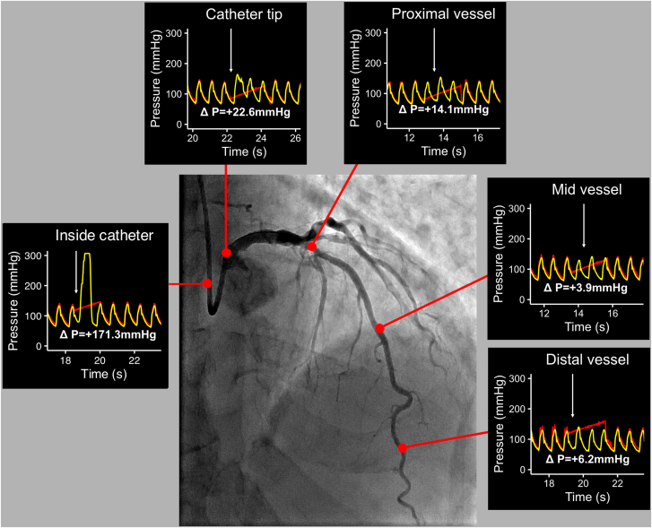
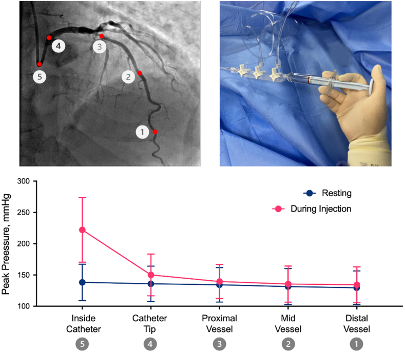
Methods: This is a prospective, single-arm, multicenter study that included patients with nonculprit, non-flow-limiting coronaries. A continuous 60-second pressure recording was taken at 5 predetermined locations during contrast injections: distal, mid, and proximal vessel, catheter tip, and inside the catheter. The primary end point was the change in intracoronary peak pressure between resting and injections in each location.

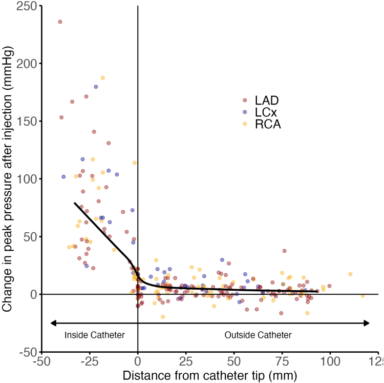
Results: A total of 269 pressure recordings (58 vessels; 52 patients) were analyzed. Injections led to a small increase in peak pressure in the distal (mean difference, +4.5 mm Hg; 95% CI, 1.5-7.4), mid (mean difference, +4.1 mm Hg; 95% CI, 1.4-6.9), and proximal (mean difference, +5.1 mm Hg; 95% CI, 2.5-7.7) vessel locations, and much higher increases at the catheter tip (mean difference, +11.7 mm Hg; 95% CI, 5.8-17.7) and inside the catheter (mean difference, +77.5 mm Hg; 95% CI, 64.5-90.4). Compared to the distal vessel, pressure changes were only significant at the catheter tip (+10 mm Hg; P < .01) and inside the catheter (+79.1 mm Hg; P < .01).

Conclusions: Contrast injections lead to negligible changes in intracoronary pressures beyond the catheter tip. Although it is sensible to minimize injections when coronary dissections are close to the catheter, it is unlikely that they would cause injuries beyond the catheter tip.

Keywords: chronic total occlusions; contrast; coronary angiography; coronary dissections; optical coherence tomography.


Figures

Figure 1
Figure 1
Pressure recording protocol. A continuous 60-second pressure recording was taken in each of the 5 predetermined locations (see text). The red tracing corresponds to the aortic pressure measured at the catheter tip (Pa). The blue tracing corresponds to the intracoronary pressure measured by the pressure wire sensor (Pd). Injections were taken after 10 seconds of resting recording and are defined by the loss of Pa waveform (orange shaded area). After injection, the recording was maintained for up to 60 seconds.
Figure 2
Figure 2
Peak pressure at resting and during injection at each coronary segment. Changes in peak pressure between resting and during injection for each analyzed recording in each vessel location. Numbers are the mean difference between resting and during injection with 95% CI.
Figure 3
Figure 3
Peak absolute pressure and pressure differences during contrast injection at each coronary segment. (A) The absolute peak pressure during injection at each coronary location. Considering the distal vessel location as a reference, significant differences in peak pressure were seen between the proximal vessel, catheter tip, and inside the catheter. (B) Differences in peak pressure from resting to during injection at each coronary location. Considering the distal vessel as a reference, significant differences in peak pressure were seen in the catheter tip and inside the catheter.
Figure 4
Figure 4
Peak pressure difference according to distance from the catheter tip. Scatterplot displaying the difference in peak pressure from resting to during injection as a function of the distance from the catheter tip. The data are fitted with a line demonstrating a smoothed moving median. LAD, left anterior descending artery; LCx, left circumflex artery; RCA, right coronary artery.
Figure 5
Figure 5
Case example demonstrating the changes in peak pressure during injection at each coronary location. Red traces are the aortic pressure (Pa) transmitted by the guiding catheter, and the yellow traces are the intracoronary pressure (Pd) transmitted by the pressure wire. The white arrow shows the point of injection. Note the significant increase of Pd during injection with loss of its normal waveform morphology when inside the catheter and the catheter tip. Note the negligible differences in the Pd during injection and at resting when the pressure sensor is in the other coronary segments.
Figure 6
Figure 6
Peak pressure at resting and during injection at each coronary segment according to the method of contrast injection. Changes in peak pressure between resting and during injection in each vessel location according to the type of injection (manual vs automatic pump injectors). Numbers are the mean difference between resting and during injection with 95% CI.
Central Illustration
Effects of contrast injections on intracoronary hemodynamics. Although contrast injections significantly increase peak pressures inside the guiding catheter, they only lead to negligible changes in pressures inside the coronary artery, beyond the guiding catheter tip.

References

    1. Hayes S.N., Kim E.S.H., Saw J., et al. Spontaneous coronary artery dissection: current state of the science: a scientific statement from the American Heart Association. Circulation. 2018;137(19):e523–e557. doi: 10.1161/CIR.0000000000000564. - DOI - PMC - PubMed
    1. Hiraide T., Sawano M., Shiraishi Y., et al. Impact of catheter-induced iatrogenic coronary artery dissection with or without postprocedural flow impairment: a report from a Japanese multicenter percutaneous coronary intervention registry. PLOS ONE. 2018;13(9) doi: 10.1371/journal.pone.0204333. - DOI - PMC - PubMed
    1. Chamié D., Bezerra H.G., Attizzani G.F., et al. Incidence, predictors, morphological characteristics, and clinical outcomes of stent edge dissections detected by optical coherence tomography. JACC Cardiovasc Interv. 2013;6(8):800–813. doi: 10.1016/j.jcin.2013.03.019. - DOI - PubMed
    1. González-Bravo D.H., Suárez-González J., Rodríguez-Vilá O. An uncommon cause of ST-segment elevation myocardial infarction: intramural coronary artery hematoma after blunt chest trauma. JACC Case Rep. 2020;2(15):2304–2309. doi: 10.1016/j.jaccas.2020.09.032. - DOI - PMC - PubMed
    1. Desai C.K., Bhatnagar U., Stys A., Jonsson O. Iatrogenic propagation of coronary dissection during diagnostic coronary angiography: an uncommon but important procedural consideration. BMJ Case Rep. 2017;2017 doi: 10.1136/bcr-2017-222463. - DOI - PMC - PubMed

LinkOut - more resources