Review
. 2025 Jan 2;14(1):14.
doi: 10.1167/tvst.14.1.14.

New Directions for Ophthalmic OCT - Handhelds, Surgery, and Robotics

Affiliations
Review

New Directions for Ophthalmic OCT - Handhelds, Surgery, and Robotics

Julia Foust et al. Transl Vis Sci Technol. .

Abstract

The introduction of optical coherence tomography (OCT) in the 1990s revolutionized diagnostic ophthalmic imaging. Initially, OCT's role was primarily in the adult ambulatory ophthalmic clinics. Subsequent advances in handheld form factors, integration into surgical microscopes, and robotic assistance have expanded OCT's utility and impact outside of its initial environment in the adult outpatient ophthalmic clinic. In this review, we cover the use of OCT in the neonatal intensive care unit (NICU) environment with a handheld OCT, recent developments in intraoperative OCT for data visualization and measurements, and recent work and demonstration of robotically aligned OCT systems outside of eye clinics. Of note, advances in these areas are a legacy of our colleague, the late Joseph Izatt. OCT has been an important innovation for ocular diagnostics, and these advances have helped it continue to extend in new directions.


Conflict of interest statement

Disclosure: J. Foust, None; M. McCloud, None; A. Narawane, None; R.M. Trout, None; X. Chen, None; A.-H. Dhalla, Theia Imaging (F, O, P), Horizon Surgical (F, P), Alcon (C), Leica (P, R); J.D. Li, Leica (E); C. Viehland, Theia Imaging (F, O, P); M. Draelos, Horizon Surgical (C); L. Vajzovic, Alcon (F, C), Novartis (C, R), Clearside Biomedical (C); R.P. McNabb, Johnson & Johnson (F), Leica (P, R); A.N. Kuo, Johnson & Johnson (F), Leica (P, R); C.A. Toth, Theia Imaging (F, O, P), Zeiss Meditec (F)

Figures

Figure 1.
Figure 1.
Multiple generations of handheld OCT probes. Our group has developed multiple generations of handheld OCT probes (references in the text). These images show one commercial (Bioptigen: left two images) and three investigational OCT and OCT angiography probes we have used for imaging infants at the bedside.
Figure 2.
Figure 2.
Research handheld OCT imaging from the BabySTEPS study NCT02887157 with a non-contact investigational handheld OCT system demonstrating the wealth of structural information in the image of the retinal response to ROP treatment. Preterm infant eyes are prior to (top row) and one week after (bottom row) anti-VEGF treatment for type 1 ROP. The cross-sectional OCT scans in (B) and € (and arrows over extraretinal neovascularization) are extracted from the site of the green line in the OCT volumes, seen in retina view in (A) and (D). The blue asterisks mark retinal locations before (A, C) and after (D, E) treatment.
Figure 3.
Figure 3.
Representative widefield handheld OCT images from an investigational swept-source handheld OCT system (Theia Imaging, Durham, NC). The investigational device used to acquire these images has not been cleared by the FDA. Images were acquired as part of a research study conducted under an abbreviated investigational device exemption and IRB-approved protocols. The red dashed box (A) and the yellow dashed box (B) in the widefield images correspond to approximate field of view of non-contact scanning (C, D, respectively). Optic nerve head is marked with red/blue asterisk and fovea with yellow. Right columns show the retinal views segmented at the RPE to highlight retinal vasculature in the top row and choroidal vasculature in the bottom row in a healthy eye (E and F, and the same as shown in the cross section) and an eye with retinal vascular disease (G, H). Red dashed circle denotes typical ROP Zone I region, the asterisk marks the fovea, and the red arrows point to the ampulla of the vortex veins.
Figure 4.
Figure 4.
Surgical 3D-OCT (top) with corresponding surgical microscopy (middle) combined to generate a single fused visualization (bottom) for enhanced guidance of surgical maneuvers. Subjects include human retinal surgery with a soft-tip cannula (column 1), finesse loop (column 2), and forceps (column 3). Column 4 is of a cornea and a partially collapsed anterior chamber (4).
Figure 5.
Figure 5.
Demonstration of quantitative measurement setup with MIOCT system used in wetlab studies (top row112) and example bleb image and segmentation in a human subject using this measurement procedure (bottom row110).
Figure 6.
Figure 6.
Robotically aligned OCT (RAOCT) system design. (A) OCT scanner mounted on a collaborative robotic arm. (B) Optical system diagram of the OCT engine used in RAOCT that includes dynamic system components for real-time beam steering. (C) Top down view of RAOCT auto-aligning to a freestanding individual. (D) Face tracking cameras detecting and segmenting the target eye. (E) View from the pupil tracking cameras. Pupil center (magenta cross), calculated center of corneal curvature (yellow cross), and gaze angle (green arrow) are projected onto the images. (F) Registered and averaged foveal b-scan of a healthy freestanding individual.
Figure 7.
Figure 7.
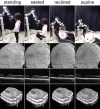
Robotically aligned OCT (RAOCT) system with scanner mounted face tracking. The original RAOCT used fixed base face tracking with a single point of view; this meant the subject's face could only be seen when directly in front of the fixed tracking cameras. By mounting the face tracking cameras to the robot and scanner, the face tracking can “see” wherever the robot and scanner can reach as seen in the various face orientations in the image. (Second row: en face OCT, third row: B-scans, and bottom row: OCT volume 3D renders.)

References

    1. Huang D, Swanson EA, Lin CP, et al. .. Optical coherence tomography. Science. 1991; 254: 1178–1181. - PMC - PubMed
    1. Swanson EA, Izatt JA, Hee MR, et al. .. In vivo retinal imaging by optical coherence tomography. Opt Lett. 1993; 18: 1864–1866. - PubMed
    1. Izatt JA, Hee MR, Swanson EA, et al. .. Micrometer-scale resolution imaging of the anterior eye in vivo with optical coherence tomography. Arch Ophthalmol. 1994; 112: 1584–1589. - PubMed
    1. Schuman JS, Hee MR, Arya AV, et al. .. Optical coherence tomography: a new tool for glaucoma diagnosis. Curr Opin Ophthalmol. 1995; 6: 89–95. - PubMed
    1. Jaffe GJ, Martin DF, Toth CA, et al. .. Macular morphology and visual acuity in the comparison of age-related macular degeneration treatments trials. Ophthalmology. 2013; 120: 1860–1870. - PMC - PubMed

MeSH terms

LinkOut - more resources