

Extensive elastofibroma dorsi with scapulo-thoracic involvement in a male laborer: A case report
- PMID: 39809056
- PMCID: PMC11782879
- DOI: 10.1016/j.ijscr.2025.110883
Extensive elastofibroma dorsi with scapulo-thoracic involvement in a male laborer: A case report
Abstract
Introduction and importance: Elastofibroma dorsi is a rare benign soft tissue lesion primarily located in the subscapular region. This distinctive lesion, with its unique radiological and histological features, poses diagnostic challenges due to its subtle presentation and overlap with other conditions.
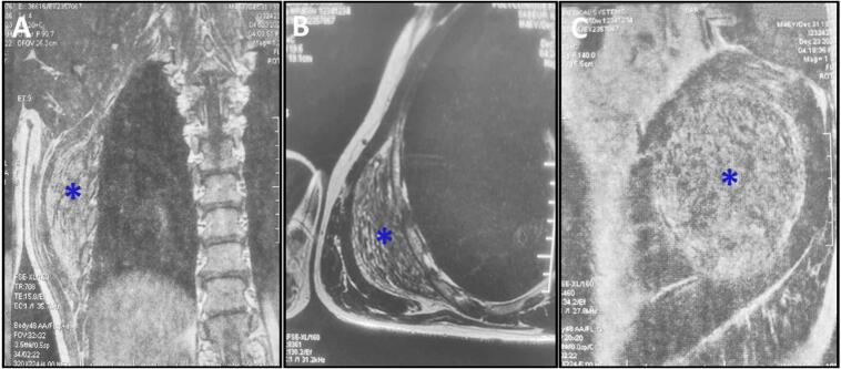
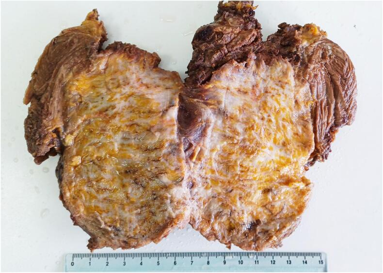
Case presentation: A 48-year-old man, manual laborer with an unremarkable medical history presented with a progressively enlarging mass below the right scapula over two years. Physical examination revealed a painless, soft, and mobile swelling near the scapular tip and along the chest wall. MRI demonstrated an elongated mass in the right posterolateral thoracic wall, characterized by smooth borders and a striped appearance on T1 and T2 sequences, consistent with elastofibroma. Surgical excision was performed, resulting in the removal of a 14 cm mass with a rubbery consistency and a gross appearance reminiscent of layered leaves. Histopathological analysis confirmed the diagnosis of elastofibroma.
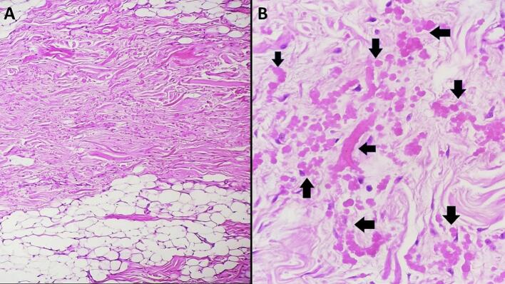
Clinical discussion: Elastofibroma, though usually asymptomatic, can present as a palpable mass in the scapular region. The characteristic striped appearance on MRI reflects alternating fibrous and fatty tissue. Histopathologically, elastofibroma is defined by abnormal elastic fibers within a fibrocollagenous matrix. Surgical excision is curative, and patients generally have an uneventful recovery with no further intervention required.
Conclusions: This case emphasizes the need to recognize elastofibroma as a differential diagnosis for palpable masses near the scapula. Awareness of its clinical, imaging, and histopathological features is essential for accurate diagnosis and effective management, often involving surgical excision with favorable outcomes.
Keywords: Benign tumor, case report; Elastofibroma; Pathology; Soft tissue mass; Surgery.
Copyright © 2025 The Authors. Published by Elsevier Ltd.. All rights reserved.
Conflict of interest statement
Declaration of competing interest None declared.
Figures

References
-
- Bartocci M., Dell’Atti C., Meacci E., Congedo M.T., Magarelli N., Bonomo L., Leone A. Clinical features, imaging findings, treatment aspects of elastofibroma dorsi and long-term outcomes after surgical resection. Eur. Rev. Med. Pharmacol. Sci. 2017;21:2061–2068. - PubMed
-
- Hoffman J.K., Klein M.H., McInerney V.K. Bilateral elastofibroma: a case report and review of the literature. Clin. Orthop. Relat. Res. 1996:245–250. - PubMed
Publication types
LinkOut - more resources
Full Text Sources
