. 2025 Jan 21;6(1):101920.
doi: 10.1016/j.xcrm.2024.101920. Epub 2025 Jan 13.

Enabling tumor-specific drug delivery by targeting the Warburg effect of cancer

Affiliations

Enabling tumor-specific drug delivery by targeting the Warburg effect of cancer

Jian Zhang et al. Cell Rep Med. .

Abstract

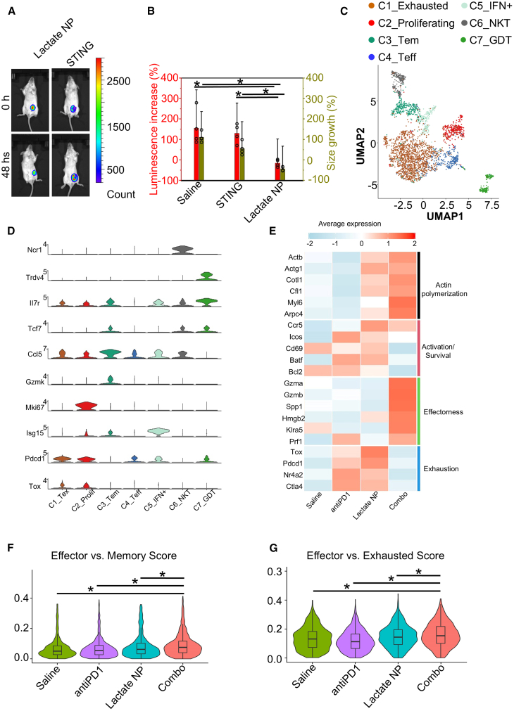
Metabolic reprogramming of tumor cells is an emerging hallmark of cancer. Among all the changes in cancer metabolism, increased glucose uptake and the accumulation of lactate under normoxic conditions (the "Warburg effect") is a common feature of cancer cells. In this study, we develop a lactate-responsive drug delivery platform by targeting the Warburg effect. We design and test a gold/mesoporous silica Janus nanoparticle system as a gated drug carrier, in which the gold particles are functionalized with lactate oxidase and the silica particles are capped with α-cyclodextrin through surface arylboronate modification. In the presence of lactate, the lactate oxidase generates hydrogen peroxide, which induces the self-immolation reaction of arylboronate, leading to uncapping and drug release. Our results demonstrate greatly improved drug delivery specificity and therapeutic efficacy with this platform for the treatment of different cancers. Our findings present an effective approach for drug delivery by metabolic targeting of tumors.

Keywords: Warburg effect; chemotherapy; drug delivery; immunotherapy; lactate; nanoparticle; tumor metabolism.


Conflict of interest statement

Declaration of interests J.Z. and X.W. are inventors for patent PCT/US2020/070052 (Lactate response system and methods). X.W. is a co-founder of Alnair Therapeutics, Inc.

Figures

None
Graphical abstract
Figure 1
Figure 1
Development of the Janus nanoparticles for lactate-inducible drug release (A) Schematic illustration of the Janus nanoparticle fabrication for tumor-specific drug delivery. CD, α-cyclodextrin; Au, gold. (B–D) Representative TEM images of the mesoporous silica (B), Au (C) nanoparticles, and the Janus nanoparticles Au/silica (D).
Figure 2
Figure 2
Lactate-responsive drug release from the Janus nanoparticles (A) Lactate can induce hydrogen peroxide-mediated uncapping and drug release of the Janus nanoparticles. After 24 h of incubation, the doxorubicin-loaded nanoparticles are presented as a stable purple colloid in phosphate-buffered saline solution (left), while the uncapped nanoparticles precipitate and doxorubicin is released to the supernatant in the presence of hydrogen peroxide (middle) or lactate (right). (B and C) Release kinetics of doxorubicin from the Janus nanoparticles at different concentrations of lactate (B) or H2O2 (C). n = 3. Data are presented as mean ± SD (standard deviation). All error bars represent SD. (D) Lactate can induce rapid release of doxorubicin from the Janus nanoparticles after 24 h incubation in PBS solution. n = 3. Data are presented as mean ± SD (standard deviation). (E) To monitor the potential uncapping and drug release in vivo, H2O2-responsive bioluminescence formulation was intraperitoneally injected to nude mice with 4T1 tumors. No luminescence signals were observed in mice without nanoparticle injection (control, left). Upon intravenous injection of the Janus nanoparticles, luminescence signals can be observed around the tumor (middle). Subcutaneous injection of lactate solution as a positive control also leads to luminescence signals around the injection site (arrow, right).
Figure 3
Figure 3
Enhanced tumor-specific drug delivery with the lactate-responsive nanocarrier (A and B) Fluorescence image (A) and quantification (B) of tumor and different organs after intravenous injection of saline (control) or doxorubicin with or without lactate-responsive nanoparticles (lactate NPs). n = 3. Data are presented as mean ± SD (standard deviation). All error bars represent SD. ∗: p < 0.05 (Student’s t test). (C–E) Quantification of doxorubicin distribution in tumor (C), plasma (D), or heart (E) after intravenous injection of free doxorubicin or doxorubicin within lactate-responsive nanoparticles or pH-responsive nanoparticles (pH NPs). The concentration of doxorubicin was evaluated by liquid chromatography/mass spectrometry. n = 3. Data are presented as mean ± SD (standard deviation). All error bars represent SD. ∗: p < 0.05 (Student’s t test).
Figure 4
Figure 4
Lactate-responsive drug carriers enhance the therapeutic efficacy of chemotherapy in vivo (A) Change in tumor size after delivery of free doxorubicin or doxorubicin within lactate NPs or pH NPs. Injection of saline serves as a control. n = 4. Data are presented as mean ± SD. All error bars represent SD. ∗ represents p < 0.05 (Student’s t test). (B) 48 h after the treatment, delivery of the drug via the lactate-responsive nanoparticles, but not free doxorubicin or delivery with pH-responsive particles, leads to a marked reduction in tumor bioluminescence. (C) Kaplan-Meier survival curve of BALB/c mice with 4T1 tumors upon different treatment as indicated. n = 8. (D) Apoptosis and proliferation of tumor cells were determined by immunohistochemistry with different antibody as indicated. (E) The bioluminescence images of Ewing’s sarcoma upon different treatments as indicated. (F) Quantification of tumor size of Ewing’s sarcoma with different treatment. n = 6 independent samples. Data are presented as mean ± SD. All error bars represent SD. (G) Kaplan-Meier survival curve of mice with Ewing’s sarcoma upon different treatments as indicated. n = 6.
Figure 5
Figure 5
Lactate-responsive drug carriers increase the therapeutic efficacy for metastatic tumors (A) The bioluminescence images of lung metastasis (4T1 model) upon different treatments as indicated. (B) Lung images and hematoxylin and eosin (H&E) staining of the tissue 8 days after different treatments as indicated. (C) Quantification of lung metastasis with different treatments (percentage of tumor area in the lung from histology analysis). n = 5. Data are presented as mean ± SD. All error bars represent SD. ∗: p < 0.05 (Student’s t test). (D) Metastatic growth in the lung was monitored by bioluminescence imaging. n = 5. Data are presented as mean ± SD. All error bars represent SD. (E) Kaplan-Meier survival curve of mice with 4T1 lung metastasis model upon different treatments as indicated. n = 5.
Figure 6
Figure 6
Delivery of immunotherapeutic agent with the lactate-responsive nanocarriers (A) Monitoring of growth of 4T1 tumors by bioluminescence imaging with different treatments as indicated. (B) Changes in bioluminescence signal and tumor size upon different treatments as indicated. n = 4. Data are presented as mean ± SD. All error bars represent SD. ∗: p < 0.05 (Student’s t test). (C) Uniform manifold approximation and projection (UMAP) embeddings of 2,931 total CD8+ T cells across four treatment conditions, colored by functional subset. Tex, T exhausted; Prolif, proliferating; Tem, T effector memory; Teff, T effector; NKT, natural killer T cell; GDT, γδ-T cell. (D) Violin plots of normalized gene expression of subset-specific markers. (E) Heatmap comparing functional gene expression across conditions in CD8+ T cells. Legend color denotes Z-scored expression. (F and G) Violin plots comparing effector gene module scores with memory gene module scores (F), and exhausted gene module scores (G) of different treatments in CD8+ T cells.
Figure 7
Figure 7
Delivery of STING agonist with the lactate-responsive nanocarrier can enhance the efficacy of immunotherapy for breast cancer (A and B) Dotplot comparing functional gene expression across conditions in proliferating (A) and exhausted (B) CD8+ T cells. (C and D) The tumor microenvironment in the combo treatment group presented significantly accumulated Gzmb+ cells and the lowest TOX level. Quantitation of the positive expression of the tumor-infiltrating granzyme B+ (C) and TOX+ (D) cells in different treatment groups. n = 4. Data are presented as mean ± SD. All error bars represent SD. ∗: p < 0.05 (Student’s t test). (E) Kaplan-Meier survival curve of mice with 4T1 lung metastasis model upon different treatments as indicated. n = 8. (F) Tumor growth was monitored by bioluminescence imaging upon different treatments as indicated. n = 8. Data are presented as mean ± SD. All error bars represent SD.

Similar articles

Cited by

References

    1. Mura S., Nicolas J., Couvreur P. Stimuli-responsive nanocarriers for drug delivery. Nat. Mater. 2013;12:991–1003. - PubMed
    1. Lu Y., Sun W., Gu Z. Stimuli-responsive nanomaterials for therapeutic protein delivery. J. Contr. Release. 2014;194:1–19. - PMC - PubMed
    1. Manzari M.T., Shamay Y., Kiguchi H., Rosen N., Scaltriti M., Heller D.A. Targeted drug delivery strategies for precision medicines. Nat. Rev. Mater. 2021;6:351–370. - PMC - PubMed
    1. Matsumoto Y., Nichols J.W., Toh K., Nomoto T., Cabral H., Miura Y., Christie R.J., Yamada N., Ogura T., Kano M.R., et al. Vascular bursts enhance permeability of tumour blood vessels and improve nanoparticle delivery. Nat. Nanotechnol. 2016;11:533–538. - PubMed
    1. Bertrand N., Wu J., Xu X., Kamaly N., Farokhzad O.C. Cancer nanotechnology: the impact of passive and active targeting in the era of modern cancer biology. Adv. Drug Deliv. Rev. 2014;66:2–25. - PMC - PubMed