

Ultrasound guided Ru106 plaque brachytherapy for treatment of exudative retinal detachment in children with diffuse choroidal haemangioma
- PMID: 39809880
- PMCID: PMC11794627
- DOI: 10.1038/s41433-024-03562-8
Ultrasound guided Ru106 plaque brachytherapy for treatment of exudative retinal detachment in children with diffuse choroidal haemangioma
Abstract
Purpose: To evaluate the efficacy of ultrasound-guided ruthenium (Ru 106) plaque brachytherapy for treatment of exudative retinal detachment in diffuse choroidal haemangioma (DCH).
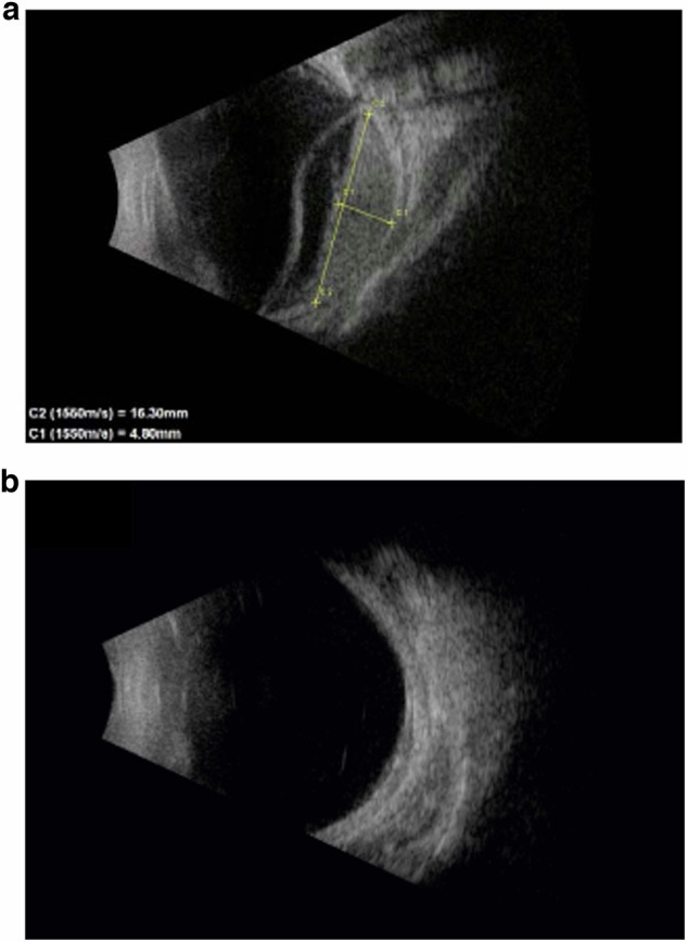
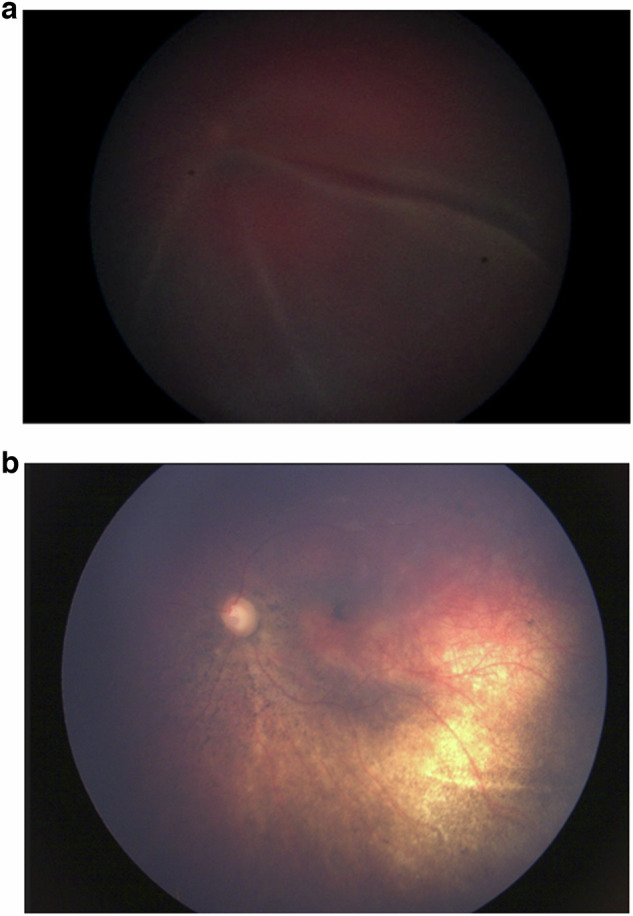
Methods: Retrospective analysis of four paediatric patients treated with ultrasound-guided Ru 106 plaque brachytherapy for DCH with total exudative retinal detachment directed to the thickest part of the DCH. A dose of 40 Gy to the tumour apex was delivered in all patients. The outcomes of treatment were regression of DCH, resolution of retinal detachment, development of neovascular glaucoma or any other radiation-associated complications which were assessed clinically and with B scan ultrasonography.
Results: There were 4 eyes included in the study, with a mean (median, range) age of 8.75 (8.4, 3-15) years. The pre-operative tumour thickness was 5.0 (5.12, 4.2-5.5) mm. The visual acuity ranged from 0.8-2.8 LogMAR and 3 of 4 eyes had only light perception at presentation. One eye had been treated with goniotomy for pre-existing secondary glaucoma and was on topical antihypertensive medications. At a mean follow-up of 14.6 months (10.5 months, 6-30 months), all patients showed regression of the tumour. The mean tumour thickness reduced to 2.05 mm (2.44 mm, 1.1-2.6 mm) post-operatively. All patients (4/4) had complete resolution of the retinal detachment. The visual acuity remained stable in all the patients with none of the patients developing neovascular glaucoma or any other radiation-related complications.
Conclusion: Ultrasound-guided Ru 106 plaque brachytherapy is an effective treatment strategy as a primary treatment in the absence of external beam radiotherapy, to achieve tumour regression and resolution of retinal detachment in DCH.
© 2025. The Author(s).
Conflict of interest statement
Competing interests: None of the other authors have any financial disclosures or conflicts of interest to declare. This manuscript has not previously been submitted for publication. The research was supported by the National Institute for Health Research (NIHR) Biomedical Research Centre based at Moorfields Eye Hospital NHS Foundation Trust and UCL Institute of Ophthalmology. The views expressed are those of the author(s) and not necessarily those of the NHS, the NIHR or the Department of Health. This paper presents a novel method of imaging with brachytherapy, in an area of ophthalmology that is both rare and important. The material here is original research, not previously published.
Figures

References
-
- Shields JA, Shields CL. Diffuse choroidal haemangioma. In Shields JA, Shields CL, eds. Intraocular Tumors: An Atlas and Textbook. 3rd ed. pp. 264–9. Philadelphia, PA: Lippincott Williams & Wilkins; 2016.
-
- Singh AD, Rundle PA, Vardy SJ, Rennie IG. Photodynamic therapy of choroidal haemangioma associated with Sturge-Weber syndrome. Eye. 2005;19:365–7. - PubMed
-
- Anand R. Photodynamic therapy for diffuse choroidal haemangioma associated with Sturge Weber syndrome. Am J Ophthalmol. 2003;136:758–60. - PubMed
-
- Thapa R, Shields CL. Oral propranolol therapy for management of exudative retinal detachment from diffuse choroidal haemangioma in Sturge-Weber syndrome. Eur J Ophthalmol. 2013;23:922–4. - PubMed
-
- Arevalo JF, Arias JD, Serrano MA. Oral propranolol for exudative retinal detachment in diffuse choroidal haemangioma. Arch Ophthalmol. 2011;129:1373–5. - PubMed
MeSH terms
Substances
LinkOut - more resources
Full Text Sources
Medical
Miscellaneous
