. 2025 Mar;242(3):563-578.
doi: 10.1007/s00213-024-06706-6. Epub 2025 Jan 15.

Captopril attenuates oxidative stress and neuroinflammation implicated in cisplatin-induced cognitive deficits in rats

Affiliations

Captopril attenuates oxidative stress and neuroinflammation implicated in cisplatin-induced cognitive deficits in rats

Fatma Mostafa et al. Psychopharmacology (Berl). 2025 Mar.

Abstract

Rationale: One of the most debilitating drawbacks of cisplatin chemotherapy is neurotoxicity which elicits memory impairment and cognitive dysfunction (chemobrain). This is primarily triggered by oxidative stress and inflammation. Captopril, an angiotensin-converting enzyme inhibitor, has been reported as a neuroprotective agent owing to its antioxidant and anti-inflammatory effects.

Objective: We examined the possible neuroprotective effect of captopril against cisplatin-induced neurological and behavioral abnormalities in rats.

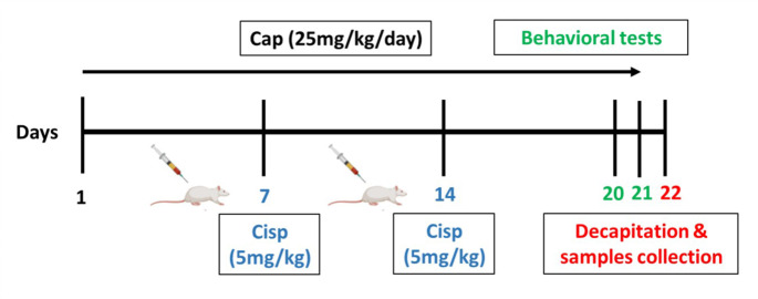
Methods: Chemobrain was induced in rats by cisplatin (5 mg/kg, i.p.) on the 7th and 14th days of the study while captopril was administered orally (25 mg/kg) daily for three weeks. The effects of captopril were assessed by performing behavioral tests, histological examination, and evaluation of oxidative stress and inflammatory markers.

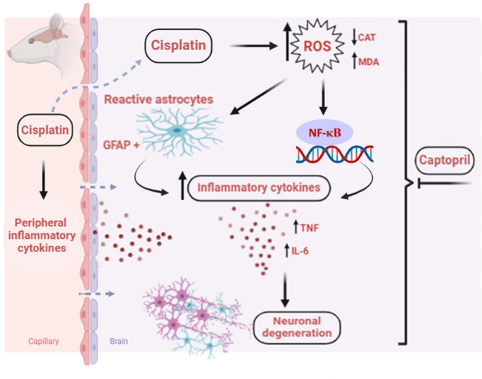
Results: Cisplatin caused learning/memory dysfunction assessed by passive avoidance and Y-maze tests, decline in locomotion, and rotarod motor balance loss which were further verified by neurodegeneration observed in histological examination. Also, cisplatin aggravated oxidative stress by elevating lipid peroxidation (MDA) levels and diminishing catalase activity. Moreover, cisplatin upregulated the neuroinflammatory markers (TNF, IL-6, GFAP, and NF-κB). Captopril successfully ameliorated cisplatin damage on the levels of neurobehavioral and histopathological changes. Mechanistically, captopril significantly diminished MDA production and preserved catalase antioxidant activity. Captopril also counteracted neuroinflammation through inhibiting NF-κB and its downstream proinflammatory cytokines besides repressing astrocyte activity by reducing GFAP expression.

Conclusion: Our findings revealed that captopril could abrogate cisplatin neurotoxicity via reducing oxidative stress and neuroinflammation thus enhancing cognitive and behavioral performance. This could suggest the repurposing of captopril as a neuroprotective agent, especially in hypertensive cancer patients receiving cisplatin.

Keywords: Captopril; Chemobrain; Cisplatin; Neuroinflammation; Oxidative stress.


Conflict of interest statement

Declarations. Ethics approval: The study procedures were conducted and accepted by the Institutional Animal Research Ethics Committee of the Faculty of Pharmacy, Ain Shams University, Cairo, Egypt (No #258). Conflict of interest: The authors proclaim that they have no known contending monetary interests or individual relationships that could have seemed to impact the work stated in this study.

Figures

Fig. 1
Fig. 1
Study timeline in terms of days of administration of Cisp and Cap; Cisp, Cisplatin; Cap, Captopril
Fig. 2
Fig. 2
Impact of captopril treatment on cisplatin-induced behavioral changes. (A) Y maze spontaneous alternation percent (SAP). Data is presented as mean ± SD (n = 10). * or #: Statistically significant from control or cisplatin group, correspondingly such that */# at P < 0.05, **/## at P < 0.01 and ***/### at P < 0.001 by means of one-way ANOVA then Tukey–Kramer post hoc test. (B) Step-through passive avoidance training session. (C) Step-through passive avoidance test session. Kruskal– Wallis test was utilized for data analysis followed by Dunn’s post hoc test represented as medians and interquartile range (n = 10). (D) Locomotor activity. Data is presented as mean ± SD (n = 10). * or #: Statistically significant from control or cisplatin group, correspondingly such that */# at P < 0.05, **/## at P < 0.01 and ***/### at P < 0.001utilizing one-way ANOVA then Tukey–Kramer post hoc test. (E) Rotarod test latency to fall. Data is presented as mean ± SD (n = 10). * or #: Statistically significant from control or cisplatin group, correspondingly such that */# at P < 0.05, **/## at P < 0.01 and ***/### at P < 0.001 via one-way ANOVA then Tukey–Kramer post hoc test
Fig. 3
Fig. 3
Impact of captopril treatment (25 mg/kg/day, p.o., for 21 days) on cisplatin-induced neurodegenerative alterations in the hippocampus and cortex using H and E staining (X40) (scale bar 25 μm, scale: 0.208 μm/ pixel). Cisplatin triggered nuclear pyknosis and deterioration in the neuronal cells of the hippocampus and the cortex. Alternatively, co-treatment with captopril (25 mg/kg) normalized the hippocampal and cortical histological architecture
Fig. 4
Fig. 4
Impact of captopril on cisplatin-induced oxidative stress in the hippocampus and cortex. MDA levels in hippocampus (A) and cortex (B). Catalase activity in hippocampus (C) and cortex (D). Data is illustrated as mean ± SD (n = 6). * or #: Statistically significant from control or cisplatin group, correspondingly such that */# at P < 0.05, **/## at P < 0.01 and ***/### at P < 0.001 applying one-way ANOVA then Tukey–Kramer post hoc test
Fig. 5
Fig. 5
Impact of captopril on GFAP-immunostaining in the hippocampus and cortex. (A) Representative photomicrographs of GFAP-immunostained sections of hippocampus and cortex (X20) (scale bar 25 μm, scale: 0.444 μm/pixel), indicating marked brown staining in the cisplatin group. (B) Bar chart representation for the optical density in the different groups. Data is indicated as mean ± SD (n = 3). * or #: Statistically significant from control or cisplatin group, correspondingly such that */# at P < 0.05, **/## at P < 0.01 and ***/### at P < 0.001 via one-way ANOVA then Tukey–Kramer post hoc test
Fig. 6
Fig. 6
Impact of captopril on NF-κB-immunostaining in the hippocampus and cortex. (A) Representative photomicrographs of NF-κB -immunostained sections of the hippocampus and cortex (X20) (scale bar 25 μm, scale: 0.444 μm/pixel), indicating remarkable brown staining in the cisplatin group. (B) Bar chart representation for the optical density of NF-κB staining in the various groups. Data is indicated as mean ± SD (n = 3). * or #: Statistically significant from control or cisplatin group, correspondingly such that */# at P < 0.05, **/## at P < 0.01 and ***/### at P < 0.001 by means of one-way ANOVA then Tukey–Kramer post hoc test
Fig. 7
Fig. 7
Impact of captopril on cisplatin-mediated alterations in TNF and IL-6 levels. TNF levels in the hippocampus (A) and cortex (B). IL-6 levels in the hippocampus (C) and cortex (D). Data is illustrated as mean ± SD (n = 6). * or #: Statistically significant from control or cisplatin group, correspondingly such that */# at P < 0.05, **/## at P < 0.01 and ***/### at P < 0.001 by means of one-way ANOVA then Tukey–Kramer post hoc test
Fig. 8
Fig. 8
A schematic diagram demonstrating the potential mechanisms for captopril’s protective effect versus cisplatin-induced chemobrain in rats. This figure was designed by means of Biorender (publication agreement number: DQ277ZOIT1)

References

    1. Abareshi A, Hosseini M, Beheshti F et al (2016) The effects of captopril on lipopolysaccharide induced learning and memory impairments and the brain cytokine levels and oxidative damage in rats. Life Sci 167:46–56. 10.1016/J.LFS.2016.10.026 - PubMed
    1. Abareshi A, Anaeigoudari A, Norouzi F et al (2019) The effects of captopril on lipopolysaccharide-induced sickness behaviors in rats. Veterinary Res Forum 10:199–205. 10.30466/VRF.2018.90760.2198 - PMC - PubMed
    1. Abdel-Aziz AK, Mantawy EM, Said RS, Helwa R (2016) The tyrosine kinase inhibitor, sunitinib malate, induces cognitive impairment in vivo via dysregulating VEGFR signaling, apoptotic and autophagic machineries. Exp Neurol 283:129–141. 10.1016/J.EXPNEUROL.2016.06.004 - PubMed
    1. Abdel-latif RG, Heeba GH, Taye A, Khalifa MMA (2018) Lixisenatide, a novel GLP-1 analog, protects against cerebral ischemia/reperfusion injury in diabetic rats. Naunyn-Schmiedeberg’s Archives Pharmacol 391:705–717. 10.1007/S00210-018-1497-1 - PubMed
    1. Abdel-Wahab WM, Moussa FI (2019) Neuroprotective effect of N-acetylcysteine against cisplatin-induced toxicity in rat brain by modulation of oxidative stress and inflammation. Drug Des Devel Ther 13:1155–1162. 10.2147/DDDT.S191240 - PMC - PubMed

MeSH terms

LinkOut - more resources