. 2025 Apr;31(4):1319-1328.
doi: 10.1038/s41591-024-03466-w. Epub 2025 Jan 14.

In vivo base editing extends lifespan of a humanized mouse model of prion disease

Affiliations

In vivo base editing extends lifespan of a humanized mouse model of prion disease

Meirui An et al. Nat Med. 2025 Apr.

Erratum in

  • Author Correction: In vivo base editing extends lifespan of a humanized mouse model of prion disease.
    An M, Davis JR, Levy JM, Serack FE, Harvey JW, Brauer PP, Pirtle CP, Berríos KN, Newby GA, Yeh WH, Kamath N, Mortberg M, Lian Y, Howard M, DeSouza-Lenz K, Guzman K, Thai A, Graffam S, Laversenne V, Coffey AA, Frei J, Pierce SE, Safar JG, Deverman BE, Minikel EV, Vallabh SM, Liu DR. An M, et al. Nat Med. 2025 Apr;31(4):1369. doi: 10.1038/s41591-025-03540-x. Nat Med. 2025. PMID: 39885360 Free PMC article. No abstract available.

Abstract

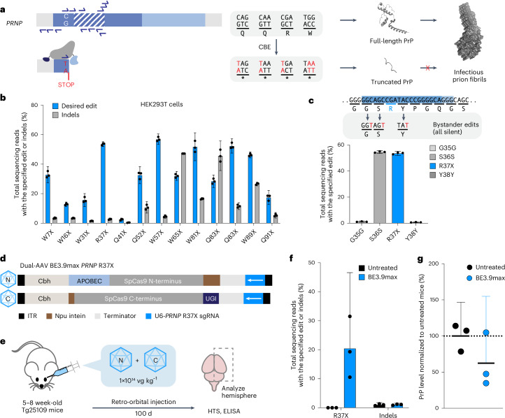
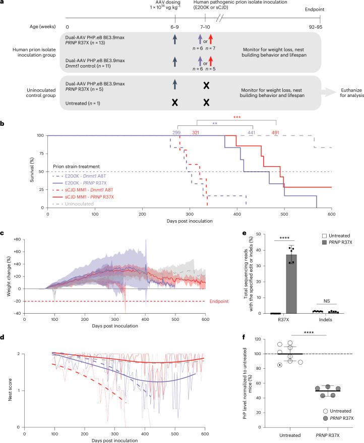
Prion disease is a fatal neurodegenerative disease caused by the misfolding of prion protein (PrP) encoded by the PRNP gene. While there is currently no cure for the disease, depleting PrP in the brain is an established strategy to prevent or stall templated misfolding of PrP. Here we developed in vivo cytosine and adenine base strategies delivered by adeno-associated viruses to permanently modify the PRNP locus to achieve PrP knockdown in the mouse brain. Systemic injection of dual-adeno-associated virus PHP.eB encoding BE3.9max and single guide RNA installing PRNP R37X resulted in 37% average installation of the desired edit, 50% reduction of PrP in the mouse brain and 52% extension of lifespan in transgenic human PRNP mice inoculated with pathogenic human prion isolates representing the most common sporadic and genetic subtypes of prion disease. We further engineered base editing systems to achieve improved in vivo potency and reduced base editor expression in nontargeting tissues, resulting in 63% average PrP reduction in the mouse brain from a 6.7-fold lower viral dose, with no detected off-target editing of anticipated clinical significance observed in either human cells or mouse tissues. These findings support the potential of in vivo base editing as one-time treatment for prion disease.


Conflict of interest statement

Competing interests: M.A., J.R.D., E.V.M., S.M.V. and D.R.L. are inventors on United States patent applications 63/700,235 and 63/718,534 relating to base editing for prion disease. D.R.L. is a consultant and/or equity owner of Prime Medicine, Beam Therapeutics, Pairwise Plants, Exo Therapeutics, Nvelop Therapeutics and Chroma Medicine, some of which are companies that use or deliver genome editing or epigenome-modulating agents. J.R.D., J.M.L. and W.-H.Y. are current employees of Prime Medicine. S.M.V. acknowledges speaking fees from Abbvie, Biogen, Eli Lilly, Illumina and Ultragenyx; consulting fees from Alnylam and Invitae; and research support from Eli Lilly, Gate Bio, Ionis and Sangamo. E.V.M. acknowledges speaking fees from Abbvie, Eli Lilly and Vertex; consulting fees from Alnylam and Deerfield; and research support from Eli Lilly, Gate Bio, Ionis and Sangamo. B.E.D. declares outside interest in Apertura Gene Therapy and Tevard Biosciences and is an inventor on US patent application US11499165B2 relating to the PHP.eB AAV capsid. All other authors declare no competing interests.

Figures

Fig. 1
Fig. 1. Development of initial base editing strategies to install stop codon in PRNP locus.
a, Schematic of the CBE-mediated stop codon installation as a strategy to knockdown cellular PrP. The PRNP locus consists of N-terminal (dark blue, amino acids 1–144) and C-terminal (light blue, amino acids 145–253) domains. The signal peptide (gray, amino acids 1–22), octapeptide repeat (OPR) region (dashed box, amino acids 51–90) and GPI signal (gray, amino acids 231–253) are highlighted. CBE may convert CAG (Gln), CAA (Gln), CGA (Arg) or TGG (Trp) codons to a stop codon. sgRNA spacers that install stop codons in PRNP evaluated in this study are shown as half-arrows. Truncated PrP no longer templates fibril formation. b, Frequency of the desired stop codon installation or indel formation from candidate sgRNAs using BE4max via plasmid transfection of HEK293T cells. c, Editing efficiency at bystander positions with BE4max and PRNP R37X sgRNA via plasmid transfection of HEK293T cells. Three silent mutations (G35G, S36S and Y38Y) are possible due to bystander editing from cytosine base editing. d, Schematic of dual-AAV PHP.eB BE3.9max with PRNP R37X sgRNA. The N-terminal AAV encodes a Cbh promoter, APOBEC deaminase domain and amino acids 1–572 of SpCas9 fused to NpuN intein. The C-terminal AAV encodes a Cbh promoter, NpuC intein, amino acids 573–1367 of SpCas9 and one copy of the UGI domain. Both AAVs contain a U6 Pol III cassette expressing the PRNP R37-targeting sgRNA. e, Experimental design for initial assessment of the effect of PRNP base editing on PrP levels. The 5–8-week-old Tg25109 mice were treated retro-orbitally with dual-AAV PHP.eB BE3.9max for installation of PRNP R37X at a dose of 1 × 1014 vg kg−1. Brain was harvested 100 d post injection to assess editing efficiency via HTS and PrP protein reduction via ELISA. f, Frequency of R37X installation in untreated (n = 3) and dual-AAV PHP.eB BE3.9max-treated mice (n = 3). g, PrP levels in dual-AAV PHP.eB BE3.9max-treated mice (n = 3) in the bulk brain hemisphere normalized to those of untreated mice (n = 3). Dots represent individual biological replicates (n = 3) and data are presented as mean ± 95% CI.
Fig. 2
Fig. 2. In vivo base editing provides protection from pathogenic human prion challenge.
a, Design of the human pathogenic prion challenge study. Tg25109 mice were divided into two cohorts: a human prion isolate inoculation group and an uninoculated control group. Among the human prion isolate inoculation group, n = 13 received dual-AAV PHP.eB BE3.9max PRNP R37X treatment and n = 11 received dual-AAV PHP.eB BE3.9max Dnmt1 control treatment. Among the uninoculated control group, n = 5 received dual-AAV PHP.eB BE3.9max PRNP R37X treatment and n = 1 remained untreated. Mice were treated with AAV at total dose of 1 × 1014 vg kg−1 at age 6–9 weeks. At 1 week after AAV treatment, mice were inoculated with either E200K or sCJD prion isolates. After prion inoculation, mice were monitored for weight loss, nest-building behavior and lifespan. Study endpoint was 600 d post prion isolate inoculation (92–95 weeks of age). The uninoculated control group was euthanized to harvest brain hemispheres for analysis via HTS and PrP ELISA. b, Kaplan–Meier curve of Tg25109 mice inoculated with either the E200K (purple) or sCJD MM1 pathogenic human prion isolate (red). Median survival from each treatment condition is marked (P = 4 × 10−4 for sCJD-inoculated cohort; P = 0.01 for E200K cohort; P = 2 × 10−6 combined). c,d, Body weight (c) (lines represent mean and shaded areas represent 95% CI) for all timepoints with ≥2 animals surviving, and nest-building score (d) (fitted to the locally estimated scatterplot smoothing (LOESS) model) of Tg25109 mice in the human prion challenge study. e,f, Frequency of the desired R37X edit (e) (P < 0.0001) and indels, and PrP protein level (f) (P < 0.0001) in the bulk brain hemisphere of mice from the uninoculated control group treated with dual-AAV PHP.eB BE3.9max with PRNP R37X sgRNA (n = 5), and in untreated mice from the uninoculated control group (n = 1, marked as a white circle with a black dot) or from additional untreated adult Tg25109 mice (n = 7, marked as white circles). Dots represent individual biological replicates and data are presented as mean ± 95% CI. Significance was calculated by two-tailed Student’s t-test; **P ≤ 0.01; ***P ≤ 0.001; ****P ≤ 0.0001. NS, not significant.
Fig. 3
Fig. 3. Optimization of CBE strategy for improved potency.
a, Frequency of R37X installation and bystander editing in HEK293T cells transfected with the specified CBEs and PRNP R37X sgRNA. b, Frequency of R37X installation in HEK293T cells transfected with TadCBEd and PRNP R37X sgRNAs with the specified scaffold modifications: canonical (sgRNA), U-to-A flip (F-sgRNA) or U-to-A flip+5-bp stem extension (F+E-sgRNA). c, Schematic of dual-AAV PHP.eB BE3.9max or TadCBEd with PRNP R37X sgRNA. The 5–8-week-old Tg25109 mice were treated retro-orbitally with AAVs at 1.5 × 1013 vg kg−1. d,e, Frequency of R37X installation (d), and PrP level in bulk brain hemisphere (e) of Tg25109 mice untreated (n = 12) or treated with dual-AAV PHP.eB packaging BE3.9max PRNP R37X sgRNA (n = 8), TadCBEd PRNP R37X sgRNA (n = 8) or TadCBEd PRNP R37X F+E-sgRNA (n = 12) at a total dose of 1.5 × 1013 vg kg−1 and harvested 35 d post injection. In d, untreated versus BE3.9max, P = 0.78; BE3.9max versus TadCBEd, P = 0.0012; TadCBEd versus TadCBEd with F+E-sgRNA, P = 0.030; BE3.9max versus TadCBEd with F+E-sgRNA, P < 0.0001. In e, untreated versus BE3.9max, P > 0.99; BE3.9max versus TadCBEd, P = 0.0031; TadCBEd versus TadCBEd with F+E-sgRNA, P = 0.045; BE3.9max versus TadCBEd with F+E-sgRNA, P < 0.0001. f, Frequency of R37X installation in Tg25109 mice 35 d (n = 8) versus 100 d (n = 6) post treatment with BE3.9max (P = 0.0042) or TadCBEd (P = 0.038). g, Scatter plot showing the relationship between R37X editing frequencies and PrP protein levels in brain tissues of Tg25109 mice treated with BE3.9max or TadCBEd, harvested at 35 d (n = 8) or 100 d (n = 6). Linear regression is shown with solid line and shaded area represents 95% CI. Dots represent individual biological replicates (n = 3 unless noted otherwise) and data are presented as mean ± 95% CI. Significance in d and e was calculated by two-way analysis of variance (ANOVA) test with Bonferroni correction; NS, P > 0.12; *P ≤ 0.033; **P ≤ 0.002; ****P ≤ 0.0001. Significance in f was calculated by two-tailed Student’s t-test; NS, P > 0.05; *P ≤ 0.05; **P ≤ 0.01. kb, kilobases.
Fig. 4
Fig. 4. Off-target analysis of R37X base editing strategy in human and mouse genome.
a, The percentage of C•G-to-T•A substitution in BE-AAV-treated samples above background (untreated samples) at 299 CIRCLE-seq nominated off-target sites in the human genome (GRCh37). Genomic DNA was extracted from HEK293T cells untreated (n = 3) or after 3 d following transfection of plasmids encoding TadCBEd and the PRNP R37X sgRNA (n = 3). Each dot represents mean of three biological replicates. b,c, The percentage of C•G-to-T•A substitution in BE-AAV-treated samples above background (untreated samples) at the top 100 CIRCLE-seq-nominated off-target sites in the mouse genome (GRCm38). Genomic DNA was extracted from the bulk brain hemisphere of Tg25109 mice untreated (n = 6), or 35 d (b) and 100 d (c) after treatment with dual-AAV PHP.eB BE3.9max with PRNP R37X sgRNA (n = 6), or dual-AAV PHP.eB TadCBEd with PRNP R37X sgRNA (n = 6) at a total dose of 1.5 × 1013 vg kg−1. Each dot represents mean of six biological replicates. d, The percentage of C•G-to-T•A substitution in BE-AAV-treated samples above background at the top 100 CIRCLE-seq-nominated off-target sites in the mouse genome (GRCm38). Genomic DNA was extracted from the bulk brain hemispheres of Tg25109 mice untreated (n = 5), or 600 d after treatment with dual-AAV PHP.eB BE3.9max with PRNP R37X sgRNA (n = 5) at a total dose of 1 × 1014 vg kg−1. Each dot represents mean of five biological replicates. In all panels, significance was calculated by one-tailed Student’s t-test. Off-target editing with P > 0.01 compared with untreated control is labeled with hollow circles, and those with P ≤ 0.01 are labeled with solid circles. Plots showing individual data points and error bars are provided in Supplementary Figs. 1–4.
Fig. 5
Fig. 5. Engineering tissue-specific expression of dual-AAV TadCBEd.
a,b, Frequency of the desired R37X edit (a), and PrP protein level in the bulk brain hemisphere (b) of Tg25109 mice treated with dual-AAV PHP.eB TadCBEd PRNP R37X F+E-sgRNA, with Cbh (n = 12), hSYN (n = 11) or EFS (n = 6) promoter driving the expression of the base editor. Data for the ‘Cbh’ condition correspond to the ‘TadCBEd+PRNP R37X F+E-sgRNA’ condition in Fig. 3d,e, and are replotted for comparison. c, Frequency of the desired R37X edit in the liver of mice untreated (n = 6) or treated with dual-AAV TadCBEd encoding Cbh promoter (Cbh; n = 6), hSYN promoter (hSYN; n = 6), hSYN promoter plus miR-183 target sites (hSYN+miR-183; n = 6), hSYN promoter plus miR-122 target sites (hSYN+miR-122; n = 5) or hSYN promoter plus miR-183 and miR-122 target sites (hSYN+miR-183+miR-122; n = 5). d, Schematic of dual-AAV PHP.eB TadCBEd PRNP R37X F+E-sgRNA with hSYN promoter driving the expression of the base editor and miR target site (TS) incorporation. ‘miR-183’ contains 4 copies of miR-183 target sites; ‘miR-122’ contains 3 copies of miR-122 target sites; ‘miR-183+miR-122’ contains 3 copies each of miR-183 and miR-122 target sites. e,f, Frequency of the desired R37X edit (e) (P < 0.0001 for all groups versus untreated), and PrP protein level (f) in the bulk brain hemisphere of Tg25109 mice harvested 35 d after treatment with dual-AAV TadCBEd, with or without the specified miR target site incorporation (untreated versus hSYN, P = 0.0003; untreated versus hSYN+miR-183, P = 0.0001; hSYN+miR-122, P = 0.0002; untreated versus hSYN+miR-183+miR-122, P < 0.0001) at a total dose of 1.5 × 1013 vg kg−1 (untreated, n = 6; hSYN, n = 11; hSYN+miR-183, n = 6; hSYN+miR-122, n = 5; hSYN+miR-183+miR-122, n = 5). Data for the ‘hSYN’ condition correspond to the ‘hSYN’ condition in a and b, and are replotted for comparison. Dots represent individual biological replicates and data are presented as mean ± 95% CI. Significance in a and b was calculated by one-way ANOVA test with Bonferroni correction; NS, P > 0.12. Significance in e and f was calculated by two-way ANOVA test with Dunnett’s correction; ***P < 0.0002; ****P < 0.0001.
Extended Data Fig. 1
Extended Data Fig. 1. In vitro validation of PrP reduction with CBE-mediated PRNP R37X installation.
a, Representative flow cytometry gating plot showing the fluorescence signal in HEK293T cells treated with BE4max, Cas9 nuclease or dead (deaminase-inactive) BE4max editor, with either PRNP R37X sgRNA (blue) or non-targeting sgRNA (red). Six days after plasmid transfection, cells were incubated with a fluorescently conjugated 6D11 antibody that binds PrP and were analyzed by flow cytometry. b, Mean fluorescence intensity (MFI) of the HEK293T cells after staining with 6D11 antibodies. MFI are plotted after normalizing the MFI of cells treated with PRNP R37X sgRNAs to MFI of cells treated with non-targeting sgRNAs. c, Frequency of the PRNP R37X edit and indels in HEK293T cells after plasmid transfection with BE4max, Cas9 nuclease or dead BE4max, with PRNP R37-targeting sgRNA. Dots represent individual biological replicates (n=3) and data are presented as mean values +/- 95% CI.
Extended Data Fig. 2
Extended Data Fig. 2. Optimization of in vivo administration route.
a, Experimental design for assessment of in vivo administration route. 4-week-old Tg66 mice were treated with dual-AAV PHP.eB BE3.9max for installation of PRNP R37X by systemic administration through retro-orbital injection or direct CNS administration through intracerebroventricular (ICV) stereotaxic injection. Mice treated with retro-orbital injection received a total of 4x10¹² vg/kg AAV (2x10¹² vg/kg each for N- and C-terminal AAV) (n=2). Mice treated with ICV injection received a total of 5.5x10¹¹ vg/kg AAV (2.5x10¹¹ vg/kg each for N- and C-terminal AAV and 5.0x10¹⁰ vg/kg for AAV encoding EGFP fused to nuclear membrane-localized Klarsicht/ANC-1/Syne-1 homology (KASH) domain (EGFP:KASH)) (n=2). Mouse brains were harvested five weeks after treatment. GFP-positive nuclei were sorted from ICV-injected samples via FACS to enrich for AAV-transduced cells. Genomic DNA extracted from the brain tissues were analyzed for editing efficiency. b, Frequency of the desired R37X edit in the bulk brain hemisphere of mice untreated (n=2) or treated with dual-AAV PHP.eB BE3.9max by retro-orbital injection (n=2), or by ICV injection (n=2). Dots represent individual biological replicates and bar graphs represent mean values.
Extended Data Fig. 3
Extended Data Fig. 3. Development of single-AAV CBE strategies.
a, Schematic of single-AAV compatible CBE-mediated stop codon installation strategy and frequency of the desired stop codon installation in HEK293T cells transfected with size-minimized CBEs and corresponding sgRNAs. Four size-minimized Cas9 domain (enCjCas9, evoCjCas9, SauriCas9, and eNme2-Cas9) were fused to TadCBEd and a UGI domain to generate size-minimized CBEs. b and c, Frequency of the desired Q91X edit (b) and R37X edit (c) in HEK293T cells transfected with the base editors and the sgRNAs with the specified modifications. Positions of the poly-U stretch in the enCjCas9 sgRNA scaffold (c) and SauriCas9 sgRNA scaffold (d) are highlighted. ‘No flip’ refers to sgRNA with canonical scaffold; UnF refers to sgRNA with the Un-An position flipped to An-Un. d, Schematic of SauriCas9-TadCBEd, composed of N-terminal NLS (N-NLS), TadCBEd domain, linker 1, SauiCas9 domain, linker 2, UGI domain, and C-terminal NLS (C-NLS). Frequency of the desired R37X edit in HEK293T cells transfected with the SauriCas9-TadCBEd containing the specified modification and the PRNP R37X F-sgRNA. e, Schematic of single-AAV PHP.eB SauriCas9-TadCBEd with PRNP R37X F-sgRNA and single-AAV PHP.eB enCjCas9-TadCBEd with PRNP Q91X F-sgRNA. AAVs (5.1 kb and 5.0 kb, respectively, including ITRs) encode an EFS promoter, TadCBEd deaminase domain, SauriCas9 or enCjCas9 domain, a UGI domain, and a U6 Pol III cassette expressing either the PRNP R37-targeting sgRNA or PRNP Q91-targeting sgRNA. f, Frequency of the specified stop codon installation in the bulk brain hemisphere of Tg25109 mice, 35 days after treatment with either single-AAV PHP.eB SauriCas9-TadCBEd PRNP R37X F-sgRNA (n=6) or single-AAV PHP.eB enCjCas9-TadCBEd PRNP Q91X F-sgRNA (n=5). AAVs were retro-orbitally administered to 5–8 weeks-old Tg25109 mice at a total dose of 1.5x10¹³ vg/kg. Dots represent individual biological replicates (n=3 unless noted otherwise) and data are presented as mean values +/- 95% CI.
Extended Data Fig. 4
Extended Data Fig. 4. Development of ABE-mediated start codon disruption strategy for PrP reduction.
a, Schematic of ABE-mediated start codon disruption strategy highlighting sgRNA spacer sequences and potential bystander sites. Frequency of M1V and bystander edits in HEK293T cells transfected with ABEs and corresponding sgRNAs. b, Schematics of AAV constructs for dual-AAV PHP.eB SpCas9-ABE8e(V106W) with A5 PRNP M1V F+E-sgRNA and single-AAV PHP.eB SauriCas9-ABE8e with A3 PRNP M1V F-sgRNA. c, Frequency of the desired M1V edit, and d, PrP protein level in the bulk brain hemisphere of Tg25109 mice untreated (n=6), or treated with dual-AAV PHP.eB SpCas9-ABE8e(V106W) A5 PRNP M1V F+E-sgRNA (n=5) or single-AAV PHP.eB SauriCas9-ABE8e A3 PRNP M1V F-sgRNA (n=6). e and f, Dose-dependent effects of single-AAV PHPe.B SauriCas9-ABE8e (5.0x10¹² vg/kg, 1.5x10¹³ vg/kg, and 4.5x10¹³ vg/kg, n=6 per dose) on M1V editing frequency (e) and PrP levels (f) in the bulk brain hemisphere of treated mice. Data for the ‘1.5x10¹³ vg/kg’ condition correspond to the ‘SauriCas9-ABE8e’ condition in Extended Data Fig. 4d and e. g, Frequency of the desired M1V edit, and h, PrP protein level in the bulk brain hemisphere of Tg25109 mice treated with single-AAV PHP.eB SauriCas9-ABE8e with A3 M1V F-sgRNA with either EFS promoter (n=6) or pCALM1 promoter (n=4) driving the expression of the SauriCas9-ABE8e. Data for the ‘EFS’ condition correspond to the ‘SauriCas9-ABE8e’ condition in Extended Data Fig. 4d and e. i, Representative allele frequency table showing editing outcome after treatment with single-AAV PHP.eB SauriCas9-ABE8e with A3 PRNP M1V F-sgRNA. Incidences where bystander edits occur without on-target editing are highlighted in red box. j, Frequency of bystander editing that leads to N3S, N3D, and N3G mutation in genomic DNA harvested from brain hemisphere of Tg25109 mice treated with single-AAV PHP.eB SauriCas9-ABE8e with A3 PRNP M1V F-sgRNA, harvested 35 days post-treatment (n=4). Dots represent individual biological replicates (n=3 unless noted otherwise) and data are presented as mean values +/- 95% CI.
Extended Data Fig. 5
Extended Data Fig. 5. Characterization of tissue-specific expression of dual-AAV PHP.eB TadCBEd with PRNP R37X F+E-sgRNA strategy.
a, Viral genome concentration normalized to the diploid genome (vg/dg) in the liver of Tg25109 mice untreated (n=6), or treated with dual-AAV PHP.eB TadCBEd PRNP R37X F+E-sgRNA with the Cbh promoter (Cbh; n=6), hSYN promoter (hSYN; n=6), hSYN promoter with miR-183 target sites incorporation (hSYN+miR-183; n=6), hSYN promoter with miR-122 target sites incorporation (hSYN+miR-122; n=5), or hSYN promoter with miR-183 and miR-122 target sites incorporation (hSYN+miR-183+miR-122; n=5) at a total dose of 1.5x10¹³ vg/kg. ddPCR was performed with probes specific for Cas9 N- and C-terminus, and Actb housekeeping gene. b and c, Cargo transgene transcript normalized to Gusb transcript in the liver (b) and DRG (c) of mice treated with dual-AAV PHP.eB TadCBEd PRNP R37X F+E-sgRNA, with Cbh promoter (Cbh; n=6), hSYN promoter (hSYN; n=6), or hSYN promoter with miR-183 and miR-122 target site incorporation (hSYN+miR183+miR122; n=4). ddPCR was performed with probes specific for Cas9 N- and C-terminus, and Gusb transcript. Dots represent individual biological replicates and data are presented as mean values +/− 95% CI.

References

    1. Prusiner, S. B. Prions. Proc. Natl Acad. Sci. USA95, 13363–13383 (1998). - PMC - PubMed
    1. Minikel, E. V. et al. Prion protein lowering is a disease-modifying therapy across prion disease stages, strains and endpoints. Nucleic Acids Res.48, 10615–10631 (2020). - PMC - PubMed
    1. Wulf, M. A., Senatore, A. & Aguzzi, A. The biological function of the cellular prion protein: an update. BMC Biol.15, 34 (2017). - PMC - PubMed
    1. Minikel, E. V. et al. Evaluating drug targets through human loss-of-function genetic variation. Nature581, 459–464 (2020). - PMC - PubMed
    1. Bueler, H. et al. Mice devoid of PrP are resistant to scrapie. Cell73, 1339–1347 (1993). - PubMed

LinkOut - more resources