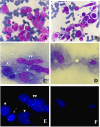
Synchronous clonally related anaplastic large cell lymphoma and malignant histiocytosis
- PMID: 39810204
- PMCID: PMC11730790
- DOI: 10.1186/s13000-025-01597-3
Synchronous clonally related anaplastic large cell lymphoma and malignant histiocytosis
Abstract
Background: Synchronous malignant histiocytoses are rare conditions that occur concurrently with another hematologic neoplasm. Most reported cases are associated with B-cell lymphoproliferative disorders, while associations with T-cell hemopathies are less common. These two diseases may share mutations and/or cytogenetic anomalies, which can lead to malignant proliferations. In such cases, the term "secondary malignant histiocytosis" can be applied.
Case description: A 26-year-old patient was diagnosed with anaplastic lymphoma kinase negative anaplastic large cell lymphoma [ALK-ALCL] associated with synchronous malignant histiocytosis. Neoplastic cells were distinguished by the exclusivity of the rearrangement of TCR genes within the lymphoma cells, whereas mutations in the KRAS and TP53 genes affected mono-histiocytic cells. However, these two cells populations shared common chromosomal abnormalities. First line treatment protocol included Brentuximab vedotin, cyclophosphamide, doxorubicin, and methylprednisolone. Despite a partial clinical and biological response after cycle 1 of treatment, the patient was refractory at the end of cycle 2. Patient died in the intensive care unit from a multiple-organ failure related to lymphohistiocytic hemophagocytosis.
Conclusion: This case represents the first documented instance of synchronous malignant histiocytosis associated with anaplastic large cell lymphoma. Notably, the uniqueness of this case lies in the absence of TCR rearrangement in the histiocytic cells, despite the presence of shared chromosomal abnormalities with the lymphomatous cells indicating a common origin for both neoplastic proliferations. Considering the rarity of such occurrences, the use of histiocytosis targeted therapy alongside conventional lymphoma treatment warrants consideration in such a context.
Keywords: Anaplastic large cell lymphoma; Case report; Secondary malignant histiocytosis; Synchronous malignant histiocytosis.
© 2025. The Author(s).
Conflict of interest statement
Declarations. Ethics approval and consent to participate: N/A. Consent for publication: available. Competing interests: The authors declare no competing interests.
Figures


Similar articles
-
Complete response in a critically ill patient with ALK-negative anaplastic large cell lymphoma treated with single agent brentuximab-vedotin.Expert Rev Anticancer Ther. 2016;16(3):279-83. doi: 10.1586/14737140.2016.1146597. Epub 2016 Feb 18. Expert Rev Anticancer Ther. 2016. PMID: 26809026
-
Therapeutic efficacy of the bromodomain inhibitor OTX015/MK-8628 in ALK-positive anaplastic large cell lymphoma: an alternative modality to overcome resistant phenotypes.Oncotarget. 2016 Nov 29;7(48):79637-79653. doi: 10.18632/oncotarget.12876. Oncotarget. 2016. PMID: 27793034 Free PMC article.
-
Brentuximab vedotin use in pediatric anaplastic large cell lymphoma.Front Immunol. 2023 May 18;14:1203471. doi: 10.3389/fimmu.2023.1203471. eCollection 2023. Front Immunol. 2023. PMID: 37275877 Free PMC article. Review.
-
Anaplastic lymphoma kinase-positive anaplastic large cell lymphoma: current and future perspectives in adult and paediatric disease.Eur J Haematol. 2014 Dec;93(6):455-68. doi: 10.1111/ejh.12360. Epub 2014 May 21. Eur J Haematol. 2014. PMID: 24766435 Review.
-
Anaplastic large cell lymphoma: pathology, genetics, and clinical aspects.J Clin Exp Hematop. 2017;57(3):120-142. doi: 10.3960/jslrt.17023. J Clin Exp Hematop. 2017. PMID: 29279550 Free PMC article. Review.
Cited by
-
Molecular Insights into the Diagnosis of Anaplastic Large Cell Lymphoma: Beyond Morphology and Immunophenotype.Int J Mol Sci. 2025 Jun 19;26(12):5871. doi: 10.3390/ijms26125871. Int J Mol Sci. 2025. PMID: 40565334 Free PMC article. Review.
References
-
- Péricart S, Waysse C, Siegfried A, et al. Subsequent development of histiocytic sarcoma and follicular lymphoma: cytogenetics and next-generation sequencing analyses provide evidence for transdifferentiation of early common lymphoid precursor—a case report and review of literature. Virchows Arch. 2020;4764:609–14. 10.1007/s00428-019-02691-w. - PubMed
-
- Congyang L, Xinggui W, Hao L, Weihua H. Synchronous histiocytic sarcoma and diffuse large B cell lymphoma involving the stomach: a case report and review of the literature. Int J Hematol. 2011;932:247–52. 10.1007/s12185-011-0773-3. - PubMed
-
- Wang E, Papalas J, Hutchinson CB, et al. Sequential development of Histiocytic Sarcoma and diffuse large B-cell lymphoma in a patient with a remote history of follicular lymphoma with genotypic evidence of a clonal relationship: a divergent [Bilineal] Neoplastic Transformation of an indolent B-cell lymphoma in a single individual. Am J Surg Pathol. 2011;353:457–63. 10.1097/PAS.0b013e3182098799. - PubMed
Publication types
MeSH terms
LinkOut - more resources
Full Text Sources
Medical
Research Materials
Miscellaneous

