. 2025 Jan 14;19(1):989-998.
doi: 10.1021/acsnano.4c12648. Epub 2024 Dec 30.

Force Nanoscopy Demonstrates Stress-Activated Adhesion between Staphylococcus aureus Iron-Regulated Surface Determinant Protein B and Host Toll-like Receptor 4

Affiliations

Force Nanoscopy Demonstrates Stress-Activated Adhesion between Staphylococcus aureus Iron-Regulated Surface Determinant Protein B and Host Toll-like Receptor 4

Telmo O Paiva et al. ACS Nano. .

Abstract

The Staphylococcus aureus iron-regulated surface determinant protein B (IsdB) has recently been shown to bind to toll-like receptor 4 (TLR4), thereby inducing a strong inflammatory response in innate immune cells. Currently, two unsolved questions are (i) What is the molecular mechanism of the IsdB-TLR4 interaction? and (ii) Does it also play a role in nonimmune systems? Here, we use single-molecule experiments to demonstrate that IsdB binds TLR4 with both weak and extremely strong forces and that the mechanostability of the molecular complex is dramatically increased by physical stress, sustaining forces up to 2000 pN, at a loading rate of 105 pN/s. We also show that TLR4 binding by IsdB mediates time-dependent bacterial adhesion to endothelial cells, pointing to the role of this bond in cell invasion. Our findings point to a function for IsdB in pathogen-host interactions, that is, mediating strong bacterial adhesion to host endothelial cells under fluid shear stress, unknown until now. In nanomedicine, this stress-dependent adhesion represents a potential target for innovative therapeutics against S. aureus-resistant strains.

Keywords: IsdB; Staphylococcus aureus; TLR4; bacterial adhesion; endothelial cells; single-cell force spectroscopy; single-molecule force spectroscopy.


Conflict of interest statement

The authors declare no competing financial interest.

Figures

Figure 1
Figure 1
IsdB mediates bacterial adhesion to TLR4-coated substrates. (a) Schematic representation of the modular structure of S. aureus IsdB. Signal sequence (S) and near iron transporter domains (NEAT1 and 2) with respective positions are represented. (b) Optical microscopy images of TLR4-coated surfaces incubated with S. aureus Δspa cells grown in RPMI and BHI (inset) media. Bacteria grown in BHI do not adhere to the surfaces, showing the role of IsdB in mediating bacterial adhesion to TLR4. For RPMI cells, images of 2 independent surfaces are shown and the scale bars are 50 μm. (c–f) Adhesion of S. aureus Δspa cells to TLR4-coated surfaces assessed by single-cell force spectroscopy (SCFS). Rupture force and rupture length histograms of (c) 2 representative cells grown in RPMI medium (n = 175 and 225 adhesive curves for cell #1 and cell #2, respectively) and (d) 1 representative cell grown in BHI medium (n = 21 adhesive curves), acquired by recording force–distance curves in PBS between single bacteria and TLR4 substrates, at a retraction speed of 1 μm/s. Schemes of the SCFS setups and representative retraction force profiles are shown as insets. (e) Box plot comparing the binding frequency of bacteria grown in RPMI and BHI culture media (n = 6 and 7 cells, respectively). (f) Box plot showing the rupture force obtained for bacteria grown in RPMI and BHI media (n = 6 and 5 cells, respectively). In both (e) and (f), means are represented by stars, medians by lines, boxes indicate the 25–75% quartiles, and whiskers the standard deviation. P-values were determined by a two-sample t-test.
Figure 2
Figure 2
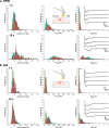
Strength of single IsdB-TLR4 interactions assessed by single-molecule force spectroscopy (SMFS). Rupture force and rupture length histograms acquired by recording force–distance curves in PBS between TLR4-functionalized AFM tips and (a) 2 representative Δspa cells grown in RPMI medium (n = 289 and 259 adhesive curves, for cell #1 and cell #2, respectively) and (b) 1 representative Δspa cell grown in BHI medium (n = 59 adhesive curves), at a retraction speed of 1 μm/s. Schemes of the SMFS setups and representative retraction force profiles are shown as insets. (c) Box plots comparing the binding frequency of bacteria grown in RPMI and BHI culture media (n = 8 and 10 cells, respectively). (d) Box plot showing the rupture force obtained for bacteria grown in RPMI and BHI media (n = 10 cells, for both cases). Means are represented by stars, medians by lines, boxes indicate the 25–75% quartiles, and whiskers the standard deviation. P-values were determined by a two-sample t-test.
Figure 3
Figure 3
IsdB-TLR4 strength confirmed by anti-TLR4 blocking and S. aureus strain lacking IsdB. (a) Rupture force histograms of 2 representative Δspa cells, cultured in RPMI medium, obtained by recording force–distance curves in PBS at a retraction speed of 1 μm/s between TLR4-functionalized AFM tips before (n = 188 and 276 adhesive curves, for cell #1 and cell #2, respectively) and after blocking with 100 μg/mL of anti-TLR4 monoclonal antibody (n = 98 and 65 adhesive curves, for cell #1 and cell #2, respectively). (b) Rupture force and rupture length histograms of a representative S. aureus cell expressing IsdB (WT, n = 200 adhesive curves) cultured in RPMI. (c) Force data of a representative S. aureus cell lacking IsdB (ΔisdB, n = 95 adhesive curves) cultured in RPMI. Schemes of the SMFS setups and representative retraction force profiles are shown as insets. Box plots comparing (d) binding frequency and (e) rupture forces obtained for Δspa cells before and after blocking with anti-TLR4 monoclonal antibody, after tip treatment with monoclonal mouse IgG as a negative control, WT and ΔisdB strains (n = 8, 8, 10, 13, and 9 cells, respectively). Means are represented by stars, medians by lines, boxes indicate the 25–75% quartiles, and whiskers the standard deviation. P-values were determined using Kruskal–Wallis test followed by post hoc Dunn’s test.
Figure 4
Figure 4
Physical stress strengthens the IsdB-TLR4 interaction. (a) Dynamic force spectroscopy plot of the IsdB-TLR4 interaction acquired on 6 Δspa cells cultured in RPMI growth medium (N = 11700). Shown are Bell-Evans fit of the single-bond data (red line) and Williams-Evans predictions for multiple simultaneous uncorrelated interactions (green dashed lines) until quadruple bonds. The blue background region includes the set of data points that are beyond the multiple bonds prediction. Bell–Evans model yielded koff = 6.52 ± 1.44 s–1 and xβ = 0.14 ± 0.01 nm, the complex off-rate constant and distance along the reaction coordinate of the transition between bound and unbound states, respectively. Solid circles represent the mean values, and error bars the standard deviations. (b) Rupture force histograms as a function of discrete loading rate ranges: LR1 < 2.7 × 10³, 2.7 × 10³ < LR2 < 6.3 × 10³, 6.3 × 10³ < LR3 < 1.5 × 10⁴, 1.5 × 10⁴ < LR4 < 3.4 × 10⁴, 3.4 × 10⁴ < LR5 < 8.0 × 10⁴, 8.0 × 10⁴ < LR6 < 1.9 × 10⁵, 1.9 × 10⁵ < LR7 < 4.3 × 10⁵ pN/s. A major shift toward high forces with increasing LRs is observed. (c) Plot of binding frequency as a function of contact time obtained for Δspa cells (n = 7 cells) cultured in RPMI medium, and associated pseudo-first-order kinetics fit (red line), yelding kon = (0.8 ± 0.2) × 104 M1·s–1, the kinetic on-rate constant of the IsdB-TLR4 complex. Means and standard deviations are indicated by solid circles and error bars, respectively.
Figure 5
Figure 5
IsdB mediates bacterial adhesion to human endothelial cells via TLR4. (a, b) Adhesion of S. aureus Δspa cells to human umbilical vein endothelial cells (HUVEC) expressing TLR4, assessed by single-cell force spectroscopy (SCFS). Rupture force, maximum adhesion force (Fmax), and work of adhesion (Wadh) histograms of three different cell pairs acquired by recording force–distance curves in HEPES, at a retraction speed of 20 μm/s, after 1 and 10 s of interaction between single bacteria grown in (a) RPMI and (b) BHI media and TLR4-expressing endothelial cells. 25 mM glucose was added to EGM-2 endothelial cell growth medium to induce TLR4 expression by HUVEC. Schemes of the SCFS setups and representative retraction force profiles are shown as insets. Data from a total of 256 curves for each cell pair.

Similar articles

References

    1. Lowy F. D. Staphylococcus Aureus Infections. N. Engl. J. Med. 1998, 339 (8), 520–532. 10.1056/NEJM199808203390806. - DOI - PubMed
    1. Tong S. Y. C.; Davis J. S.; Eichenberger E.; Holland T. L.; Fowler V. G. S. aureus Infections: Epidemiology, Pathophysiology, Clinical Manifestations, and Management. Clin. Microbiol. Rev. 2015, 28 (3), 603–661. 10.1128/CMR.00134-14. - DOI - PMC - PubMed
    1. de Jong N. W. M.; van Kessel K. P. M.; van Strijp J. A. G. Immune Evasion by S. aureus. Microbiol. Spectrum 2019, 7 (2), 006110.1128/microbiolspec.GPP3-0061-2019. - DOI - PMC - PubMed
    1. Tam K.; Torres V. J.. S. aureus Secreted Toxins and Extracellular Enzymes Microbiol. Spectrum 2019; Vol. 7 (2), 10.1128/microbiolspec.GPP3-0039-2018. - DOI - PMC - PubMed
    1. Foster T. J.; Geoghegan J. A.; Ganesh V. K.; Höök M. Adhesion, invasion and evasion: the many functions of the surface proteins of S. aureus. Nat. Rev. Microbiol. 2014, 12 (1), 49–62. 10.1038/nrmicro3161. - DOI - PMC - PubMed

Publication types

Substances

LinkOut - more resources