Review
. 2025 Jan 6;16(5):2105-2135.
doi: 10.1039/d4sc06762b. eCollection 2025 Jan 29.

Multi-target-directed therapeutic strategies for Alzheimer's disease: controlling amyloid-β aggregation, metal ion homeostasis, and enzyme inhibition

Affiliations
Review

Multi-target-directed therapeutic strategies for Alzheimer's disease: controlling amyloid-β aggregation, metal ion homeostasis, and enzyme inhibition

Jeasang Yoo et al. Chem Sci. .

Abstract

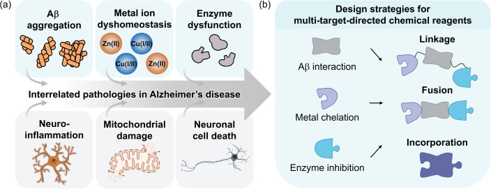
Alzheimer's disease (AD) is the most prevalent neurodegenerative dementia, marked by progressive cognitive decline and memory impairment. Despite advances in therapeutic research, single-target-directed treatments often fall short in addressing the complex, multifactorial nature of AD. This arises from various pathological features, including amyloid-β (Aβ) aggregate deposition, metal ion dysregulation, oxidative stress, impaired neurotransmission, neuroinflammation, mitochondrial dysfunction, and neuronal cell death. This review illustrates their interrelationships, with a particular emphasis on the interplay among Aβ, metal ions, and AD-related enzymes, such as β-site amyloid precursor protein cleaving enzyme 1 (BACE1), matrix metalloproteinase 9 (MMP9), lysyl oxidase-like 2 (LOXL2), acetylcholinesterase (AChE), and monoamine oxidase B (MAOB). We further underscore the potential of therapeutic strategies that simultaneously inhibit Aβ aggregation and address other pathogenic mechanisms. These approaches offer a more comprehensive and effective method for combating AD, overcoming the limitations of conventional therapies.


Conflict of interest statement

There are no conflicts to declare.

Figures

Fig. 1
Fig. 1. Pathological features in AD and design strategies for multi-target-directed chemical reagents. (a) Multifaceted pathology associated with AD. (b) Schematic illustration of design strategies (linkage, fusion, and incorporation) to develop small molecules against Aβ aggregation, metal ion dyshomeostasis, and enzyme dysfunction simultaneously.
Fig. 2
Fig. 2. Recent drug development against Aβ species or BACE1. (a) Illustration of the proteolytic cleavage of APP. Non-amyloidogenic and amyloidogenic pathways occur depending on the combinational action of α-, β-, and γ-secretases. (b) Amino acid sequences of Aβ40 and Aβ42, with side views of 3D reconstructed Aβ fibrils, and top views of their cross-sections from cryo-EM (for Aβ40, PDB 6SHS; for Aβ42, PDB 5OQV). Amino acid residues responsible for the self-recognition sites are highlighted in bold and underlined, while those forming salt bridges in fibrillar structures are indicated in red and depicted as sticks. Core structures of Aβ fibrils are also shown in stick representation. Reproduced with permission from ref. and . Copyright© 2019 Springer Nature and 2017 The American Association for the Advancement of Science. (c) Schematic description of Aβ aggregation and Aβ-lowering monoclonal antibodies. (d) Relative safety and efficacy of Aβ-lowering monoclonal antibodies in patients with sporadic early-symptomatic AD. Monoclonal antibodies approved by the U.S. FDA for AD are shown in red dots, while other monoclonal antibodies are presented in black. Reproduced with permission from ref. . Copyright© 2023 Springer Nature. (e) Structure of BACE1 (PDB 1W50 ), zoom-in view of the target site, and BACE1 inhibitors that have reached phase III clinical trials. The catalytic dyad is presented in red and depicted as sticks. (f) Structure of γ-secretase (PDB 5A63 ), illustration of the binding pocket, and γ-secretase inhibitors that have progressed to clinical trials. The catalytic dyad is indicated in red with sticks.
Fig. 3
Fig. 3. Metal-related pathological factors associated with AD and therapeutic interventions against these components. (a) Examples of metal chelators. (b) Representative coordination modes of Cu(i/ii), Zn(ii), and Fe(ii) to Aβ with a chemical structure of metal-bound L2-b. L2-b, N1,N1-dimethyl-N4-(pyridin-2-ylmethyl)benzene-1,4-diamine. (c) Structures of MMP9 (PDB 1L6J) and its inhibitor (prinomastat). The Zn(ii)-coordination residues are highlighted in red and depicted as sticks. (d) Structures of LOXL2 (PDB 5ZE3 ) and its inhibitor (PXS-5505). The Cu(ii)-coordination residues and catalytic lysine are indicated in red with sticks. Note that Cu(ii) occupies the Zn(ii)-binding site in human LOXL2, contrary to the crystallographic illustration.
Fig. 4
Fig. 4. AChE, MAOB, and examples of their inhibitors. (a) Structure of AChE (PDB 4EY4 ), binding mode of donepezil at its active site (PDB 7E3H), and three U.S. FDA-approved AChE inhibitors (donepezil, rivastigmine, and galantamine). The amino acid residues involved in donepezil binding are highlighted in red and depicted as sticks. (b) Structure of MAOB (PDB 1GOS), interaction with selegiline at its catalytic domain (PDB 2BYB), and three representative inhibitors (lazabemide, selegiline, and safinamide). The amino acid residues involved in selegiline binding are indicated in red with sticks.
Fig. 5
Fig. 5. Examples of chemical reagents controlling Aβ and BACE1. (a) Small peptide-based compounds (GPA-1 and GPA-2) and their effects on the cognition of mice models after inducing impaired memory with scopolamine on days 12 to 14. GPA-1, NH2-Gly-Pro-Ala-OMe; GPA-2, Cbz-Gly-Pro-Ala-O-cinnamyl. Reproduced with permission from ref. . Copyright© 2024 American Chemical Society. (b) A flavonoid-based natural product (YCC31) and its inhibitory impact on the activity of BACE1 and the aggregation of Aβ42. The interactions between YCC31 and BACE1 at the active site, the morphology of the resultant Aβ42 species, and its inhibitory effect on Aβ42 aggregation were analyzed by MD simulations, TEM, and the ThT assay, respectively. YCC31, quercetin-3-O-[β-d-glucopyranosyl-(1→3)-O-α-l-rhamnopyranosyl-(1→6)-O-β-d-glucopyranoside]. Reproduced with permission from ref. . Copyright© 2024 Elsevier. (c) A phenothiazine-derived compound (19c) and its effect against the activity of BACE1 and the aggregation of Aβ40. The interactions between 19c and BACE1 at the active site were obtained from docking studies and its inhibitory effect on the formation of β-sheet-rich Aβ40 aggregates was quantitatively measured by the ThT assay. 19c, 4-amino-N-[(4-chlorobenzyl)]-2-[methyl({2-[2-(10H-phenothiazin-10-yl)ethoxy]ethyl})amino]butanamide. Reproduced with permission from ref. . Copyright© 2023 Elsevier.
Fig. 6
Fig. 6. Examples of chemical reagents regulating Aβ peptides and metal ions. (a) Structure of the d-enantiomeric decapeptide RTHLVFFARK-NH2 (rk10) and inhibitory effects of rk10 and its l-enantiomer (RK10) on the generation of ROS and metal-free and Cu(ii)–Aβ42 aggregation. The ROS level produced by Cu(ii)–Aβ42 in the absence and presence of rk10 or RK10 in SH-SY5Y cell was measured by a fluorescence assay with 2′,7′-dichlorofluorescin diacetate (DCFH-DA). The statistical significance level is expressed by the pound (control as Aβ42 + Cu(ii), ###P < 0.001) and asterisk (control as Aβ42 + Cu(ii) + RK10, *P < 0.05). The morphology of Aβ42 aggregates generated with and without Cu(ii) and rk10 or RK10 was analyzed by AFM. Reproduced with permission from ref. . Copyright© 2019 American Chemical Society. (b) Structure of PPD and its impact on the aggregation of both metal-free and metal-added Aβ40. The size distribution of Aβ40 aggregates produced with and without PPD or metal ions was investigated by gel/western blot. The formation of the covalent adduct between Aβ40 and benzoquinone was detected by ESI–MS. PPD, p-phenylenediamine. Reproduced with permission from ref. . Copyright© 2020 American Chemical Society. (c) Structure of L1 and its influence on Cu(ii)-added Aβ40 aggregation. The size distribution and morphology of the resultant Aβ40 aggregates produced with and without L1 or Cu(ii) were investigated by gel/western blot and TEM, respectively. Chemical modifications onto the Cu(ii)-coordination sphere of Aβ40 by L1 were monitored by ESI–MS. L1, N1,N1-dimethyl-N4-(thiophen-2-ylmethyl)benzene-1,4-diamine. Reproduced with permission from ref. . Copyright© 2020 United States National Academy of Sciences.
Fig. 7
Fig. 7. Examples of chemical reagents controlling Aβ and MMP9 or LOXL2. (a) Structure of doxycycline [DOX; (4S,4aR,5S,5aR,6R,12aS)-4-(dimethylamino)-3,5,10,12,12a-pentahydroxy-6-methyl-1,11-dioxo-1,4,4a,5,5a,6,11,12a-octahydrotetracene-2-carboxamide]. (b) Intermolecular interactions between MMP9 (PDB 1L6J) and DOX. The Zn(ii)-coordination and DOX-interacting residues are indicated as sticks, and highlighted in red and green, respectively. (c) Interactions of DOX with Aβ42 fibrils (PDB 2MXU). The morphology of the resultant Aβ42 species was analyzed with TEM. Reproduced with permission from ref. and . Copyright© 2019 Multidisciplinary Digital Publishing Institute and 2001 John Wiley and Sons. (d) Structure of curcumin (CUR). CUR, (1E,6E)-1,7-bis(4-hydroxy-3-methoxyphenyl)hepta-1,6-diene-3,5-dione. (e) Intermolecular interactions between LOXL2 (PDB 5ZE3 ) and CUR. The Cu(ii)-coordination and CUR-interacting residues are shown as sticks, and depicted in red and green, respectively. (f) Interactions of CUR with Aβ42 fibrils (PDB 2MXU). The formation of the smaller Aβ42 species was detected by TEM. Reproduced with permission from ref. and . Copyright© 2019 Multidisciplinary Digital Publishing Institute and 2017 Elsevier.
Fig. 8
Fig. 8. Examples of chemical reagents regulating metal-free or metal-bound Aβ and AChE. (a) Structures of flavonoids. Isoflavone, 3-phenyl-4H-chromen-4-one; isoflavone-1, 3-(3,4-dihydroxyphenyl)-5,7-dihydroxy-4H-chromen-4-one; isoflavone-2, 5,6,7-trihydroxy-3-phenyl-4H-chromen-4-one; isoflavone-3, 3-(3,4-dihydroxyphenyl)-5,6,7-trihydroxy-4H-chromen-4-one; flavone, 2-phenyl-4H-chromen-4-one; flavone-1, 2-(3,4-dihydroxyphenyl)-3,5,7-trihydroxy-4H-chromen-4-one; flavone-2, 2-(3,4-dihydroxyphenyl)-5,7-dihydroxy-4H-chromen-4-one; flavone-3, 2-(4-hydroxyphenyl)-3,5,7-trihydroxy-4H-chromen-4-one. (b) Intermolecular interactions between the flavonoids and AChE. Reproduced with permission from ref. and . Copyright© 2023 Royal Society of Chemistry and 2020 Royal Society of Chemistry. (c) Impact of isoflavone-3 on the formation of metal-free Aβ42 and metal–Aβ42 aggregates. The morphology of the resultant Aβ42 species was analyzed by TEM. Reproduced with permission from ref. . Copyright© 2023 Royal Society of Chemistry. (d) Structures of emodin and its derivatives with different functional groups at the R position. Emodin, 1,3,8-trihydroxy-6-methylanthracene-9,10-dione; emodin-1, 1,8-dihydroxy-3-methyl-6-(2-(piperidin-1-yl)ethoxy)anthracene-9,10-dione; emodin-2, 1,8-dihydroxy-3-methyl-6-(2-(pyrrolidin-1-yl)ethoxy)anthracene-9,10-dione; emodin-3, 1,8-dihydroxy-3-methyl-6-(2-(dimethylamino)ethoxy)anthracene-9,10-dione; emodin-4, 1,8-dihydroxy-3-methyl-6-(2-morpholinoethoxy)anthracene-9,10-dione. (e) Binding of emodin-1 against AChE. Reproduced with permission from ref. . Copyright© 2024 Elsevier. (f) Effect of emodin on Aβ42 aggregation. The inhibitory activity of emodin on the production of β-sheet-rich Aβ42 aggregates was quantitatively measured by the ThT assay. Reproduced with permission from ref. . Copyright© 2021 John Wiley and Sons. (g) Impact of emodin-1 and emodin-2 on Cu(ii)-induced Aβ42 aggregation. The influence of emodin-1 and emodin-2 on the generation of β-sheet-rich Cu(ii)–Aβ42 aggregates was analyzed by the ThT assay. Reproduced with permission from ref. . Copyright© 2023 Springer Nature.
Fig. 9
Fig. 9. Examples of chemical reagents controlling Aβ and MAOB. (a) Structures of rasagiline, clorgyline, and RC-6j. The inhibitory effect of RC-6j against the activity of MAOB was represented in Lineweaver–Burk reciprocal plots, with its impact on the formation Aβ42 aggregates. The catalytic rate of MAOB was measured through the amount of H2O2 generated by catalytic oxidation of p-tyramine, observed by the Amplex Red H2O2/peroxidase assay. The morphology of the resultant Aβ42 species was monitored by TEM. RC-6j, 7-((5-(methyl(prop-2-yn-1-yl)amino)pentyl)oxy)chroman-4-one. Reproduced with permission from ref. . Copyright© 2020 Elsevier. (b) Structures of Schiff base derivatives and 3b, binding mode of 3b towards MAOB, and its effects on Cu(ii)-added Aβ42 aggregation. The morphology of the Cu(ii)-induced Aβ42 aggregates produced with and without 3b was investigated by TEM. 3b, 2,2′-((1E,1′E)-hydrazine-1,2-diylidenebis(methanylylidene))diphenol. Reproduced with permission from ref. . Copyright© 2020 Springer Nature.
Fig. 10
Fig. 10. Examples of chemical reagents regulating Aβ, AChE, and MAOB. (a) Structures of chromone and its derivatives, binding of chromone-1 to MAOB, and its impact on AChE activity and Aβ42 aggregation. The inhibitory effect of chromone-1 against the activity of AChE was performed using Ellman's method and represented in a double reciprocal Lineweaver–Burk plot. The morphology of Aβ42 aggregates generated with or without chromone-1 was obtained by FE-SEM. Reproduced with permission from ref. . Copyright© 2024 American Chemical Society. Chromone-1, 2-(4-(2-(pyrrolidin-1-yl)ethoxy)phenyl)-4H-chromen-4-one; chromone-2, 2-(3-methoxy-4-(2-(pyrrolidin-1-yl)ethoxy)phenyl)-4H-chromen-4-one; chromone-3, 2-(3-(2-morpholinoethoxy)phenyl)-4H-chromen-4-one. (b) Structure of BV-14, its binding mode towards MAOB, and effects on the Aβ42 aggregation and AChE activity. The extent of the inhibition of Aβ42 aggregation was assessed using the ThT assay. The AChE inhibition by BV-14 was measured using Ellman's method and shown in an overlaid Lineweaver–Burk reciprocal plot. BV-14, (Z)-4-(1-phenylprop-1-en-2-yl)-6-(4-(prop-2-yn-1-yloxy)phenyl)pyrimidine. Reproduced with permission from ref. . Copyright© 2024 Royal Society of Chemistry. (c) Structure of CF-4, its binding to MAOB (PDB 1GOS), and impact on the production of Aβ42 aggregates. The morphology of the resultant Aβ42 species was analyzed by TEM. CF-4, (E)-4-(3-(3,4-dihydroisoquinolin-2(1H)-yl)-3-oxoprop-1-en-1-yl)-2-methoxyphenyl ethyl(methyl)carbamate. Reproduced with permission from ref. . Copyright© 2020 Elsevier.

Similar articles

Cited by

References

    1. Zhang Y. Chen H. Li R. Sterling K. Song W. Amyloid β-based therapy for Alzheimer's disease: Challenges, successes and future. Signal Transduct. Targeted Ther. 2023;8:1–26. doi: 10.1038/s41392-022-01259-6. - DOI - PMC - PubMed
    1. Nguyen P. H. Ramamoorthy A. Sahoo B. R. Zheng J. Faller P. Straub J. E. Dominguez L. Shea J.-E. Dokholyan N. V. De Simone A. Ma B. Nussinov R. Najafi S. Ngo S. T. Loquet A. Chiricotto M. Ganguly P. McCarty J. Li M. S. Hall C. Wang Y. Miller Y. Melchionna S. Habenstein B. Timr S. Chen J. Hnath B. Strodel B. Kayed R. Lesné S. Wei G. Sterpone F. Doig A. J. Derreumaux P. Amyloid oligomers: A joint experimental/computational perspective on Alzheimer's disease, Parkinson's disease, Type II diabetes, and amyotrophic lateral sclerosis. Chem. Rev. 2021;121:2545–2647. doi: 10.1021/acs.chemrev.0c01122. - DOI - PMC - PubMed
    1. Benek O. Korabecny J. Soukup O. A perspective on multitarget drugs for Alzheimer's disease. Trends Pharmacol. Sci. 2020;41:434–445. doi: 10.1016/j.tips.2020.04.008. - DOI - PubMed
    1. Leng F. Edison P. Neuroinflammation and microglial activation in Alzheimer disease: Where do we go from here? Nat. Rev. Neurol. 2021;17:157–172. doi: 10.1038/s41582-020-00435-y. - DOI - PubMed
    1. Wang W. Zhao F. Ma X. Perry G. Zhu X. Mitochondria dysfunction in the pathogenesis of Alzheimer's disease: Recent advances. Mol. Neurodegener. 2020;15:30. doi: 10.1186/s13024-020-00376-6. - DOI - PMC - PubMed

LinkOut - more resources